Review
. 2014:773:435-70.
doi: 10.1007/978-1-4899-8032-8_20.

Nuclear mechanics in cancer

Affiliations
Review

Nuclear mechanics in cancer

Celine Denais et al. Adv Exp Med Biol. 2014.

Abstract

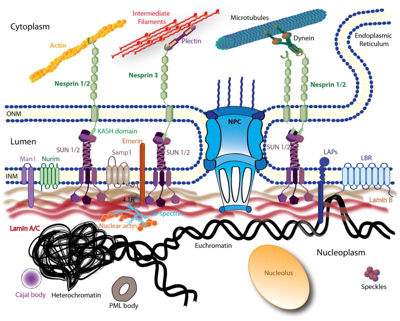
Despite decades of research, cancer metastasis remains an incompletely understood process that is as complex as it is devastating. In recent years, there has been an increasing push to investigate the biomechanical aspects of tumorigenesis, complementing the research on genetic and biochemical changes. In contrast to the high genetic variability encountered in cancer cells, almost all metastatic cells are subject to the same physical constraints as they leave the primary tumor, invade surrounding tissues, transit through the circulatory system, and finally infiltrate new tissues. Advances in live cell imaging and other biophysical techniques, including measurements of subcellular mechanics, have yielded stunning new insights into the physics of cancer cells. While much of this research has been focused on the mechanics of the cytoskeleton and the cellular microenvironment, it is now emerging that the mechanical properties of the cell nucleus and its connection to the cytoskeleton may play a major role in cancer metastasis, as deformation of the large and stiff nucleus presents a substantial obstacle during the passage through the dense interstitial space and narrow capillaries. Here, we present an overview of the molecular components that govern the mechanical properties of the nucleus, and we discuss how changes in nuclear structure and composition observed in many cancers can modulate nuclear mechanics and promote metastatic processes. Improved insights into this interplay between nuclear mechanics and metastatic progression may have powerful implications in cancer diagnostics and therapy and may reveal novel therapeutic targets for pharmacological inhibition of cancer cell invasion.


Figures

Figure 1
Figure 1. Schematic overview of the nuclear structure and the LINC complex
The nuclear envelope is composed of the inner nuclear membrane (INM) and the outer nuclear membrane (ONM) punctuated by nuclear pore complexes (NPC). The ONM is continuous with the endoplasmic reticulum (ER). Several structures of the nuclear interior are depicted here, including the nucleolus, Cajal bodies, promyelocytic leukemia bodies (PML) and speckles. Chromatin is shown in its two states, very condensed (heterochromatin) and loosely organized (euchromatin). Only a subset of nuclear membrane proteins are portrayed in this picture: lamin B receptor (LBR), emerin, MAN1, and nurim. The schematic also illustrates some of the interactions between these proteins with the lamina meshwork (lamins B and A/C). The LINC complex is represented by nesprins, Sad1p/UNC-84 (SUN) proteins and Samp1. On the outer membrane, nesprin-1 and -2 can directly bind to actin filaments or indirectly interact with microtubules through motors proteins (dyneins or kinesin). Nesprin-3 is shown interacting with intermediate filaments via plectin.
Figure 2
Figure 2. Invasive cancer cell MDA-MB-231 squeezing into 8 μm width constriction
Image sequences of a cancer cell being perfused through 8 μm-wide constriction at a pressure (ΔP) of 10 psi. The viscoelastic deformation as the nucleus flows through the constriction is clearly visible.
Figure 3
Figure 3. Migration of cancer cell in a constrained environment
(A) Fibrosarcoma cell (HT1080 cell line) migrating through a dense collagen fiber matrix. The rat tail collagen matrix was imaged by reflection microscopy; the nucleus is visible in red (DAPI), F-actin in green (phalloidin). The cell body has already advanced in the direction of migration (yellow arrow), while the nucleus is still in the process of squeezing through constrictions in the collagen matrix (red arrow head). Image courtesy of Katarina Wolf, University of Nijmegen. (B) Fibrosarcoma cells (HT1080) migrating through 2 μm × 5 μm and 5 μm × 5 μm constrictions in a microfluidic channel. The cytoplasm is visible in green, the nucleus in blue, and the nuclear lamina (lamin B2) in red. (C) Time-lapse series of MDA-MB-231 breast cancer cell expressing a green fluorescent protein migrating through a 5 μm-wide constriction in a microfluidic channel. The nucleus is outlined in red (dashed line).

Similar articles

Cited by

References

    1. Fawcett DW. An Atlas of fine structure (The Cell) 2. W. B. Saunders Company; 1966.
    1. Hanahan D, Weinberg RA. Hallmarks of cancer: the next generation. Cell. 2011;144(5):646–674. doi: 10.1016/j.cell.2011.02.013. - DOI - PubMed
    1. Suresh S. Biomechanics and biophysics of cancer cells. Acta Biomater. 2007;3(4):413–438. doi: 10.1016/j.actbio.2007.04.002. - DOI - PMC - PubMed
    1. Guck J, Schinkinger S, Lincoln B, Wottawah F, Ebert S, Romeyke M, Lenz D, Erickson HM, Ananthakrishnan R, Mitchell D, Käs J, Ulvick S, Bilby C. Optical deformability as an inherent cell marker for testing malignant transformation and metastatic competence. Biophys J. 2005;88(5):3689–3698. doi: 10.1529/biophysj.104.045476. - DOI - PMC - PubMed
    1. Remmerbach TW, Wottawah F, Dietrich J, Lincoln B, Wittekind C, Guck J. Oral cancer diagnosis by mechanical phenotyping. Cancer Res. 2009;69(5):1728–1732. doi: 10.1158/0008-5472.can-08-4073. - DOI - PubMed

Publication types