

Oxidation of endogenous N-arachidonoylserotonin by human cytochrome P450 2U1
- PMID: 24563460
- PMCID: PMC4036169
- DOI: 10.1074/jbc.M114.550004
Oxidation of endogenous N-arachidonoylserotonin by human cytochrome P450 2U1
Abstract
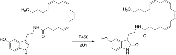
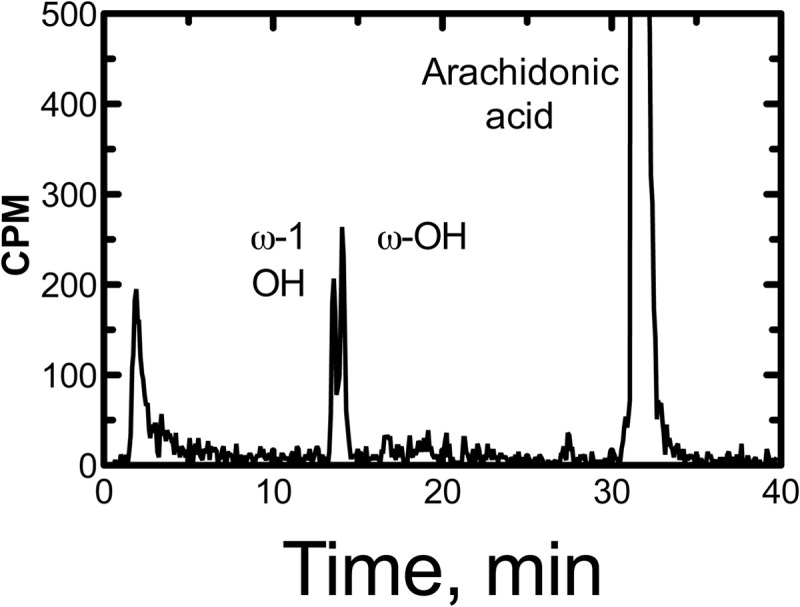
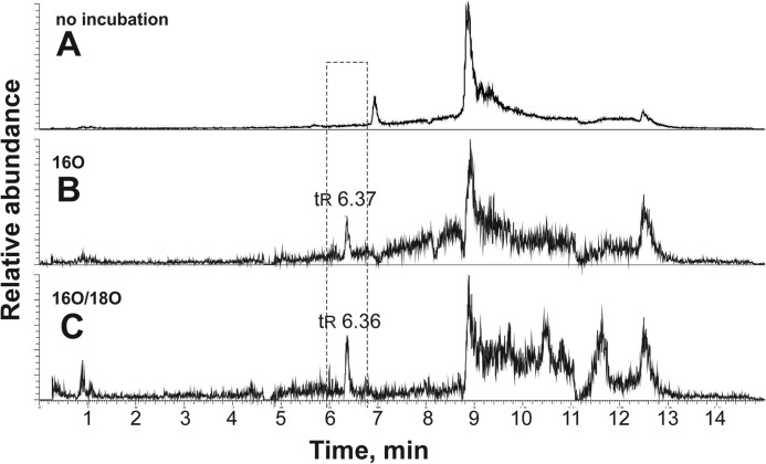
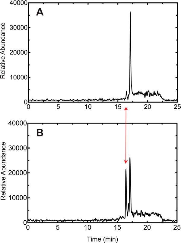
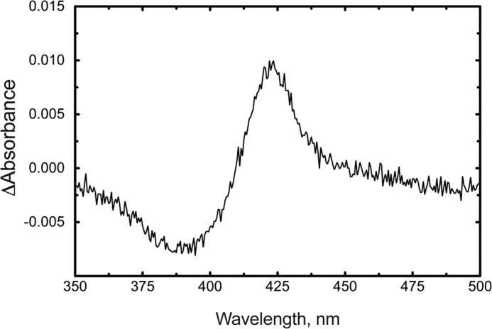
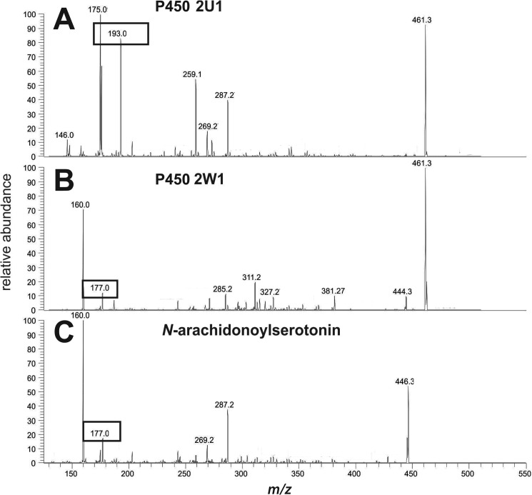
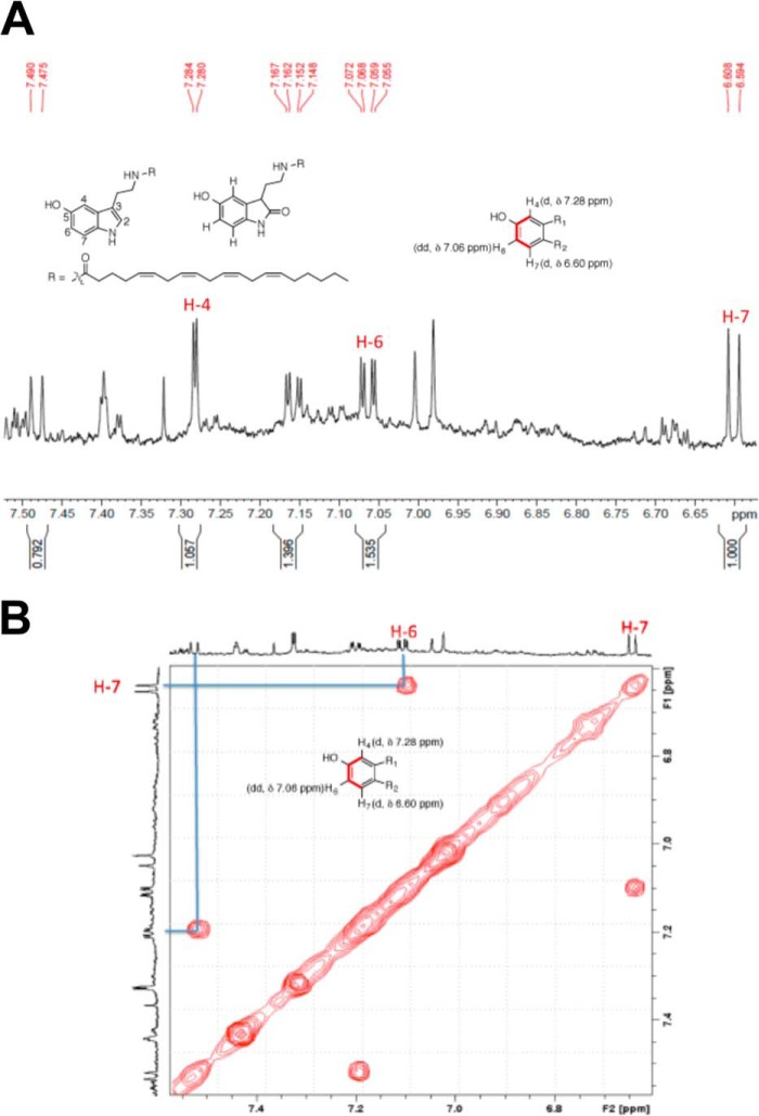
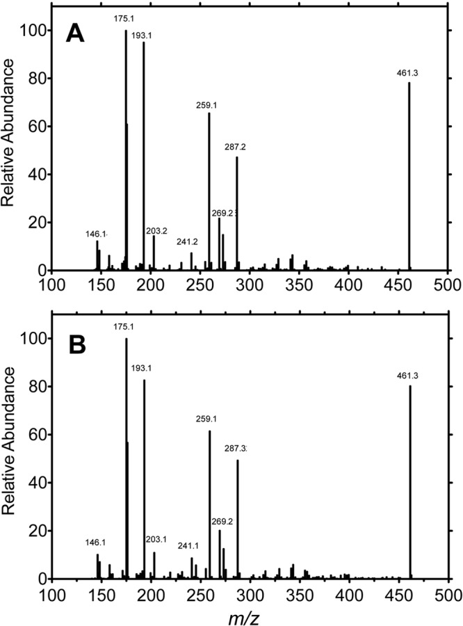
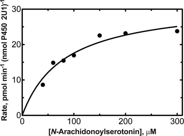
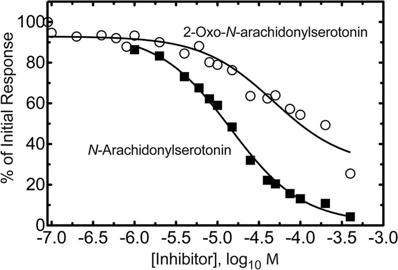
Cytochrome P450 (P450) 2U1 has been shown to be expressed, at the mRNA level, in human thymus, brain, and several other tissues. Recombinant P450 2U1 was purified and used as a reagent in a metabolomic search for substrates in bovine brain. In addition to fatty acid oxidation reactions, an oxidation of endogenous N-arachidonoylserotonin was characterized. Subsequent NMR and mass spectrometry and chemical synthesis showed that the main product was the result of C-2 oxidation of the indole ring, in contrast to other human P450s that generated different products. N-Arachidonoylserotonin, first synthesized chemically and described as an inhibitor of fatty acid amide hydrolase, had previously been found in porcine and mouse intestine; we demonstrated its presence in bovine and human brain samples. The product (2-oxo) was 4-fold less active than N-arachidonoylserotonin in inhibiting fatty acid amide hydrolase. The rate of oxidation of N-arachidonoylserotonin was similar to that of arachidonic acid, one of the previously identified fatty acid substrates of P450 2U1. The demonstration of the oxidation of N-arachidonoylserotonin by P450 2U1 suggests a possible role in human brain and possibly other sites.
Keywords: Arachidonic Acid; Cytochrome P450; Eicosanoid; Lipid Oxidation; Mass Spectrometry (MS).
Figures
Similar articles
-
Cytochrome P450 2U1, a very peculiar member of the human P450s family.Cell Mol Life Sci. 2017 May;74(10):1859-1869. doi: 10.1007/s00018-016-2443-3. Epub 2017 Jan 12. Cell Mol Life Sci. 2017. PMID: 28083596 Free PMC article. Review.
-
Spectral and 3D model studies of the interaction of orphan human cytochrome P450 2U1 with substrates and ligands.Biochim Biophys Acta Gen Subj. 2017 Jan;1861(1 Pt A):3144-3153. doi: 10.1016/j.bbagen.2016.07.018. Epub 2016 Jul 25. Biochim Biophys Acta Gen Subj. 2017. PMID: 27456766
-
Expression in yeast, new substrates, and construction of a first 3D model of human orphan cytochrome P450 2U1: Interpretation of substrate hydroxylation regioselectivity from docking studies.Biochim Biophys Acta. 2015 Jul;1850(7):1426-37. doi: 10.1016/j.bbagen.2015.03.014. Epub 2015 Apr 7. Biochim Biophys Acta. 2015. PMID: 25857771
-
CYP2U1, a novel human thymus- and brain-specific cytochrome P450, catalyzes omega- and (omega-1)-hydroxylation of fatty acids.J Biol Chem. 2004 Feb 20;279(8):6305-14. doi: 10.1074/jbc.M311830200. Epub 2003 Dec 3. J Biol Chem. 2004. PMID: 14660610
-
Differential behavior of the sub-sites of cytochrome 450 active site in binding of substrates, and products (implications for coupling/uncoupling).Biochim Biophys Acta. 2007 Mar;1770(3):360-75. doi: 10.1016/j.bbagen.2006.09.018. Epub 2006 Oct 5. Biochim Biophys Acta. 2007. PMID: 17134838 Review.
Cited by
-
Cytochrome P450 2U1 Is a Novel Independent Prognostic Biomarker in Breast Cancer Patients.Front Oncol. 2020 Aug 5;10:1379. doi: 10.3389/fonc.2020.01379. eCollection 2020. Front Oncol. 2020. PMID: 32850442 Free PMC article.
-
Cytochrome P450 2U1, a very peculiar member of the human P450s family.Cell Mol Life Sci. 2017 May;74(10):1859-1869. doi: 10.1007/s00018-016-2443-3. Epub 2017 Jan 12. Cell Mol Life Sci. 2017. PMID: 28083596 Free PMC article. Review.
-
Roles of Individual Human Cytochrome P450 Enzymes in Drug Metabolism.Pharmacol Rev. 2024 Oct 16;76(6):1104-1132. doi: 10.1124/pharmrev.124.001173. Pharmacol Rev. 2024. PMID: 39054072 Review.
-
Lipids in the Physiopathology of Hereditary Spastic Paraplegias.Front Neurosci. 2020 Feb 28;14:74. doi: 10.3389/fnins.2020.00074. eCollection 2020. Front Neurosci. 2020. PMID: 32180696 Free PMC article. Review.
-
Ninety-eight semesters of cytochrome P450 enzymes and related topics-What have I taught and learned?J Biol Chem. 2024 Feb;300(2):105625. doi: 10.1016/j.jbc.2024.105625. Epub 2024 Jan 5. J Biol Chem. 2024. PMID: 38185246 Free PMC article.
References
-
- Ortiz de Montellano P. R. (ed.) (2005) Cytochrome P450: Structure, Mechanism, and Biochemistry, 3rd Ed., Kluwer Academic/Plenum Publishers, New York
-
- Coon M. J. (2005) Cytochrome P450: nature's most versatile biological catalyst. Annu. Rev. Pharmacol. Toxicol. 45, 1–25 - PubMed
-
- Guengerich F. P. (2005) Human cytochrome P450 enzymes. In Cytochrome P450: Structure, Mechanism, and Biochemistry, 3rd Ed., Ortiz de Montellano P. R. (ed), pp. 377–530, Kluwer Academic/Plenum Publishers, New York
-
- Mast N., Norcross R., Andersson U., Shou M., Nakayama K., Bjorkhem I., Pikuleva I. A. (2003) Broad substrate specificity of human cytochrome P450 46A1 which initiates cholesterol degradation in the brain. Biochemistry 42, 14284–14292 - PubMed
Publication types
MeSH terms
Substances
Grants and funding
LinkOut - more resources
Full Text Sources
Other Literature Sources
Molecular Biology Databases
Miscellaneous
