. 2014 Apr 11;289(15):10582-10591.
doi: 10.1074/jbc.M114.550608. Epub 2014 Feb 22.

Direct regulation of osteocytic connexin 43 hemichannels through AKT kinase activated by mechanical stimulation

Affiliations

Direct regulation of osteocytic connexin 43 hemichannels through AKT kinase activated by mechanical stimulation

Nidhi Batra et al. J Biol Chem. .

Abstract

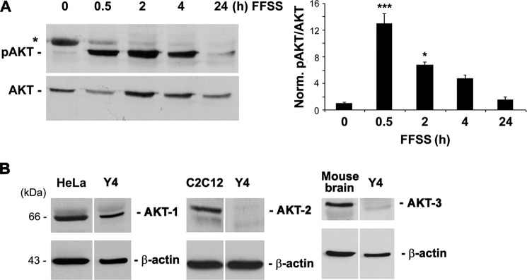
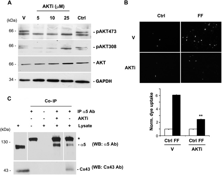
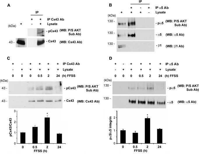
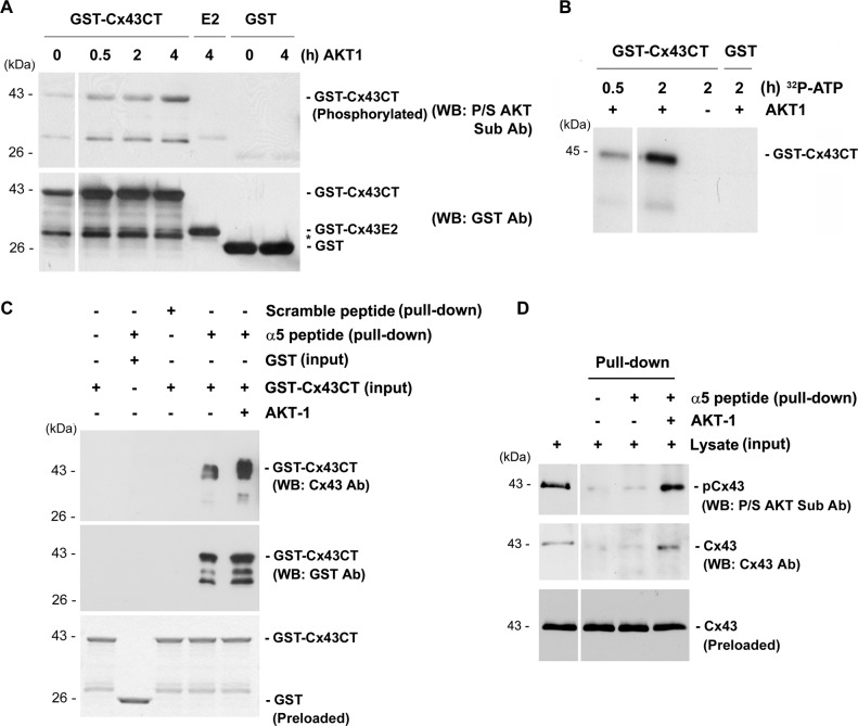
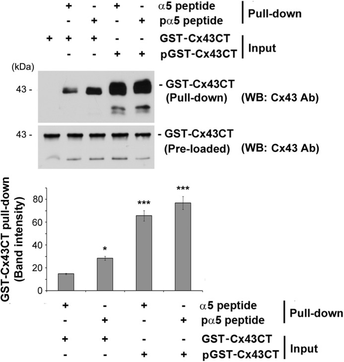
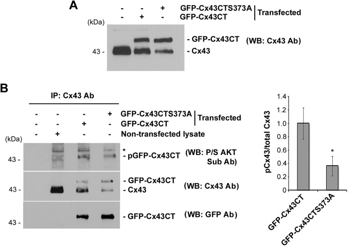
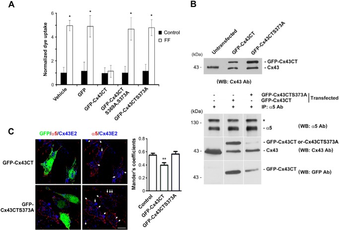
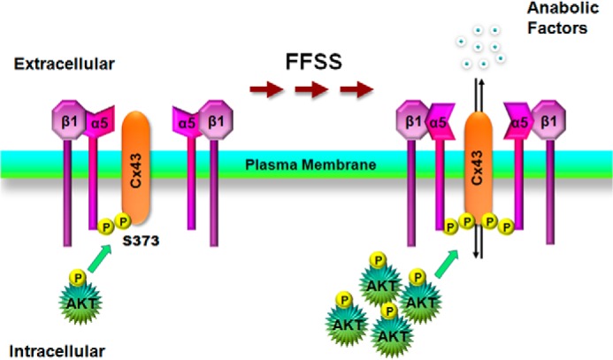
Connexin (Cx) 43 hemichannels in osteocytes are thought to play a critical role in releasing bone modulators in response to mechanical loading, a process important for bone formation and remodeling. However, the underlying mechanism that regulates the opening of mechanosensitive hemichannels is largely unknown. We have recently shown that Cx43 and integrin α5 interact directly with each other, and activation of PI3K appears to be required for Cx43 hemichannel opening by mechanical stimulation. Here, we show that mechanical loading through fluid flow shear stress (FFSS) increased the level of active AKT, a downstream effector of PI3K, which is correlated with the opening of hemichannels. Both Cx43 and integrin α5 are directly phosphorylated by AKT. Inhibition of AKT activation significantly reduced FFSS-induced opening of hemichannels and disrupted the interaction between Cx43 and integrin α5. Moreover, AKT phosphorylation on Cx43 and integrin α5 enhanced their interaction. In contrast to the C terminus of wild-type Cx43, overexpression of the C-terminal mutant containing S373A, a consensus site previously shown to be phosphorylated by AKT, failed to bind with α5 and hence could not inhibit hemichannel opening. Together, our results suggest that AKT activated by FFSS directly phosphorylates Cx43 and integrin α5, and Ser-373 of Cx43 plays a predominant role in mediating the interaction between these two proteins and Cx43 hemichannel opening, a crucial step to mediate the anabolic function of mechanical loading in the bone.

Keywords: Akt; Connexin; Integrin; Mechanotransduction; Phosphorylation.


Figures

FIGURE 1.
FIGURE 1.
FFSS activated AKT kinase, and AKT-1 is a major isoform expressed in osteocytic MLO-Y4 cells. A, FF increases AKT phosphorylation. MLO-Y4 cells subjected to continuous FFSS for 30 min, 2, 4, and 24 h were analyzed for activation of AKT by immunoblotting the cell lysates with anti-phospho-AKT (Ser-473) antibody. A graph plot using mean band densities of pAKT and total AKT shows that pAKT levels were significantly increased after 30 min of FFSS and gradually declined at 2, 4, and 24 h (right panel). 0.5 h versus 0 h; ***, p < 0.001, n = 3. B, MLO-Y4 cells primarily express AKT-1 isoform. Lysates of MLO-Y4 cells were immunoblotted with the different anti-AKT isoform antibodies (AKT-1, AKT-2, and AKT-3). Lysates from HeLa cells, C2C12 cells, and mouse brain tissue were used as positive controls for AKT-1, AKT-2, and AKT-3 expression, respectively. β-Actin was used as a protein loading control.
FIGURE 2.
FIGURE 2.
Activated AKT is required for the opening of HCs and the interaction between Cx43 and integrin α5. A, AKT inhibitor blocked the AKT activation. AKT was activated in MLO-Y4 cells by addition of fresh media with serum and treated with an inhibitor which blocked activation of AKT, AKTi at different concentrations ranging from 5 to 25 μm for 1 h. Cell lysates were then immunoblotted with antibodies to the two phosphorylated residues on AKT, Ser-473 and Thr-308, total AKT, and GAPDH. AKTi completely inhibited the phosphorylation on the two sites (n = 3). B, AKTi blocked FFSS-induced HC opening. LY dye uptake was performed on MLO-Y4 cells under two conditions, treated with AKTi for 1 h (AKTi) and the other not treated with AKTi (Vehicle). Cells treated with either of these conditions were further subjected to FFSS for 30 min (FF) or not (Ctrl). MLO-Y4 cells treated with AKTi and followed by FFSS showed a significant resistance to HC opening in presence of AKTi compared with cells subjected to FFSS directly. FFSS (AKTi) versus FFSS (vehicle, V). **, p < 0.05, n = 3. C, AKTi disrupts the interaction between Cx43 and integrin α5. MLO-Y4 cells were treated with and without AKTi for 1 h, and immunoprecipitation assay was performed using anti-integrin α5 antibody. The immunoprecipitates were immunoblotted with anti-Cx43 or α5 antibody. Cx43 was absent in immunoprecipitates of AKTi-treated cells using anti-α5 antibody (n = 3).
FIGURE 3.
FIGURE 3.
FFSS increases phosphorylation of Cx43 and integrin α5, and both proteins are direct substrates of AKT. A, AKT phosphorylates Cx43 in MLO-Y4 cells. Cx43 was immunoprecipitated from cell lysates using mouse monoclonal anti-Cx43 antibody. Immunoprecipitates were immunoblotted with anti-Pro/Ser AKT substrate or Cx43 antibody. Cx43 in the MLO-Y4 cells was phosphorylated by AKT (n = 3). B, AKT phosphorylates integrin α5 in MLO-Y4 cells. Integrin α5 from cell lysates was immunoprecipitated using integrin α5 antibody (IP α5 Ab), and the immunoprecipitates were immunoblotted (WB) using anti-Pro/Ser AKT substrate, integrin α5, or integrin β1 antibody. Immunoprecipitated integrin α5 was a substrate for phosphorylation by AKT (n = 3). C and D, FFSS increases phosphorylation of Cx43 and integrin α5 in MLO-Y4 cells. Cells were subjected to FFSS for different time periods (0, 0.5, 2, and 24 h) and Cx43 and integrin α5 were immunoprecipitated from cell lysates with anti-Cx43 or integrin α5 antibody, respectively, and immunoprecipitates were immunoblotted with Pro/Ser AKT substrate and with Cx43 antibody (C) or with integrin α5 antibody (D). An increase in phosphorylation of both Cx43 and integrin α5 by AKT was observed after 2 h of FFSS. Graphs were plotted using the ratio of mean band densities of AKT-phosphorylated Cx43 or integrin α5 to total Cx43 or integrin α5, respectively. 2 h versus 0 h; *, p < 0.05, n = 3.
FIGURE 4.
FIGURE 4.
Phosphorylation of the C termini of integrin α5 and Cx43 enhances their interaction. A, in vitro phosphorylation of GST-Cx43CT using AKT-1 kinase showed increase in phosphorylation from 30 min to 4 h in GST-Cx43CT, but not in GST-Cx43 (E2) or GST alone (n = 3). B, in vitro phosphorylation of GST-Cx43CT using AKT-1 kinase and [γ-32P]ATP also showed an increase in phosphorylation from 30 min to 2 h. C, a dramatic increase in the interaction between GST-Cx43CT and the peptide containing the C terminus of integrin α5 was observed when GST-Cx43CT was phosphorylated by AKT. Protein pulldown was conducted by first treating GST-Cx43CT or GST with AKT-1 kinase for 4 h and then incubating with magnetic beads conjugated with a peptide containing the C terminus of integrin α5 (n = 3). D, phosphorylation of Cx43 by AKT-1 increased its association with the C terminus of integrin α5. Lysates of MLO-Y4 cell were treated with AKT-1 kinase for 1 h and then incubated with magnetic beads conjugated with a peptide containing C terminus of α5 for pulldown assay.
FIGURE 5.
FIGURE 5.
Phosphorylation of the C terminus of Cx43 by AKT plays a predominant role in its interaction with integrin α5. Purified GST-Cx43CT and/or a peptide containing the C terminus of α5 (α5 peptide) conjugated with magnetic beads was incubated with and without AKT-1 kinase for 4 h. Elutes and inputs from the pulldown assay were immunoblotted (WB) with anti-Cx43 antibody. The band intensity was quantified showing a drastic increase in the interaction between GST-Cx43CT and the peptide containing the C terminus of integrin α5 when GST-Cx43CT was phosphorylated by AKT-1. Non-AKT-1-treated (first column) versus all other conditions; *, p < 0.05; ***, p < 0.001, n = 3.
FIGURE 6.
FIGURE 6.
Ser-373 on the C terminus of Cx43 is a major site for phosphorylation of AKT on Cx43. A, exogenous GFP fusion proteins containing Cx43CT and Cx43CTS373A mutants were comparably expressed in MLO-Y4 cells. Cell lysates transfected with Cx43CT or Cx43CTS373A were immunoblotted (WB) with anti-Cx43 antibody. B, Ser-373 on Cx43 is a major site for AKT phosphorylation. Either GFP-Cx43CT or GFP-Cx43CTS373A was exogenously expressed in MLO-Y4 cells. Cell lysates were immunoprecipitated using anti-Cx43 antibody. Immunoprecipitates were immunoblotted with antibodies to P/S AKT substrate (top panel), Cx43 (middle panel), or GFP (bottom panel). Intensity of the band was quantified (right panel). GFP-Cx43CT versus GFP-Cx43CTS373A; *, p < 0.05, n = 3.
FIGURE 7.
FIGURE 7.
Ser-373 on Cx43 is critical for the interaction of Cx43 with integrin α5 and HC opening. A, S373A mutation on Cx43 failed to inhibit HC opening induced by FFSS. MLO-Y4 cells transfected with DNA constructs containing GFP-Cx43CT, GFP-Cx43CTS373A, GFP-Cx43CTS369A,S373A, GFP, or vector alone were subjected to FFSS for 15 min or non-FFSS static controls. Uptake assay was performed with Alexa Fluor 350 dye and RD. The number of cells with GFP signals that took up the dye were counted and quantified. Cells expressing GFP-Cx43CT, but not GFP-Cx43CTS373A, GFP-Cx43CTS369A,S373A, or vehicle control, showed reduction in dye uptake in response to FFSS. GFP-Cx43CT versus all other conditions; *, p < 0.05, n = 3. B, S373A mutation reduced Cx43CT binding to integrin α5. Lysates of MLO-Y4 cells exogenously expressing GFP-Cx43CT or GPF-Cx43CTS373A were immunoblotted using anti-Cx43 antibody. Cell lysates were immunoprecipitated with anti-α5 antibody, and immunoprecipitates were immunoblotted (WB) with antibody to α5 (top panel), Cx43 (middle panel), or GFP (lower panel) n = 3. C, reduction of the co-localization between integrin α5 and Cx43 with the expression of GFP-Cx43CT, but not GFP-Cx43CTS373A. MLO-Y4 cells transfected with DNA constructs containing GFP-Cx43CT or GFP-Cx43CTS373A were co-immunofluorescence labeled with anti-integrin α5 (red) and Cx43E2 (against E2 domain) (blue) antibodies (left panels). The transfected cells are indicated by GFP fluorescence. The co-localized signals are indicated by white arrows. The extent of co-localization on the cell surface was analyzed using ImageJ (right panel). GFP-Cx43CT versus control (untransfected) and GFP-Cx43CTS373A; **, p < 0.01, n = 20. Scale bar, 10 μm.
FIGURE 8.
FIGURE 8.
Illustration of the role of AKT activation by FFSS regulates Cx43 interaction with integrin α5 and HC opening. Under non-FFSS static conditions, there are low levels of basal AKT which mediates phosphorylation on Cx43 and integrin α5 and promotes weak interaction between these two proteins. FFSS activates AKT which leads to the phosphorylation of Cx43 and α5 and enhancement of the interaction between these two proteins and a possible conformational activation of α5 (25). The interaction and activation lead to the opening of HCs in osteocytes which may associate with the release of small autocrine/paracrine factors important for bone formation and remodeling in response to mechanical loading.

References

    1. Srinivasan S., Gross T. S., Bain S. D. (2012) Bone mechanotransduction may require augmentation in order to strengthen the senescent skeleton. Ageing Res. Rev. 11, 353–360 - PMC - PubMed
    1. Aarden E. M., Burger E. H., Nijweide P. J. (1994) Function of osteocytes in bone. J. Cell. Biochem. 55, 287–299 - PubMed
    1. Burger E. H., Klein-Nulend J. (1999) Mechanotransduction in bone: role of the lacunocanalicular network. FASEB J. 13, S101–112 - PubMed
    1. Cowin S. C., Moss-Salentijin L., Moss M. L. (1991) Candidates for the mechanosensory system in bone. J. Biomed. Eng. 113, 191–197 - PubMed
    1. Jiang J. X., Siller-Jackson A. J., Burra S. (2007) Roles of gap junctions and hemichannels in bone cell functions and in signal transmission of mechanical stress. Front. Biosci. 12, 1450–1462 - PMC - PubMed

Publication types