Review
. 2014 Mar 1;192(5):2019-26.
doi: 10.4049/jimmunol.1302426.

True grit: programmed necrosis in antiviral host defense, inflammation, and immunogenicity

Affiliations
Review

True grit: programmed necrosis in antiviral host defense, inflammation, and immunogenicity

Edward S Mocarski et al. J Immunol. .

Abstract

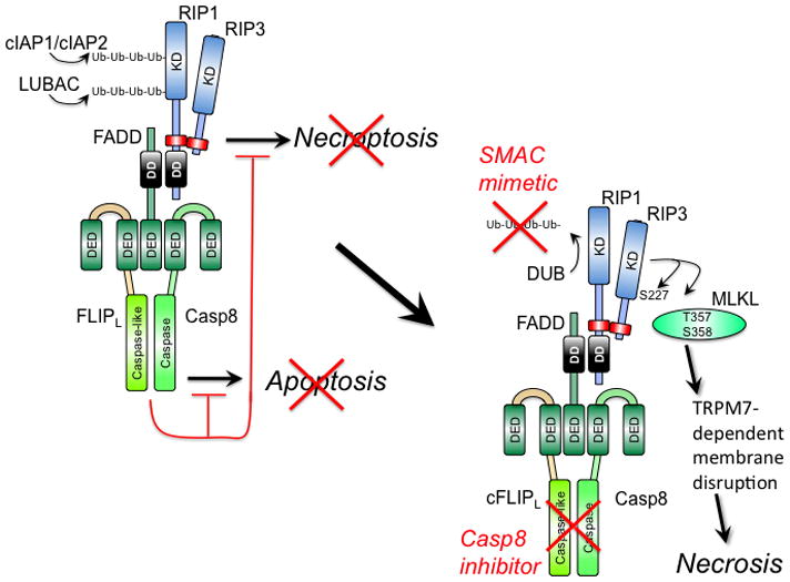
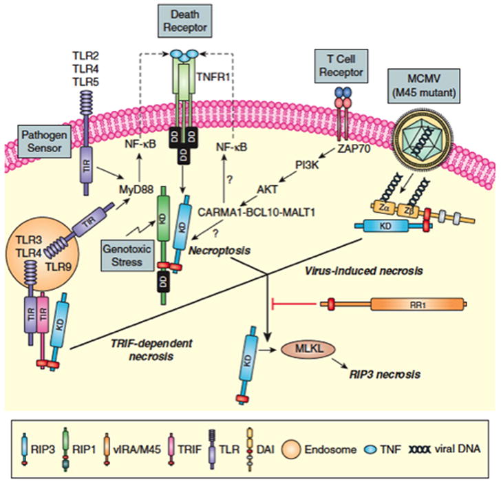
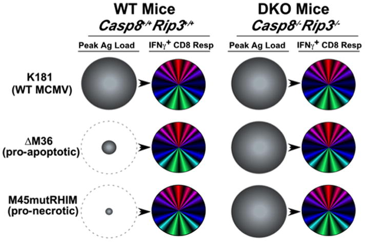
Programmed necrosis mediated by receptor interacting protein kinase (RIP)3 (also called RIPK3) has emerged as an alternate death pathway triggered by TNF family death receptors, pathogen sensors, IFNRs, Ag-specific TCR activation, and genotoxic stress. Necrosis leads to cell leakage and acts as a "trap door," eliminating cells that cannot die by apoptosis because of the elaboration of pathogen-encoded caspase inhibitors. Necrotic signaling requires RIP3 binding to one of three partners-RIP1, DAI, or TRIF-via a common RIP homotypic interaction motif. Once activated, RIP3 kinase targets the pseudokinase mixed lineage kinase domain-like to drive cell lysis. Although necrotic and apoptotic death can enhance T cell cross-priming during infection, mice that lack these extrinsic programmed cell death pathways are able to produce Ag-specific T cells and control viral infection. The entwined relationship of apoptosis and necrosis evolved in response to pathogen-encoded suppressors to support host defense and contribute to inflammation.


Figures

Figure 1
Figure 1. Regulation of extrinsic apoptosis and RIP3 necrosis by a ‘Necrosome’ or ‘Ripoptosome’ complex
(Left) Cytoprotection. Signal transduction via death receptors (e.g. TNF) (–39), pathogen sensors (e.g. TLR3 signaling) (27, 51), TCR activation (32, 33) or intracellular genotoxic stress (34) supports FADD association with the FLIP-Casp8 heterodimer via DED as well as RIP1 via DD interaction. RIP1 orchestrates recruitment of RIP3 via a RHIM interaction (red rectangle). The FLIP-Casp8 association prevents self-cleavage activation of Casp8 and maintains sufficient basal protease activity to also prevent necroptosis. E3 ubiquitin ligases cIAP1/cIAP2 and LUBAC also prevent necroptosis by maintaining K63 or linear polyubiquitination (Ub-Ub) of RIP1 and other targets (34, 100, 101). (Right) Activation of necroptosis. When Casp8 activity is blocked by an inhibitor or E3 ubiquitin ligases are compromised (red “X”) by a mimetic of second mitochondria-derived activator of caspases (SMAC), RIP3 kinase autophosphorylates at S277 and targets MLKL (42) for phosphorylation at T357 and S358 (41). These modifications drive trimerization of MLKL and membrane disruption associated with Ca2+ influx via a TRPM7-dependent plasma membrane channel (43). Deubiquitinase (DUB) activity removes polyubiquitin chains in the presence of SMAC mimetic, sensitizing to necrosis when Casp8 activity is compromised.
Figure 2
Figure 2. Three distinct RHIM complexes trigger RIP3 necrosis
RIP1-RIP3 necroptosis first characterized downstream of death receptor activation via RIP1-RIP3 complex formation (–39) is also induced by pathogen sensor (e.g. TLR2, TLR4, TLR5 or TLR9 MyD88-dependent signaling) (27, 28), TCR activation (32, 33), intracellular genotoxic stress (34) and vaccinia virus infection (37). Virus-induced DAI-RIP3 necrosis (3, 25, 26) is activated by MCMV M45 mutant virus infection. TRIF-RIP3 necrosis in fibroblasts is activated TLR3 or TRL4 ligands (27, 28). RIP3 complexes with RIP1, DAI or TRIF depend on RHIM interactions that activate RIP3 kinase-dependent modification of MLKL (7, 41, 42) (see Fig. 1). MCMV M45-encoded vIRA functions as a dominant RHIM-inhibitor preventing RIP3 association with RIP1, DAI or TRIF.
Figure 3
Figure 3. Model: Viral Ag load and cell death pathways collaborate in cross-presentation to drive CD8 T cell immunity during infection
Model derived from studies on the impact of apoptotic and necrotic cell death pathways on cross-presentation in the CD8 T cell response (50, 106, 107) as well as developing understanding of MCMV immune response parameters. Relative peak viral load (shaded grey circles) at day 3 pi and peak CD8 T cell response at day 7–10 pi (multicolored circles) with WT MCMV (K181 strain), pro-apoptotic mutant ΔM36 or pro-necrotic mutant M45mutRHIM. The benefit of enhanced cross-presentation from either pro-apoptotic or pro-necrotic viruses is depicted by the dashed grey circles.

Similar articles

Cited by

References

    1. Lamkanfi M, V, Dixit M. Manipulation of host cell death pathways during microbial infections. Cell Host Microbe. 2010;8:44–54. - PubMed
    1. Galluzzi L, Brenner C, Morselli E, Touat Z, Kroemer G. Viral control of mitochondrial apoptosis. PLoS Pathog. 2008;4:e1000018. - PMC - PubMed
    1. Mocarski ES, Upton JW, Kaiser WJ. Viral infection and the evolution of caspase 8-regulated apoptotic and necrotic death pathways. Nat Rev Immunol. 2011;12:79–88. - PMC - PubMed
    1. Castanier C, Arnoult D. Mitochondrial localization of viral proteins as a means to subvert host defense. Biochim Biophys Acta. 2011;1813:575–583. - PubMed
    1. Kaiser WJ, Upton JW, Mocarski ES. Viral modulation of programmed necrosis. Curr Opin Virol. 2013;3:296–306. - PMC - PubMed

Publication types

MeSH terms