. 2014 Apr;20(4):568-79.
doi: 10.1261/rna.043513.113. Epub 2014 Feb 21.

RNase E forms a complex with polynucleotide phosphorylase in cyanobacteria via a cyanobacterial-specific nonapeptide in the noncatalytic region

RNase E forms a complex with polynucleotide phosphorylase in cyanobacteria via a cyanobacterial-specific nonapeptide in the noncatalytic region

Ju-Yuan Zhang et al. RNA. 2014 Apr.

Abstract

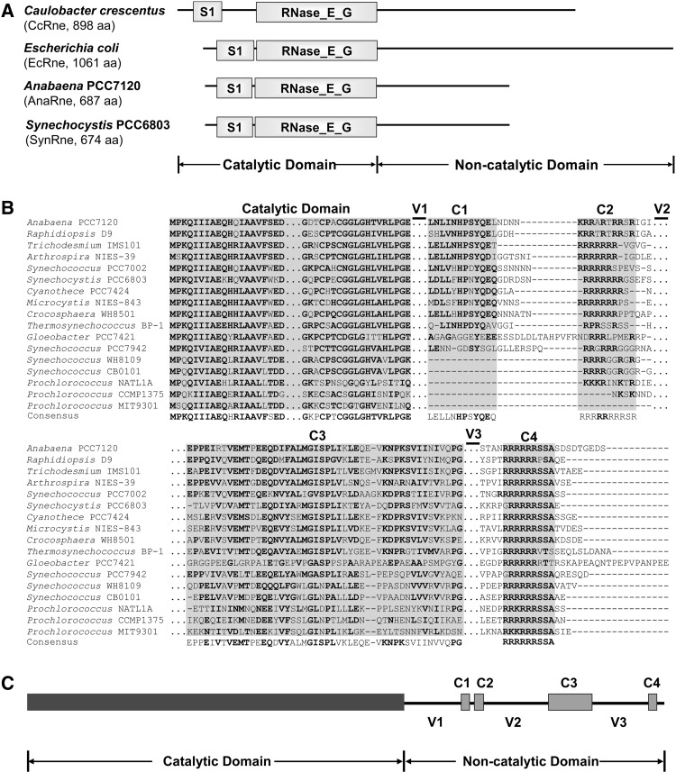
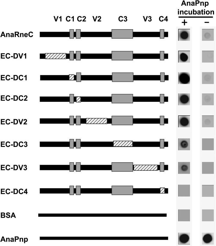
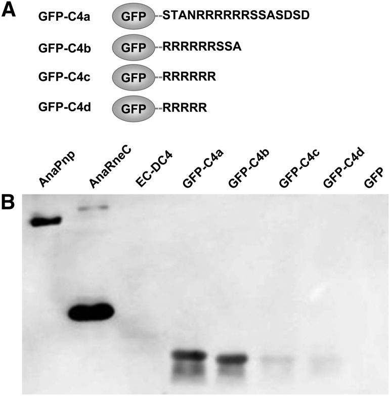
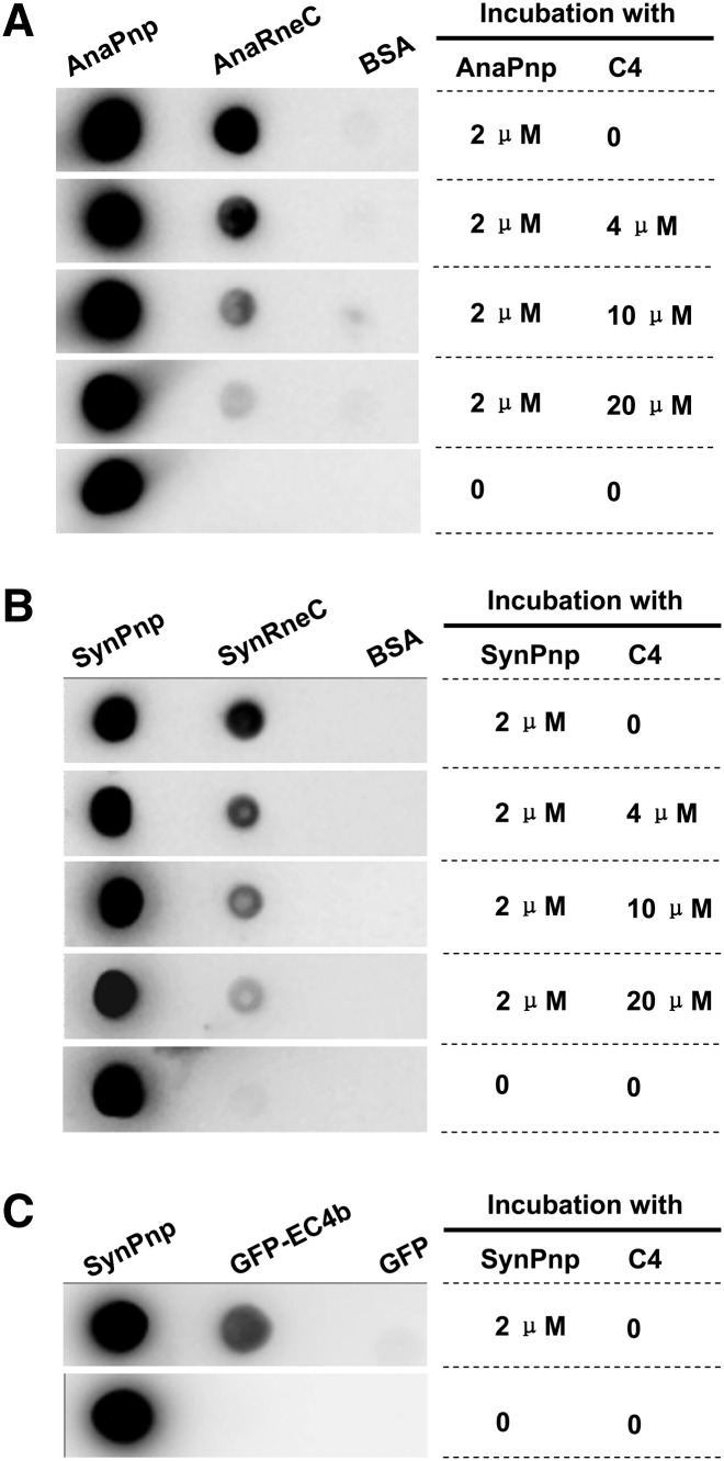
RNase E, a central component involved in bacterial RNA metabolism, usually has a highly conserved N-terminal catalytic domain but an extremely divergent C-terminal domain. While the C-terminal domain of RNase E in Escherichia coli recruits other components to form an RNA degradation complex, it is unknown if a similar function can be found for RNase E in other organisms due to the divergent feature of this domain. Here, we provide evidence showing that RNase E forms a complex with another essential ribonuclease-the polynucleotide phosphorylase (PNPase)-in cyanobacteria, a group of ecologically important and phylogenetically ancient organisms. Sequence alignment for all cyanobacterial RNase E proteins revealed several conserved and variable subregions in their noncatalytic domains. One such subregion, an extremely conserved nonapeptide (RRRRRRSSA) located near the very end of RNase E, serves as the PNPase recognition site in both the filamentous cyanobacterium Anabaena PCC7120 and the unicellular cyanobacterium Synechocystis PCC6803. These results indicate that RNase E and PNPase form a ribonuclease complex via a common mechanism in cyanobacteria. The PNPase-recognition motif in cyanobacterial RNase E is distinct from those previously identified in Proteobacteria, implying a mechanism of coevolution for PNPase and RNase E in different organisms.

Keywords: PNPase recognition site; RNA degradosome; RNase E; cyanobacteria.


Figures

FIGURE 1.
FIGURE 1.
(A) The domain structures of the RNase E proteins from Caulobacter crescentus, Escherichia coli, Anabaena PCC7120, and Synechocystis PCC6803. The catalytic domain of each RNase E is composed of a S1 domain and an RNase_E_G domain. The C-terminal noncatalytic domains of the two cyanobacterial strains show no similarity to those of other bacteria including the two proteobacterial strains listed here. (B) Alignment of RNase E proteins from 17 representative cyanobacterial strains. The alignment was performed using MEGA5 (Tamura et al. 2011) and refined manually. Four short conserved subregions (C1–C4) and three variable subregions (V1–V3) were revealed in the noncatalytic domains. The regions not shown are depicted with triple dots (…). Gaps in the alignment are depicted with short horizontal lines (-). The residues with high conservation are in bold. (C) Schematic structure of the Anabaena RNase E based on the alignment shown above. The variable and conserved regions of AnaRne revealed by the sequence alignment are depicted as lines and filled boxes, respectively.
FIGURE 2.
FIGURE 2.
Comparison of the catalytic properties of EcRneN (the catalytic domain of E. coli RNase E), AnaRne (the full length of Anabaena RNase E), and AnaRneN (the catalytic domain of Anabaena RNase E) on the 9S RNA as substrate. (CK-) 9S RNA incubated for 60 min in the reaction buffer alone. In each of the other reactions, an equal amount of substrate and enzyme were used. The electrophoresis was performed in a 6% PAGE gel containing 7 M urea, and the gel was stained with ethidium bromide. The bands of 9S RNA and its end product p5S RNA are indicated by arrows.
FIGURE 3.
FIGURE 3.
Copurification of AnaPnp with the His-tagged GFP-AnaRneC fusions expressed in Anabaena cells using Ni-NTA beads. (A) Schematic presentation of the His-tagged GFP and GFP-AnaRneC fusions expressed in Anabaena cells. (B) Total proteins from Anabaena cells expressing the His-tagged recombinant proteins were applied to the Ni-NTA columns for purification. Proteins eluted were analyzed by SDS-PAGE gels, followed by silver staining or immunodetection using polyclonal antibodies against AnaPnp. Lanes 13, samples from Anabaena cells expressing hGFP, hGFP-EC, and EC-GFPh, respectively.
FIGURE 4.
FIGURE 4.
(A) Far-Western dot-blot assay investigating the interaction between the recombinant AnaRneC and AnaPnp purified from E. coli cells with or without the treatment with micrococcal nuclease (MNase). AnaPnp and BSA were dotted onto the membranes as a positive control and a negative control, respectively. (B) Determining the complex of AnaRneC and AnaPnp by gel electrophoresis. AnaRneC and AnaPnp were incubated under native conditions for 30 min, then the mixture was separated on the native-PAGE gel (the middle lane in the left panel). The band of protein complex (indicated by an arrow) in the native gel was excised and subsequently subjected to SDS-PAGE electrophoresis (the middle lane in the right panel). Purified AnaRneC and AnaPnp were included as size markers in both the native-PAGE gel and the SDS-PAGE gel. Note that AnaRneC did not migrate as a sharp band in the native-PAGE gel. (C) Bacterial two-hybrid assay investigating the interaction between AnaRneC and AnaPnp. The E. coli XL1-Blue MRF′ Kan cells cotransformed by the indicated two-hybrid plasmid pairs were spotted on the selective and the nonselective plates, respectively. Cells cotransformed with pBT-LGF2 and pTRG-Gal11P (supplied in the kit) were used as the positive control (CK+), and cells cotransformed with the empty vectors of pBT and pTRG were used as the negative control. Note that, due to unclear reasons, cells containing pTRG-AnaPnp/pBT or pTRG-AnaPnp/pBT showed weak growth.
FIGURE 5.
FIGURE 5.
Far-Western dot-blot assay studying the role of each subregion of AnaRneC in AnaPnp–AnaRneC interaction. AnaRneC and its subregion deletion variants were dotted onto duplicate nitrocellulose membranes. Subsequently, one of the membranes was incubated with 2 µM AnaPnp and the other one was not. Both membranes were finally subjected to immunodetection with the antibody against AnaPnp (see Materials and Methods for details). The deleted regions in AnaRneC variants are illustrated with boxes with gray stripes. BSA and AnaPnp were used as the negative control and the positive control, respectively.
FIGURE 6.
FIGURE 6.
Investigation of the interaction between AnaPnp and GFP-C4 variants. (A) Schematic illustration of GFP-C4 variants. (B) Far-Western blot assay demonstrating the interaction between AnaPnp and GFP-C4 variants. All proteins were separated on a 12% SDS-PAGE gel and transferred onto a nitrocellulose membrane. The membrane was then incubated with AnaPnp, followed by immunodetection with the antibody against AnaPnp (see Materials and Methods for details). AnaPnp and AnaRneC were included as the positive controls; EC-DC4 and GFP were included as the negative controls. The amount of protein in each lane was 10 µg, except that 100 ng was loaded for AnaPnp.
FIGURE 7.
FIGURE 7.
(A) Competition assay of the synthetic peptide C4 on the interaction between AnaRneC and AnaPnp by Far-Western dot-blot. AnaRneC, AnaPnp (the positive control), and BSA (the negative control) were dotted onto nitrocellulose membranes. After incubation with the indicated amount of AnaPnp and C4, the membranes were immunodetected with the antibody against AnaPnp (see Materials and Methods for details). (B) Effect of the C4 peptide on the interaction between SynRneC and SynPnp. The experiment was performed as described in A. (C) Interaction between SynPnp and GFP-C4b. The experiment was also performed as described in A. SynPnp and GFP proteins were dotted on the membranes as the positive control and the negative control, respectively.

References

    1. Aït-Bara S, Carpousis AJ 2010. Characterization of the RNA degradosome of Pseudoalteromonas haloplanktis: Conservation of the RNase E-RhlB interaction in the gammaproteobacteria. J Bacteriol 192: 5413–5423 - PMC - PubMed
    1. Arraiano CM, Andrade JM, Domingues S, Guinote IB, Malecki M, Matos RG, Moreira RN, Pobre V, Reis FP, Saramago M, et al. 2010. The critical role of RNA processing and degradation in the control of gene expression. FEMS Microbiol Rev 34: 883–923 - PubMed
    1. Baginsky S, Shteiman-Kotler A, Liveanu V, Yehudai-Resheff S, Bellaoui M, Settlage RE, Shabanowitz J, Hunt DF, Schuster G, Gruissem W 2001. Chloroplast PNPase exists as a homo-multimer enzyme complex that is distinct from the Escherichia coli degradosome. RNA 7: 1464–1475 - PMC - PubMed
    1. Blum E, Carpousis AJ, Higgins CF 1999. Polyadenylation promotes degradation of 3′-structured RNA by the Escherichia coli mRNA degradosome in vitro. J Biol Chem 274: 4009–4016 - PubMed
    1. Callaghan AJ, Aurikko JP, Ilag LL, Günter Grossmann J, Chandran V, Kühnel K, Poljak L, Carpousis AJ, Robinson CV, Symmons MF, et al. 2004. Studies of the RNA degradosome-organizing domain of the Escherichia coli ribonuclease RNase E. J Mol Biol 340: 965–979 - PubMed

Publication types

MeSH terms

LinkOut - more resources