. 2014 Feb 7;9(2):e88383.
doi: 10.1371/journal.pone.0088383. eCollection 2014.

Role of tryptophan side chain dynamics on the Trp-cage mini-protein folding studied by molecular dynamics simulations

Affiliations

Role of tryptophan side chain dynamics on the Trp-cage mini-protein folding studied by molecular dynamics simulations

Srinivasaraghavan Kannan et al. PLoS One. .

Abstract

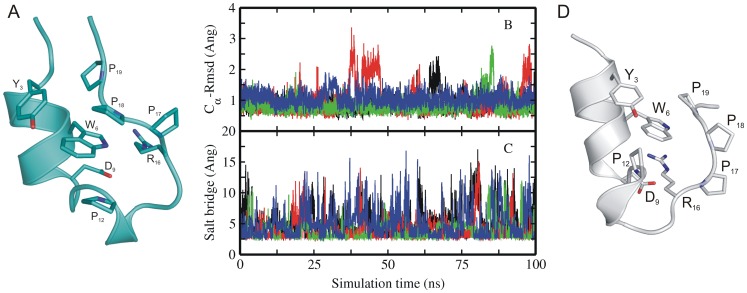
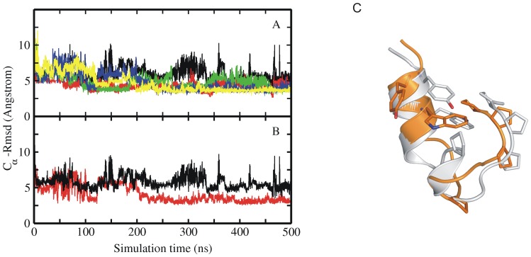
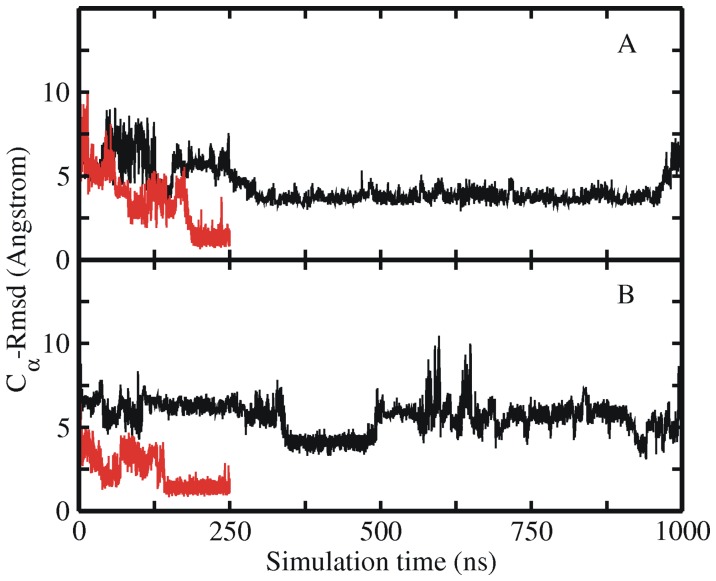
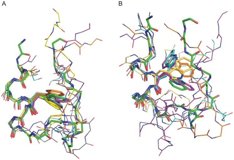
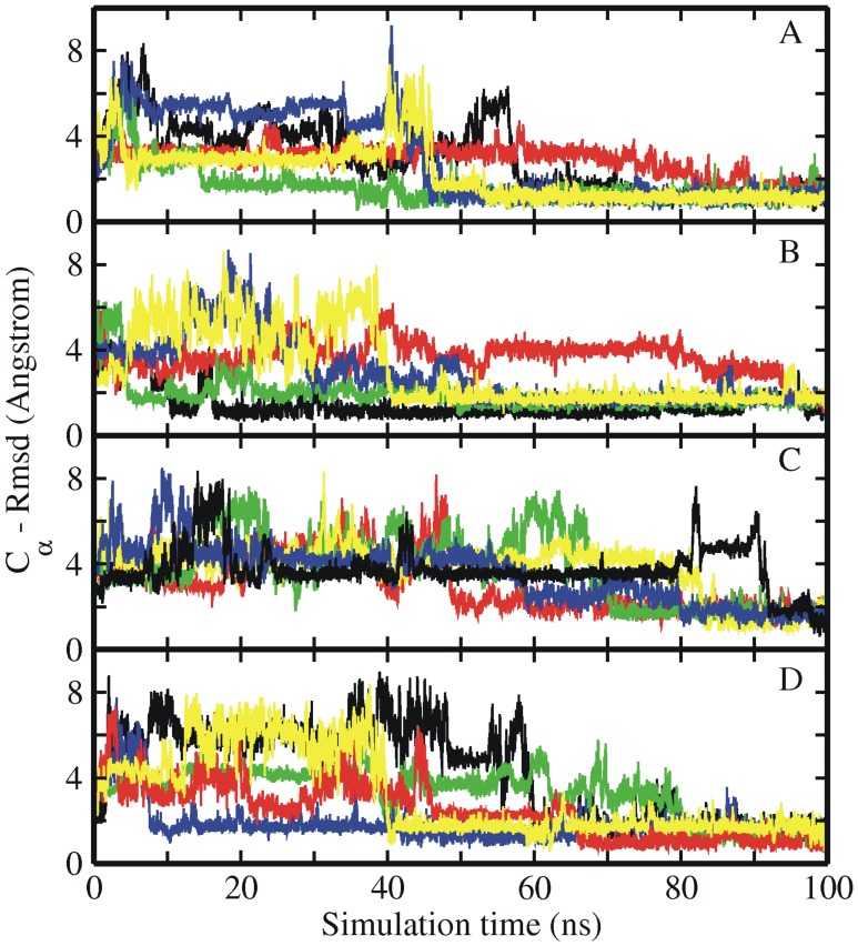
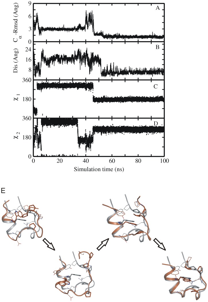
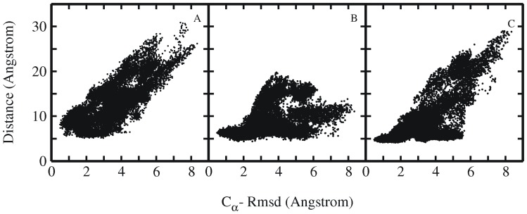
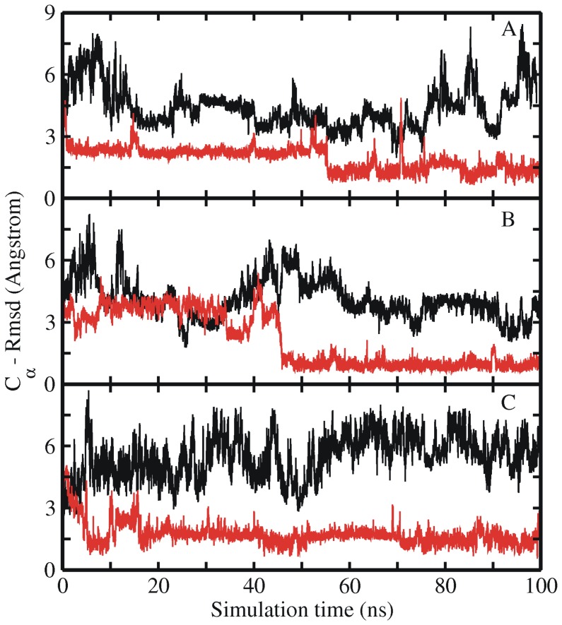
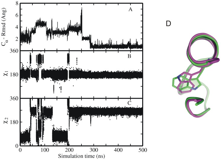
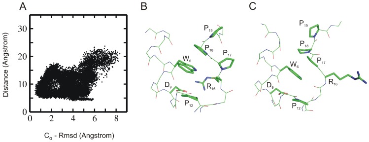
The 20 residue Trp-cage mini-protein is one of smallest proteins that adopt a stable folded structure containing also well-defined secondary structure elements. The hydrophobic core is arranged around a single central Trp residue. Despite several experimental and simulation studies the detailed folding mechanism of the Trp-cage protein is still not completely understood. Starting from fully extended as well as from partially folded Trp-cage structures a series of molecular dynamics simulations in explicit solvent and using four different force fields was performed. All simulations resulted in rapid collapse of the protein to on average relatively compact states. The simulations indicate a significant dependence of the speed of folding to near-native states on the side chain rotamer state of the central Trp residue. Whereas the majority of intermediate start structures with the central Trp side chain in a near-native rotameric state folded successfully within less than 100 ns only a fraction of start structures reached near-native folded states with an initially non-native Trp side chain rotamer state. Weak restraining of the Trp side chain dihedral angles to the state in the folded protein resulted in significant acceleration of the folding both starting from fully extended or intermediate conformations. The results indicate that the side chain conformation of the central Trp residue can create a significant barrier for controlling transitions to a near native folded structure. Similar mechanisms might be of importance for the folding of other protein structures.


Conflict of interest statement

Competing Interests: The authors have declared that no competing interests exist.

Figures

Figure 1
Figure 1. Experimental structure and MD simulations of native Trp-cage miniprotein.
(A) Cartoon representation of the folded experimental structure of Trp-cage mini protein . A subset of side chains important for folding are labeled (stick representation). (B) Root-mean –square deviation (RMSD) of Cα–atoms with respect to native structure and (C) distance between Asp9-Arg16 (salt bridge) of sampled Trp-cage conformations in explicit solvent starting from the native structure (1st entry of pdb 1L2Y) versus simulation time. Simulations were performed with four different force fields encoded as different line colors (black:ff03, red:ff99SB, green:ff99SB_ILDN,blue:ff99SB_NMR). (D) Partially unfolded Trp-cage structures sampled during simulation starting from native state (with the C-terminal PolyProII motive transiently dissociated from the central Trp6 residue).
Figure 2
Figure 2. MD simulations of extended Trp-cage structure.
(A) RMSD of sampled conformations during five cMD simulations with different atomic velocities starting from an extended Trp-cage structure (A). Simulation results in A were obtained using the ff03 force field. (B) Comparison of the RMSD of sampled structures for one simulation with respect to a partially unfolded conformation (red curve) and with respect to the native Trp-cage (black curve). The partially unfolded conformation was obtained in simulations starting from the native Trp-cage structure and contains a partially dissociated C-terminal segment (see Figure 2C). (C) Example snapshot of a collapsed structure (orange Cartoon) that came close to within 2.5 Å RMSD with respect to the partially unfolded structure (grey Cartoon in C). Residues important for folding (Tyr3, Trp6, Pro12, Pro17, Pro18 and Pro19) are shown as sticks.
Figure 3
Figure 3. Free and restrained MD simulations of extended Trp-cage structure.
RMSD of sampled Trp-cage conformations in an unrestraint explicit solvent MD simulation starting from an extended Trp-cage conformation (black line) and including dihedral angle restraints of the Trp6 residue (red line). (A) Both simulations were started from the same start structure and initial velocities employing the ff03 force field. (B) Same as in (A) but using the ff99SB force field (instead of ff03).
Figure 4
Figure 4. Structural superposition of Set1 and Set2 intermediate start structures.
Superposition of (A) set 1 and (B) set2 intermediate start structures (superimposed on α-helix of the native Trp-cage structure indicated as atom colored stick representation). The intermediate start structures are shown as thin sticks using different colors. For clarity only the side chain of the central Trp6 residue in each structure is shown explicitly (stick representation).
Figure 5
Figure 5. MD simulations of intermediate Trp-cage structures.
RMSD of sampled Trp-cage conformations in explicit solvent starting from a set of intermediate structures (a subset of intermediate structures of set2 shown in Figure 5B,) vs. simulation time with different Amber force fields (A) ff03 (B) ff99SB, (C) ff99SB_ILDN, (D) ff99SB_NMR. All these examples eventually reached a near-native conformation with RMSD <2 Å from the native Trp-cage structure.
Figure 6
Figure 6. Time-dependence of backbone Rmsd and Trp6 side chain dihedral angles.
(A) RMSD from native structure and (B) Asp9-Arg16 salt bridge distance as well as (C, D) side chain dihedral angels (χ1 and χ2) of Trp-6 residue of sampled Trp-cage conformations along one folding trajectory starting from a set2 intermediate structure vs. simulation time (force field ff03). The dihedral angles of the native Trp6 side chain correspond to χ1 in trans (∼180°) and χ2 in –gauche (∼270°) (E) MD simulation snapshots showing the various steps in the folding process of the protein, as in the order of occurrence. Simulation snapshots (gold ribbons) are superimposed onto the native structure (grey ribbons), and key residues (Tyr3, Trp6, Ala9, Pro12, Arg16 and Pro17-19) are shown as sticks (same color coding as for ribbons).
Figure 7
Figure 7. Rmsd and residue pair distance distribution.
Distribution plots showing the correlation between RMSD and distance between the residue pairs (A) Tyr3-Pro19, (B) Trp6-Pro12 and (C) Trp6-Pro18. Each point represents one sampled state in all simulations starting from intermediate Trp-cage structures.
Figure 8
Figure 8. Free and restrained MD simulations of intermediate Trp-cage structure.
RMSD with respect to the native Trp-cage structure starting from an intermediate state (of set 2) with (red lines) and without (black lines) side chain dihedral angle restraints on Trp6 residue vs. simulation time. Results are shown for three different force fields (A) ff03, (B) ff99SB and (C) ff99SB_NMR.
Figure 9
Figure 9. MD simulation of native Trp-cage structure with non-native Trp6 orientation.
(A) Root-mean –square deviation (RMSD) and (B, C) side chain dihedral angels of Trp6 residue of sampled Trp-cage conformations in explicit solvent starting from native structure with Trp6 residue in incorrect orientation versus simulation time with force field ff03. (D)The right panel shows the superposition of two examples of Trp-cage structures with the Trp6 side chain in the native conformation (green, χ2∶270°) and the in a flipped (incorrect, χ2∶60°) conformation (magenta). Average values of side chain dihedrals angels (χ1 and χ2) of Trp-6 residue of sampled Trp-cage conformations correspond to 180° and 270°.
Figure 10
Figure 10. Rmsd and salt bridge distribution.
(A) Distribution plot showing the correlation between RMSD (from native structure) and the salt-bridge distance between residues Asp9– Arg16 of sampled Trp-cage conformations (starting from intermediate states simulated using Amber 03 force field). (B) Example snapshot with a small near native salt bridge distance (side chains of residues near Trp6 are shown as sticks with labels, backbone as thin sticks). In this conformation the Pro18 stacks on the side chain of Trp6. (C) Example of a near-native sampled state with large Asp9-Arg16 distance and non-native stacking of Pro17 (instead of Pro18) onto the Trp6 side chain.

References

    1. Neidigh JW, Fesinmeyer MR, Andersen HN (2002) Designing a 20-residue protein. Nat. Struct. Biol. 9: 425–430. - PubMed
    1. Qiu L, Pabit SA, Roitberg AE, Hagen SJ (2002) Smaller and faster: the 20-residue Trp-cage protein folds in 4 micros. J. Am. Chem. Soc. 124: 12952–12953. - PubMed
    1. Scian M, Lin JC, Le Trong I, Makhatadze GI, Stenkamp RE, et al. (2012) Crystal and NMR structures of a Trp-cage mini-protein benchmark for computational fold prediction. Proc. Natl. Acad. Sci. USA. 109: 12521–12525. - PMC - PubMed
    1. Streicher WW, Makhatadze GI (2007) Unfolding thermodynamics of Trp-cage, a 20 residue miniprotein, studied by differential scanning calorimetry and circular dichroism spectroscopy. Biochemistry. 46: 2876–2880. - PubMed
    1. Neuweiler H, Doose S, Sauer M (2005) A microscopic view of miniprotein folding: enhanced folding efficiency through formation of an intermediate. Proc. Natl. Acad. Sci. USA 102: 16650–16655. - PMC - PubMed

Publication types

LinkOut - more resources