

Association of reticular pseudodrusen and early onset drusen
- PMID: 24563787
- PMCID: PMC3914185
- DOI: 10.1155/2013/273085
Association of reticular pseudodrusen and early onset drusen
Abstract
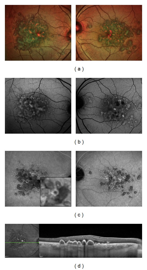
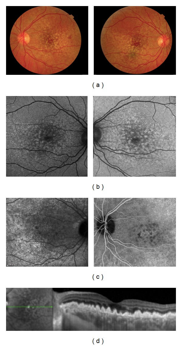
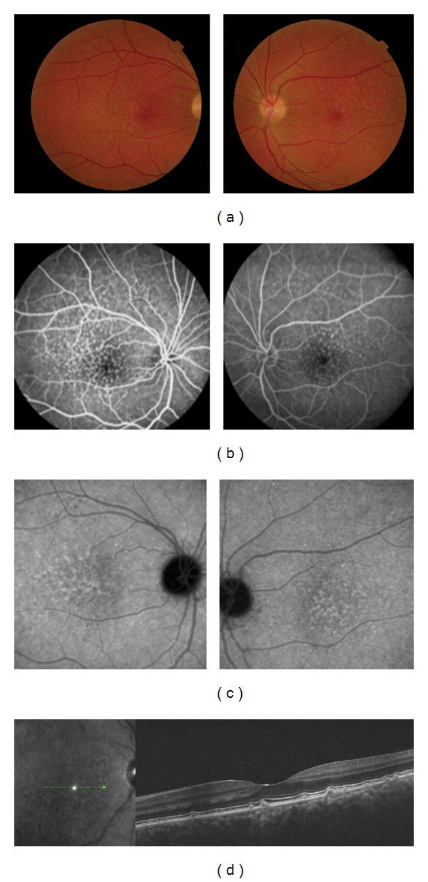
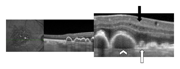
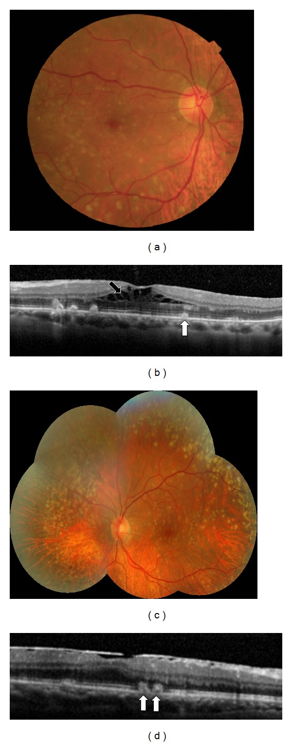
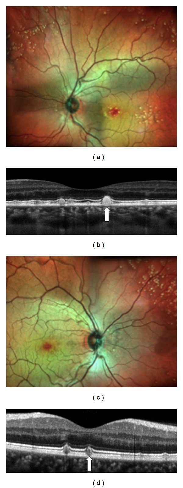
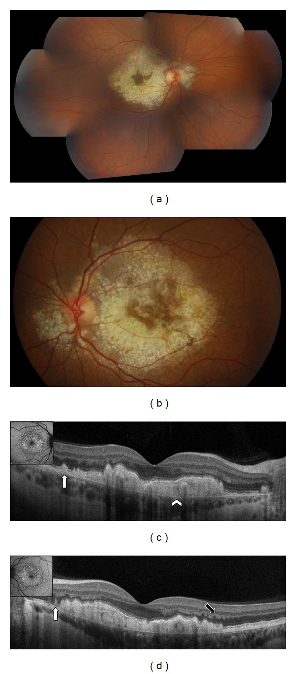
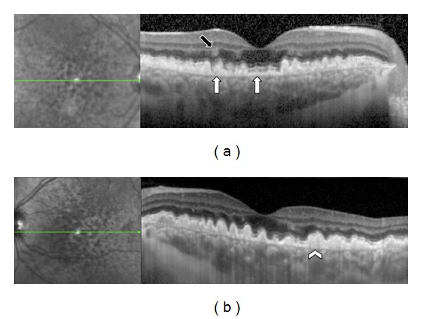
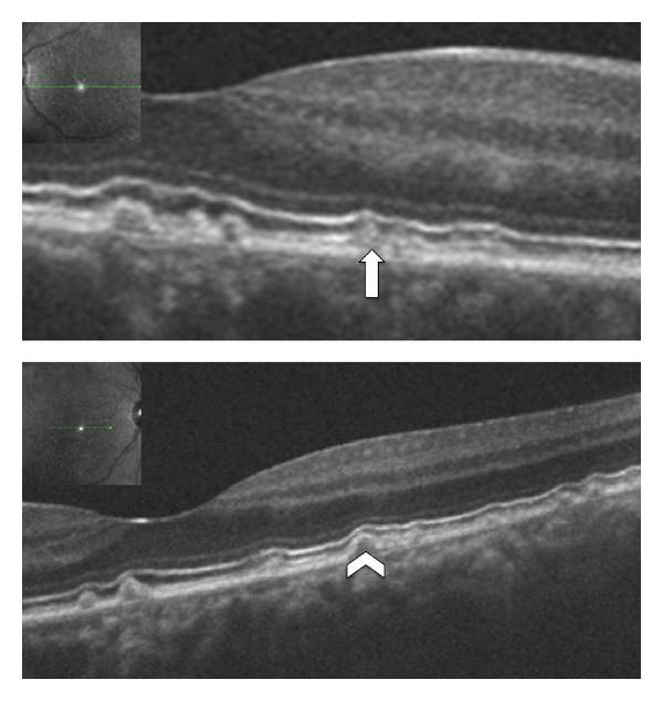
Purpose. To report an association between reticular pseudodrusen, located above the retinal pigment epithelium (RPE), and Early Onset Drusen (EOD) as described using Spectral-Domain Optical Coherence Tomography (SD-OCT). Methods. Eight patients (16 eyes) with EOD were examined. EOD were classified into three entities called Large Colloid Drusen (LCD), Malattia Leventinese (ML), and Cuticular Drusen (CD). Best-corrected visual acuity, fundus examination, color fundus photographs, fundus autofluorescence (FAF), fluorescein angiography (FA), indocyanine green angiography (ICGA), and SD-OCT were performed in all study patients. Results. Four patients had LCD, 2 had ML, and 2 had CD. Reticular pseudodrusen were observed with SD-OCT in all study patients; all these patients had hyperreflective lesions above and below the RPE. Conclusion. Early Onset Drusen appear to be associated with reticular pseudodrusen. SD-OCT is helpful in distinguishing the location of the deposits that are above and below the RPE in EOD. Further studies are needed to understand the role of reticular pseudodrusen in the pathophysiology of EOD.
Figures
Similar articles
-
Multimodal morphological and functional characterization of Malattia Leventinese.Graefes Arch Clin Exp Ophthalmol. 2013 Mar;251(3):705-14. doi: 10.1007/s00417-012-2106-5. Epub 2012 Jul 20. Graefes Arch Clin Exp Ophthalmol. 2013. PMID: 22814526
-
Insights into pathology of cuticular drusen from integrated confocal scanning laser ophthalmoscopy imaging and corresponding spectral domain optical coherence tomography.Graefes Arch Clin Exp Ophthalmol. 2011 Nov;249(11):1617-25. doi: 10.1007/s00417-011-1702-0. Epub 2011 May 10. Graefes Arch Clin Exp Ophthalmol. 2011. PMID: 21556939
-
Multimodal imaging of autosomal dominant drusen.Klin Monbl Augenheilkd. 2012 Apr;229(4):399-402. doi: 10.1055/s-0031-1299404. Epub 2012 Apr 11. Klin Monbl Augenheilkd. 2012. PMID: 22496012
-
Clinical Manifestations of Cuticular Drusen: Current Perspectives.Clin Ophthalmol. 2021 Sep 21;15:3877-3887. doi: 10.2147/OPTH.S272345. eCollection 2021. Clin Ophthalmol. 2021. PMID: 34584401 Free PMC article. Review.
-
Subretinal drusenoid deposits AKA pseudodrusen.Surv Ophthalmol. 2018 Nov-Dec;63(6):782-815. doi: 10.1016/j.survophthal.2018.05.005. Epub 2018 May 31. Surv Ophthalmol. 2018. PMID: 29859199 Review.
Cited by
-
Age-related macular degeneration eyes presenting with cuticular drusen and reticular pseudodrusen.Sci Rep. 2022 Apr 5;12(1):5681. doi: 10.1038/s41598-022-09608-9. Sci Rep. 2022. PMID: 35383241 Free PMC article.
-
Prevalence of Subretinal Drusenoid Deposits in Older Persons with and without Age-Related Macular Degeneration, by Multimodal Imaging.Ophthalmology. 2016 May;123(5):1090-100. doi: 10.1016/j.ophtha.2015.12.034. Epub 2016 Feb 10. Ophthalmology. 2016. PMID: 26875000 Free PMC article.
-
Quantitative analysis of photoreceptor layer reflectivity on en-face optical coherence tomography as an estimator of cone density.Graefes Arch Clin Exp Ophthalmol. 2017 Nov;255(11):2119-2126. doi: 10.1007/s00417-017-3761-3. Epub 2017 Aug 8. Graefes Arch Clin Exp Ophthalmol. 2017. PMID: 28791546
-
Haller's vessels patterns in non-neovascular age-related macular degeneration.Graefes Arch Clin Exp Ophthalmol. 2020 Oct;258(10):2163-2171. doi: 10.1007/s00417-020-04769-7. Epub 2020 Jun 13. Graefes Arch Clin Exp Ophthalmol. 2020. PMID: 32535671
-
Spotlight on reticular pseudodrusen.Clin Ophthalmol. 2017 Sep 20;11:1707-1718. doi: 10.2147/OPTH.S130165. eCollection 2017. Clin Ophthalmol. 2017. PMID: 29033536 Free PMC article. Review.
References
-
- Gass JDM. Drusen and disciform macular detachment and degeneration. Archives of Ophthalmology. 1973;90(3):206–217. - PubMed
-
- Guigui B, Leveziel N, Martinet V, et al. Angiography features of early onset drusen. British Journal of Ophthalmology. 2011;95(2):238–244. - PubMed
-
- Querques G, Guigui B, Leveziel N, et al. Insights into pathology of cuticular drusen from integrated confocal scanning laser ophthalmoscopy imaging and corresponding spectral domain optical coherence tomography. Graefe's Archive for Clinical and Experimental Ophthalmology. 2011;249(11):1617–1625. - PubMed
-
- Guigui B, Querques G, Leveziel N, et al. Spectral-domain optical tomography of early onset large colloid drusen. Retina. 2013 - PubMed
LinkOut - more resources
Full Text Sources
Other Literature Sources