. 2013 Dec 6:4:203.
doi: 10.4172/2157-7412.1000203.

Microtubule defects & Neurodegeneration

Affiliations

Microtubule defects & Neurodegeneration

Fiona J Baird et al. J Genet Syndr Gene Ther. .

Abstract

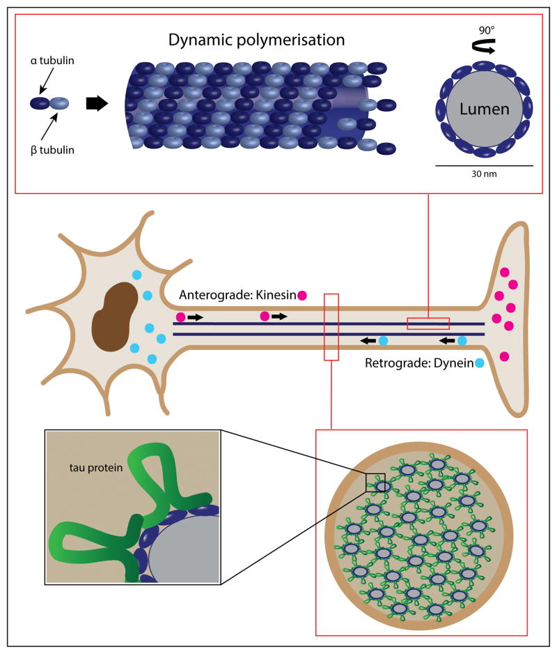
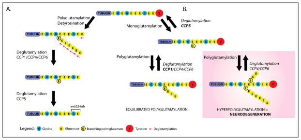
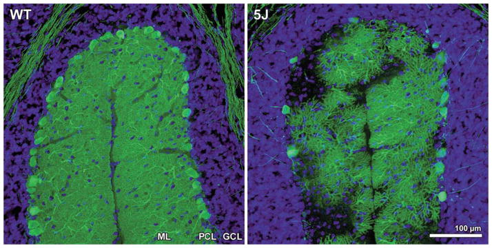
One of the major challenges facing the long term survival of neurons is their requirement to maintain efficient axonal transport over long distances. In humans as large, long-lived vertebrates, the machinery maintaining neuronal transport must remain efficient despite the slow accumulation of cell damage during aging. Mutations in genes encoding proteins which function in the transport system feature prominently in neurologic disorders. Genes known to cause such disorders and showing traditional Mendelian inheritance have been more readily identified. It has been more difficult, however, to isolate factors underlying the complex genetics contributing to the more common idiopathic forms of neurodegenerative disease. At the heart of neuronal transport is the rail network or scaffolding provided by neuron specific microtubules (MTs). The importance of MT dynamics and stability is underscored by the critical role tau protein plays in MT-associated stabilization versus the dysfunction seen in Alzheimer's disease, frontotemporal dementia and other tauopathies. Another example of the requirement for tight regulation of MT dynamics is the need to maintain balanced levels of post-translational modification of key MT building-blocks such as α-tubulin. Tubulins require extensive polyglutamylation at their carboxyl-terminus as part of a novel post-translational modification mechanism to signal MT growth versus destabilization. Dramatically, knock-out of a gene encoding a deglutamylation family member causes an extremely rapid cell death of Purkinje cells in the ataxic mouse model, pcd. This review will examine a range of neurodegenerative conditions where current molecular understanding points to defects in the stability of MTs and axonal transport to emphasize the central role of MTs in neuron survival.


Figures

Figure 1
Figure 1. The microtubule is a dynamic structure essential to axonal transport and neuron survival
The center panel depicts the basic neuron structure, highlighting the role of kinesin family proteins for anterograde transport from the cell body; and the dynein molecular motor, essential for retrograde transport back from the axon terminal. The upper panel represents and expansion from one isolated microtubule showing α-tubulin and β-tubulin as dynamic building blocks of the microtubule (side view and cross-section). The lower panel is an expansion of the axon in cross-section, underscoring the role of tau protein in microtubule stabilization.
Figure 2
Figure 2. The role of α-tubulin glutamate post-translational modification is depicted here, and can be appreciated by the rapid Purkinje Cell neurodegeneration which occurs in pcd mice via functional loss of the deglutamylation enzyme, CCP1
At the top of the figure is the eleven carboxyl-terminal amino-acids of mouse α-tubulin. The terminal tyrosine reside, which is cleavable, is indicated in red. (A), to the left, detyrosination and polyglutamylation generate an α-tubulin isoform that can be acted upon by cytosolic carboxy-Peptidases, CCP1, CCP4, and CCP6. Each of these enzymes can cleave glutamate residues from the polyE side-chain down to the ‘branch point’, and cleave the second last gene-encoded glutamate, creating the Δ2-Tub isomer. The role of CCP5 alone is to specifically cleave the branch glutamate. (B), on the right side, addition of the branch point glutamate initiates these modification. Thereafter, the situation where CCP1 or Nna1 is absent (pink shading), hyperglutamylation – neurodegeneration; is contrasted with the normal CCP1 present situation (no shading), defined by equilibrated polyglutamylation. Redundant function of CCP4 and CCP6 does not compensate for CCP1 loss, as studies have shown CCP1 is the enzyme predominantly expressed in the neuron subset which undergoes degeneration in pcd mice. This illustration was modified from figure 7 and supplemental material 1st reported by Rogowski et al., (2010).
Figure 3
Figure 3. Purkinje cell degeneration in pcd5J homozygous mice
Here we see cerebellar sections for matched folia from P25 wild-type DBA/2J (WT) and P25 pcd5J homozygous mice (5J) immunostained with an anti-calbindin antibody and counterstained with DAPI. Although WT P25 cerebellum displays numerous Purkinje cell neurons with extensive dendritic arborization visible in the molecular layer (ML), the pcd5J P25 cerebellum reveals pronounced loss of Purkinje cells and dramatic degeneration of surviving Purkinje cells. The degree of Purkinje cell loss and degeneration observed in pcd5J homozygous mice is consistent with the disease’s natural history in pcd5J homozygous mice, suggesting that 5J is a severe allele. ML = molecular layer; PCL = Purkinje cell layer; GCL = granule cell layer. Reproduced with permission from the corresponding author Albert R. La Spada (Chakrabarti et al., 2006).

References

    1. Audebert S, Koulakoff A, Berwald-Netter Y, Gros F, Denoulet P, Edde B. Developmental regulation of polyglutamylated alpha- and beta-tubulin in mouse brain neurons. J Cell Sci. 1994;107 (Pt 8):2313–2322. - PubMed
    1. Barra HS, Arce CA, Argarana CE. Posttranslational tyrosination/detyrosination of tubulin. Mol Neurobiol. 1988;2 (2):133–153. - PubMed
    1. Ben Othmane K, Middleton LT, Loprest LJ, Wilkinson KM, Lennon F, Rozear MP, Stajich JM, Gaskell PC, Roses AD, Pericak-Vance MA, et al. Localization of a gene (CMT2A) for autosomal dominant Charcot-Marie-Tooth disease type 2 to chromosome 1p and evidence of genetic heterogeneity. Genomics. 1993;17 (2):370–375. - PubMed
    1. Byrne BJ. Pathway for approval of a gene therapy orphan product: treading new ground. Mol Ther. 2013;21 (8):1465–1466. - PMC - PubMed
    1. Center for Drug Evaluation R, & Center for Biologics Evaluation and Research. Guidance for Industry Expedited Programs for Serious Conditions – Drugs and Biologics. F. a. D. A. U. S. A. U.S. Department of Health and Human Services; 2013.

LinkOut - more resources