. 2013;14 Suppl 16(Suppl 16):S13.
doi: 10.1186/1471-2105-14-S16-S13. Epub 2013 Oct 22.

MetaSel: a metaphase selection tool using a Gaussian-based classification technique

MetaSel: a metaphase selection tool using a Gaussian-based classification technique

Ravi Uttamatanin et al. BMC Bioinformatics. 2013.

Abstract

Background: Identification of good metaphase spreads is an important step in chromosome analysis for identifying individuals with genetic disorders. The process of finding suitable metaphase chromosomes for accurate clinical analysis is, however, very time consuming since they are selected manually. The selection of suitable metaphase chromosome spreads thus represents a major bottleneck for conventional cytogenetic analysis. Although many algorithms have been developed for karyotyping, none have adequately addressed the critical bottleneck of selecting suitable chromosome spreads. In this paper, we present a software tool that uses a simple rule-based system to efficiently identify metaphase spreads suitable for karyotyping.

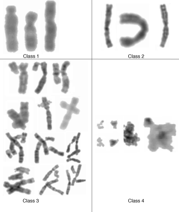
Results: The chromosome shapes can be classified by the software into four main classes. The first and the second classes refer to individual chromosomes with straight and skewed shapes, respectively. The third class is characterized as those chromosomes with overlapping bodies and the fourth class is for the non-chromosome objects. Good metaphase spreads should largely contain chromosomes of the first and the second classes, while the third class should be kept minimal. Several image parameters were examined and used for creating rule-based classification. The threshold value for each parameter is determined using a statistical model. We observed that the Gaussian model can represent the empirical probability density function of the parameters and, hence, the threshold value can be easily determined. The proposed rules can efficiently and accurately classify the individual chromosome with > 90% accuracy.

Conclusions: The software tool, termed MetaSel, was developed. Using the Gaussian-based rules, the tool can be used to quickly rank hundreds of chromosome spread images so as to assist cytogeneticists to perform karyotyping effectively. Furthermore, MetaSel offers an intuitive, yet comprehensive, workflow to assist karyotyping, including tools for editing chromosome (split, merge and fix) and a karyotyping editor (moving, rotating, and pairing homologous chromosomes). The program can be freely downloaded from "http://www4a.biotec.or.th/GI/tools/metasel".


Figures

Figure 1
Figure 1
Metaphase image and Karyotyping image. (A) The metaphase image with good metaphase spread that is proper for karyotyping (B) The karyotyping image with ordered/labelled chromosomes.
Figure 2
Figure 2
Types of chromosome classification. Chromosomes from Class-1 and Class-2 are individually separated. Both classes are differentiated by their straightness, i.e., Class-1 is straight individual chromosome while Class-2 is individually separable but with bended or skewed structure. Class-3 chromosomes are those that appear touching/overlapping with other chromosomes. Finally, Class-4 is characterized as non-chromosome residues and to be excluded in future analyses.
Figure 3
Figure 3
Image parameters for chromosome image classification. The segmented chromosomal objects in the left metaphase image are rotated into vertical orientation and calculated image parameters. Wrect and Hrect are width (pixel) and height (pixel) of the smallest enclosing rectangle of the segmented object respectively.
Figure 4
Figure 4
Empirical and Gaussian probability density functions of the area ratio. Gaussian model was used to determine the threshold value of the area ratio for classification. When the area ratio is greater than 67.84%, the chromosome can be classified as Class-1, straight object.
Figure 5
Figure 5
Empirical and Gaussian probability density functions of the Wrectratio. The experimental studies of this ratio were performed using 222, 327 and 500 samples of small, large residual objects and straight individual chromosomes respectively. When 0.9897 ≤ Wrectratio ≤ 1.5597, the object is classified as straight individual chromosome (Class-1) while if Wrectratio <0.9897 indicates that the object is potentially a small non-chromosome residue (Class-4). The object is considered to be a large residue (Class-4) when Wrectratio > 1.5597.
Figure 6
Figure 6
Empirical and Gaussian probability density functions of the height ratio. The statistical analysis was performed to determine the threshold value of the height ratio for eliminating residual objects. It can be observed that the empirical probability density function can be approximated by Gaussian model. From the Gaussian model, the objects are classified as residual objects when Hiratio < 0.7507. When Hiratio ≥ 0.7507, the objects are classified as mixing between skewed objects and touching/overlapping chromosomes.
Figure 7
Figure 7
Empirical and Gaussian probability density functions of the maximum width ratio. Gaussian model was used to approximate the empirical model for threshold calculation. The threshold for separating skewed individuals and overlapping chromosomes was chosen to be 2.3453 (the intercept between the two Gaussian curves). In other words, the objects will be classified as overlapping chromosomes when Wmaxratio is greater than this selected threshold. When Wmaxratio is less than or equal to the threshold, objects will be classified as skewed individuals.
Figure 8
Figure 8
Chromosomal image classification rule. (A) The proposed decision rule is used to classify chromosome images into four classes, straight individual chromosome (Class-1), skewed individual chromosome (Class-2), touching/overlapping chromosomes (Class-3) and non-chromosome residues (Class-4). (B) Pseudo-code of the previous rule-based flowchart.
Figure 9
Figure 9
Demonstration: Selecting metaphase image input directory. Users first choose the input directory containing metaphase images (Metaphase Analysis →Metaphase Directory tab).
Figure 10
Figure 10
Demonstration: Performing metaphase selection. After choosing the input directory, users can click the menu "Metaphase Selection" to perform the metaphase selection process.
Figure 11
Figure 11
Demonstration: window showing metaphase selection output. The metaphase images will be grouped into four classes and ranked according to the total number of individual chromosomes, which is calculated by combining the number of objects in Class-1 and Class-2.
Figure 12
Figure 12
Demonstration: Classification results of input Metaphase spreads. MetaSel lined up the individual chromosomes from Class-1, and Class-2. Users can observe the detail of the metaphase images and choose the analyzable image to perform karyotyping.
Figure 13
Figure 13
Demonstration: Semi-automatic karyotyping. MetaSel arranged all individual chromosomes by their lengths automatically. The touching/overlapping chromosomes were manually edited to separate them into individual chromosomes before karyotyping.

References

    1. Wang X, Li S, Liu H, Wood M, Chen W, Zheng B. Automated identification of analyzable metaphase chromosomes depicted on microscopic digital images. Journal of biomedical informatics. 2008;14(2):264–271. doi: 10.1016/j.jbi.2007.06.008. - DOI - PMC - PubMed
    1. Castleman Kenneth R. The PSI Automatic MetaphaseFinder. Journal of Radiation Research. 1992;14(Suppl 1):124–128. - PubMed
    1. Huber R, Kulka U, Lörch T, Braselmann H, Bauchinger M. Automated metaphase finding: an assessment of the efficiency of the METAFER2 system in a routine mutagenicity assay. Mutation Research/ Environmental Mutagenesis and Related Subjects. 1995;14:(1) 97–102. - PubMed
    1. Popescu M, Gader P, Keller J, Klein C, Stanley J, Caldwell C. Automatic karyotyping of metaphase cells with overlapping chromosomes. Computers in biology and medicine. 1999;14(1):61–82. doi: 10.1016/S0010-4825(98)00040-7. - DOI - PubMed
    1. Wang X, Zheng B, Li S, Mulvihill J, Wood C, Liu H. Automated classification of metaphase chromosomes: optimization of an adaptive computerized scheme". Journal of biomedical informatics. 2009;14(1):22–31. doi: 10.1016/j.jbi.2008.05.004. - DOI - PMC - PubMed

Publication types

LinkOut - more resources