Review
. 2013;4 Suppl 1(Suppl 1):S18.
doi: 10.1186/scrt379. Epub 2013 Dec 20.

Neurovascular unit on a chip: implications for translational applications

Review

Neurovascular unit on a chip: implications for translational applications

Donald J Alcendor et al. Stem Cell Res Ther. 2013.

Abstract

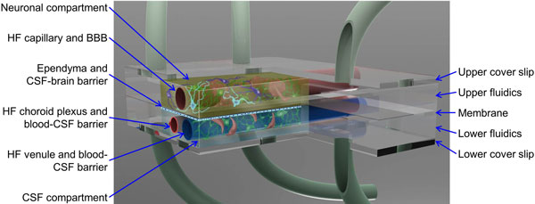
The blood-brain barrier (BBB) dynamically controls exchange between the brain and the body, but this interaction cannot be studied directly in the intact human brain or sufficiently represented by animal models. Most existing in vitro BBB models do not include neurons and glia with other BBB elements and do not adequately predict drug efficacy and toxicity. Under the National Institutes of Health Microtissue Initiative, we are developing a three-dimensional, multicompartment, organotypic microphysiological system representative of a neurovascular unit of the brain. The neurovascular unit system will serve as a model to study interactions between the central nervous system neurons and the cerebral spinal fluid (CSF) compartment, all coupled to a realistic blood-surrogate supply and venous return system that also incorporates circulating immune cells and the choroid plexus. Hence all three critical brain barriers will be recapitulated: blood-brain, brain-CSF, and blood-CSF. Primary and stem cell-derived human cells will interact with a variety of agents to produce critical chemical communications across the BBB and between brain regions. Cytomegalovirus, a common herpesvirus, will be used as an initial model of infections regulated by the BBB. This novel technological platform, which combines innovative microfluidics, cell culture, analytical instruments, bioinformatics, control theory, neuroscience, and drug discovery, will replicate chemical communication, molecular trafficking, and inflammation in the brain. The platform will enable targeted and clinically relevant nutritional and pharmacologic interventions for or prevention of such chronic diseases as obesity and acute injury such as stroke, and will uncover potential adverse effects of drugs. If successful, this project will produce clinically useful technologies and reveal new insights into how the brain receives, modifies, and is affected by drugs, other neurotropic agents, and diseases.


Figures

Figure 1
Figure 1
Microphysiological model of the neurovascular unit that supports blood, brain, and cerebral spinal fluid compartments. The system, under development by Vanderbilt University, Meharry Medical College, and the Cleveland Clinic Foundation, utilizes two rectangular, microfabricated compartments representing the brain and the cerebral spinal fluid (CSF) that are separated by a planar ependymal layer that forms the brain-CSF barrier. The upper chamber contains the neurons (purple and blue) and an artificial, hollow fiber (HF) capillary that carries blood to the brain surrounded by the cells that make up the blood-brain barrier (BBB). Endothelial cells line the luminal surface of the HF, and astrocytes (pink) and pericytes (green) cover the abluminal surface. The lower chamber is filled with CSF and contains a small HF that serves as an artificial choroid plexus (red) that produces CSF, and a larger HF venule (blue) that carries blood away from the brain and controls entry of immune cells into the CSF. Each HF, with inner diameters ranging from 200 to 600 μm, is lined with endothelial cells and surrounded by the appropriate cells for choroid plexus and venule function. The lower compartment thereby supports a collection of cells that form the blood-CSF and CSF-brain barriers. Collectively, all the cells will reproduce the neurovascular microenvironment found in the brain. Leukocytes can circulate in the blood-surrogate medium. Microdialysis fibers (not shown) in each compartment will enable near-real-time monitoring of metabolites and signaling molecules. The geometry is suitable for massive parallelization as required for high-content screening, and for daisy-chaining different brain regions to allow the study, for example, of chemical communication in the developing brain.
Figure 2
Figure 2
Benefit of translating various components of the neurovascular unit on a chip to pharmaceutical industry/clinic. The initial work represents largely uncoupled biological development (green shading) of well-plate triculture of endothelial cells, pericytes, and astrocytes and technological development (blue) of microfluidic micropumps and microvalves. Later, these technologies enable hollow fiber (HF) multiculture bioreactors that also include neurons and microglia. The integrated organ microfluidics technologies support studies with platforms containing relevant cell populations. Each of these four components has immediate translational potential to science, industry, and national security. Ultimately, the biology and technology are totally merged to create a fully instrumented neurovascular unit (NVU) suitable for translation to industry and medicine. The near-term and ultimate biological and technological returns from the development of a fully instrumented human NVU on a chip should improve our understanding of the physiology of the blood-brain interactions and the development and assessment of the safety and toxicity of both new central nervous system (CNS) drugs and systemic drugs that might affect the CNS. As a result, the approach outlined here could contribute to the diagnosis, treatment, and even prevention of traumatic brain injury, obesity, aging and neurodevelopmental abnormalities, cancer, stroke, epilepsy, Alzheimer's disease, schizophrenia, Parkinson's disease, HIV-associated neurocognitive disorders, neurotropic viruses and parasites, and drug addiction. A low-cost NVU model could be useful in studies of brain tissue regeneration, drug interference toxicity screening, personalized medicine, neural regenerative medicine, and brain-tissue stem-cell technologies.

References

    1. Esch MB, King TL, Shuler ML. The role of body-on-a-chip devices in drug and toxicity studies. Annu Rev Biomed Engr. 2011;4:55–72. doi: 10.1146/annurev-bioeng-071910-124629. - DOI - PubMed
    1. van der Meer AD, van den Berg A. Organs-on-chips: breaking the in vitro impasse. Integr Biol. 2012;4:461–470. doi: 10.1039/c2ib00176d. - DOI - PubMed
    1. Huh D, Torisawa YS, Hamilton GA, Kim HJ, Ingber DE. Microengineered physiological biomimicry: organs-on-chips. Lab Chip. 2012;4:2156–2164. doi: 10.1039/c2lc40089h. - DOI - PubMed
    1. Wikswo JP, Block FE III, Cliffel DE, Goodwin CR, Marasco CC, Markov DA, McLean DL, McLean JA, McKenzie JR, Reiserer RS, Samson PC, Schaffer DK, Seale KT, Sherrod SD. Engineering challenges for instrumenting and controlling integrated organ-on-a-chip systems. IEEE Trans Biomed Eng. 2013;4:682–690. - PMC - PubMed
    1. Integrated Microphysiological Systems for Drug Efficacy and Toxicity Testing in Human Health and Disease (UH2/UH3) http://grants.nih.gov/grants/guide/rfa-files/RFA-RM-11-022.html

Publication types

MeSH terms

Substances