. 2014 Feb 24;5(1):28.
doi: 10.1186/scrt417.

Human Wharton's jelly mesenchymal stem cells promote skin wound healing through paracrine signaling

Human Wharton's jelly mesenchymal stem cells promote skin wound healing through paracrine signaling

Anna I Arno et al. Stem Cell Res Ther. .

Abstract

Introduction: The prevalence of nonhealing wounds is predicted to increase due to the growing aging population. Despite the use of novel skin substitutes and wound dressings, poorly vascularized wound niches impair wound repair. Mesenchymal stem cells (MSCs) have been reported to provide paracrine signals to promote wound healing, but the effect of human Wharton's jelly-derived MSCs (WJ-MSCs) has not yet been described in human normal skin.

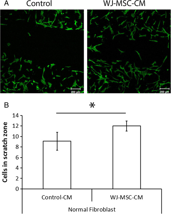
Methods: Human WJ-MSCs and normal skin fibroblasts were isolated from donated umbilical cords and normal adult human skin. Fibroblasts were treated with WJ-MSC-conditioned medium (WJ-MSC-CM) or nonconditioned medium.

Results: Expression of genes involved in re-epithelialization (transforming growth factor-β2), neovascularization (hypoxia-inducible factor-1α) and fibroproliferation (plasminogen activator inhibitor-1) was upregulated in WJ-MSC-CM-treated fibroblasts (P≤0.05). WJ-MSC-CM enhanced normal skin fibroblast proliferation (P≤0.001) and migration (P≤0.05), and promoted wound healing in an excisional full-thickness skin murine model.

Conclusions: Under our experimental conditions, WJ-MSCs enhanced skin wound healing in an in vivo mouse model.


Figures

Figure 1
Figure 1
Upregulation of wound healing genes by human Wharton's jelly-derived mesenchymal stem cell conditioned medium in human normal skin fibroblasts. (A-L) mRNA transcript expression relative to 18S after 7 days of culture of human normal skin fibroblasts with Wharton's jelly-derived mesenchymal stem cell conditioned medium (WJ-MSC-CM; treatment group) or nonconditioned medium (control group) from five different patients (but four patients for FGF-2 and three patients for collagen I, collagen III and decorin). Overall, WJ-MSC-CM enhanced a wound healing promoting phenotype in human normal skin fibroblasts in our culture conditions. *P ≤ 0.05. COL, collagen; CTGF, connective tissue growth factor; FGF-2, fibroblast growth factor-2; HIF-1α, hypoxia-inducible factor-1α; PAI-1, plasminogen activator inhibitor-1; TGF-β, transforming growth factor beta; VEGF, vascular endothelial growth factor.
Figure 2
Figure 2
Wharton's jelly-derived mesenchymal stem cell conditioned medium increases normal skin fibroblast proliferation, but does not affect apoptosis. Cell proliferation was examined using Ki67 staining. (A) Wharton's jelly-derived mesenchymal stem cell conditioned medium (WJ-MSC-CM)-treated normal fibroblasts showed enhanced proliferative rates compared with the control group (quantified in (B)). (C) Terminal transferase TdT-mediated dUTP biotin end-labeling (TUNEL) staining of WJ-MSC-CM-treated human normal skin fibroblasts versus control (non-WJ-MSC-CM-treated) normal skin fibroblasts showed no significant difference in induction of apoptosis (quantified in (E)). Note that the total number of viable cells was significantly higher in the WJ-MSC-CM-treated cells compared with the non-WJ-MSC-CM-treated cells (D). n = 3 samples per group. *P ≤ 0.05, ***P ≤ 0.001. DAPI, 4′,6-diamidino-2-phenylindole.
Figure 3
Figure 3
Wharton's jelly-derived mesenchymal stem cell conditioned medium accelerates wound closure in vitro. (A), (B) Scratch wound assay was performed to examine migration properties of Wharton's jelly-derived mesenchymal stem cell conditioned medium (WJ-MSC-CM)-treated and untreated normal skin fibroblasts. The treated group showed significantly enhanced migration rates and coapted wound borders faster than the control group. *P ≤ 0.05.
Figure 4
Figure 4
Wharton's jelly-derived mesenchymal stem cell conditioned medium enhances wound healing in an in vivo mouse model. A murine wound healing model was used, and animals were wounded and treated as described previously. Images corresponded to histological sections of wounds and the satellite donut area (10×) from BALB-c mice, after 1 week of full-thickness excisional skin wounding and reconstruction with Wharton's jelly-derived mesenchymal stem cell conditioned medium (WJ-MSC-CM) and vehicle (Matrigel; BD Biosciences, San Jose, CA, USA) (B), or vehicle alone (A). Photomicrographs were taken after Masson’s Trichrome staining. BALB-c mice WJ-MSC-CM-treated wounds showed enhanced wound healing rates compared to the control mice (P ≤ 0.05) (C). *P ≤ 0.05. Error bars represent the 95% confidence interval.
Figure 5
Figure 5
Wharton's jelly-derived mesenchymal stem cell conditioned medium promotes cell proliferation in an in vivo mouse wound healing model. A BALB-c mouse wound healing model was used and animals were wounded and treated as described previously. Animals received one dose of bromodeoxyuridine (BrdU) intraperitoneally 24 hours before harvesting of wounds. Four animals were included in each group, and four wounds were performed per animal (total of 16 wounds in each group). Cutaneous tissue specimens were stained for BrdU in both groups, control (A) and treatment (B). Enhanced magnification (40×) of the above microscopic images were included for nonconditioned medium-treated (C) and Wharton's jelly-derived mesenchymal stem cell conditioned medium (WJ-MSC-CM)-treated normal skin fibroblasts (D) to examine in further detail the increase in cell number or stained nuclei (black arrows, BrdU-positive cells) in the WJ-MSC-CM-treated wounds, compared with controls. This denoted that WJ-MSC-CM stimulated cell proliferation in vivo(E, F). Together, these results suggest that WJ-MSC promoted wound healing and repair by one-way paracrine signaling in an in vivo preclinical model. *P ≤ 0.05, **P ≤ 0.01. Error bars represent the 95% confidence interval. Arrows, BrdU-positive nuclei; arrowheads, BrdU-negative nuclei. HPF, high-power field.

References

    1. Caspersen CJ, Thomas GD, Boseman LA, Beckles GL, Albright AL. Aging, diabetes, and the public health system in the United States. Am J Public Health. 2012;102:1482–1497. doi: 10.2105/AJPH.2011.300616. - DOI - PMC - PubMed
    1. Khosrotehrani K. Mesenchymal stem cell therapy in skin: why and what for? Exp Dermatol. 2013;22:307–310. doi: 10.1111/exd.12141. - DOI - PubMed
    1. Liu S, Yuan M, Hou K, Zhang L, Zheng X, Zhao B, Sui X, Xu W, Lu S, Guo Q. Immune characterization of mesenchymal stem cells in human umbilical cord Wharton’s jelly and derived cartilage cells. Cell Immunol. 2012;278:35–44. doi: 10.1016/j.cellimm.2012.06.010. - DOI - PubMed
    1. Hanson SE, Bentz ML, Hematti P. Mesenchymal stem cell therapy for nonhealing cutaneous wounds. Plast Reconstr Surg. 2010;125:510–516. doi: 10.1097/PRS.0b013e3181c722bb. - DOI - PMC - PubMed
    1. Nekanti U, Rao VB, Bahirvani AG, Jan M, Totey S, Ta M. Long-term expansion and pluripotent marker array analysis of Wharton’s jelly-derived mesenchymal stem cells. Stem Cells Dev. 2010;19:117–130. doi: 10.1089/scd.2009.0177. - DOI - PubMed

Publication types

MeSH terms

Substances