. 2014 Feb 24:13:35.
doi: 10.1186/1476-4598-13-35.

MicroRNA-26b suppresses the NF-κB signaling and enhances the chemosensitivity of hepatocellular carcinoma cells by targeting TAK1 and TAB3

Affiliations

MicroRNA-26b suppresses the NF-κB signaling and enhances the chemosensitivity of hepatocellular carcinoma cells by targeting TAK1 and TAB3

Na Zhao et al. Mol Cancer. .

Abstract

Background: Abnormal activation of the NF-κB pathway is closely related to tumorigenesis and chemoresistance. Therefore, microRNAs that possess the NF-κB inhibitory activity may provide novel targets for anti-cancer therapy. miR-26 family members have been shown to be frequently downregulated in hepatocellular carcinoma (HCC) and correlated with the poor survival of HCC patients. To date, there is no report disclosing the regulatory role of miR-26 on the NF-κB pathway and its biological significance.

Methods: The effects of miR-26b on the NF-κB signaling pathway and the chemosensitivity of cancer cells were examined in two HCC cell lines, QGY-7703 and MHCC-97H, using both gain- and loss-of-function studies. The correlation between miR-26b level and apoptosis rate was further investigated in clinical HCC specimens.

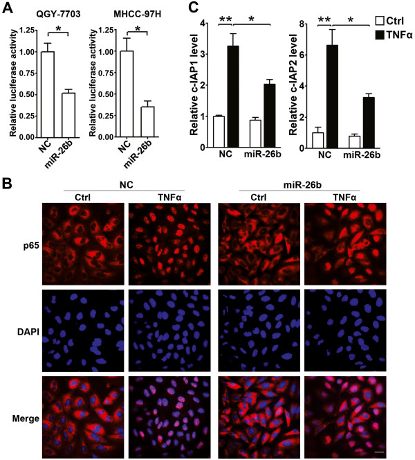
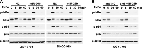
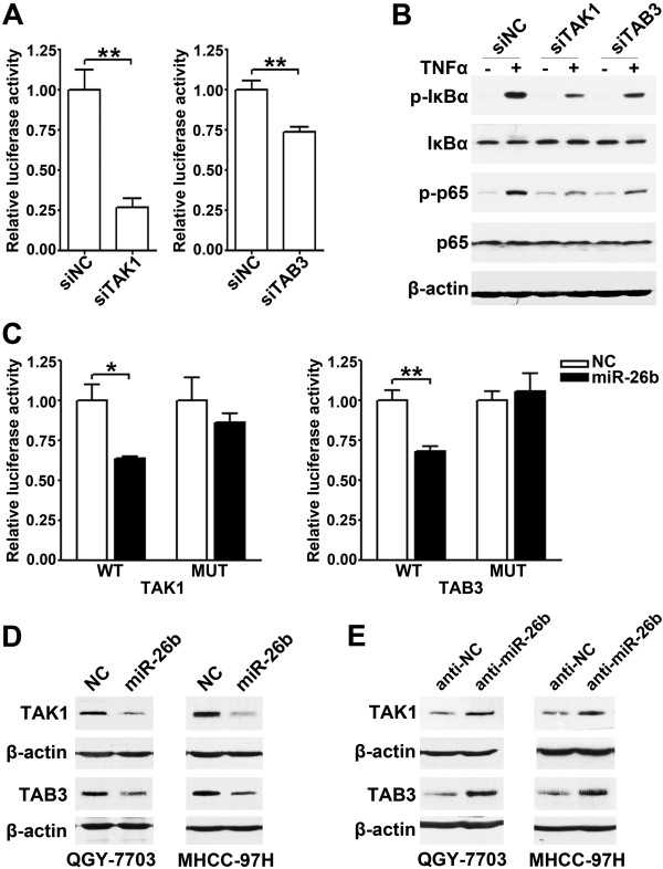
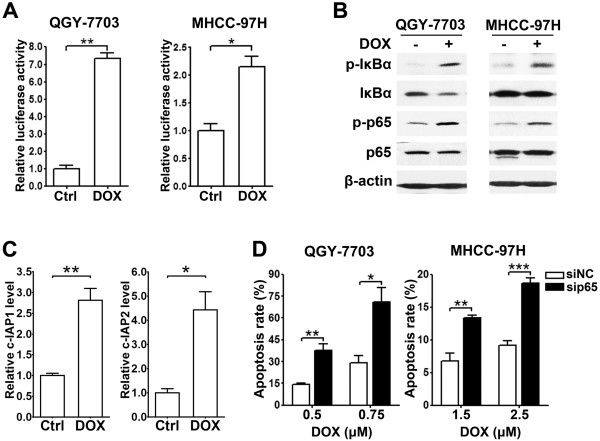
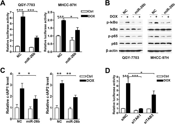
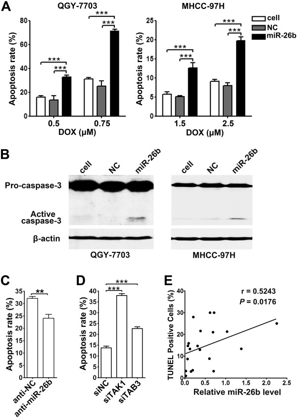
Results: Both TNFα and doxorubicin treatment activated the NF-κB signaling pathway in HCC cells. However, the restoration of miR-26b expression significantly inhibited the phosphorylation of IκBα and p65, blocked the nuclear translocation of NF-κB, reduced the NF-κB reporter activity, and consequently abrogated the expression of NF-κB target genes in TNFα or doxorubicin-treated HCC cells. Furthermore, the ectopic expression of miR-26b dramatically sensitized HCC cells to the doxorubicin-induced apoptosis, whereas the antagonism of miR-26b attenuated cell apoptosis. Consistently, the miR-26b level was positively correlated with the apoptosis rate in HCC tissues. Subsequent investigations revealed that miR-26b inhibited the expression of TAK1 and TAB3, two positive regulators of NF-κB pathway, by binding to their 3'-untranslated region. Moreover, knockdown of TAK1 or TAB3 phenocopied the effects of miR-26b overexpression.

Conclusions: These data suggest that miR-26b suppresses NF-κB signaling and thereby sensitized HCC cells to the doxorubicin-induced apoptosis by inhibiting the expression of TAK1 and TAB3. Our findings highlight miR-26b as a potent inhibitor of the NF-κB pathway and an attractive target for cancer treatment.


Figures

Figure 1
Figure 1
miR-26b suppresses the TNFα-stimulated NF-κB signaling. (A) miR-26b inhibited the TNFα-induced NF-κB reporter activity. HCC cells were first transfected with NC or miR-26b duplexes, followed by co-transfection of pRL-TK and pNF-κB-Luc or pTAL-Luc, treatment with TNFα, and analysis for luciferase activity. (B) miR-26b blocked the TNFα-induced nuclear translocation of NF-κB. QGY-7703 cells transfected with NC or miR-26b were untreated (Ctrl) or treated with TNFα before immunofluorescent staining for p65 (red). The nuclei were stained blue with DAPI. Scale bar, 10 μm. (C) miR-26b suppressed the TNFα-induced expression of NF-κB target genes. QGY-7703 cells transfected with NC or miR-26b were treated with 20 ng/ml TNFα for 3 hours before qPCR analysis. *, P < 0.05; **, P < 0.01.
Figure 2
Figure 2
miR-26b inhibits the TNFα-induced phosphorylation of IκBα and p65. (A) Introduction of miR-26b attenuated the TNFα-induced phosphorylation of IκBα and p65. HCC cells transfected with NC or miR-26b duplexes were treated with 20 ng/ml TNFα for the indicated time before immunoblotting. (B) Antagonism of miR-26b enhanced the TNFα-induced phosphorylation of IκBα and p65. QGY-7703 cells transfected with anti-NC or anti-miR-26b were treated with 2 ng/ml TNFα for the indicated time.
Figure 3
Figure 3
miR-26b suppresses NF-κB signaling by targeting TAK1 and TAB3. (A) Knockdown of TAK1 or TAB3 inhibited the TNFα-induced NF-κB reporter activity. QGY-7703 cells were treated and analyzed as in Figure  1A. (B) Knockdown of TAK1 or TAB3 attenuated the TNFα-induced phosphorylation of IκBα and p65. QGY-7703 cells transfected with siNC (lanes 1, 2), siTAK1 (lanes 3, 4) or siTAB3 (lanes 5, 6) were untreated (-) or treated with 20 ng/ml TNFα (+) for 3 minutes before immunoblotting. (C) miR-26b repressed the activity of the luciferase reporter containing the wild-type 3’UTR of TAK1 or TAB3. QGY-7703 cells were co-transfected with NC or miR-26b duplexes, pRL-TK and a firefly luciferase reporter plasmid carrying the wild-type (WT) or the mutant (MUT) 3’UTR of TAK1 or TAB3 before luciferase activity analysis. (D) Expression of miR-26b reduced the protein levels of cellular TAK1 and TAB3. HCC cells were transfected with NC or miR-26b duplexes for 48 hours before immunoblotting. (E) Antagonism of endogenous miR-26b enhanced the levels of TAK1 and TAB3 proteins. HCC cells were transfected with anti-NC or anti-miR-26b for 48 hours before immunoblotting. *, P < 0.05; **, P < 0.01.
Figure 4
Figure 4
Doxorubicin activates NF-κB signaling and knockdown of NF-κB promotes the doxorubicin-induced apoptosis. (A) Doxorubicin enhanced NF-κB reporter activity. HCC cells transfected with pRL-TK and pNF-κB-Luc or pTAL-Luc were untreated (Ctrl) or treated (DOX) with doxorubicin for 12 hours before luciferase activity analysis. (B) Doxorubicin treatment increased the phosphorylation of IκBα and p65. HCC cells were untreated (-) or treated (+) with doxorubicin for 6 hours before immunoblotting. (C) Doxorubicin stimulated the expression of NF-κB target genes. QGY-7703 cells were untreated (Ctrl) or treated (DOX) with doxorubicin for 24 hours before qPCR analysis. (D) Knockdown of p65 sensitized HCC cells to the doxorubicin-triggered apoptosis. HCC cells transfected with siNC or sip65 duplex were treated with indicated concentrations of doxorubicin for 48 hours before the morphological analysis for apoptosis by DAPI staining. *, P < 0.05; **, P < 0.01; ***, P < 0.001.
Figure 5
Figure 5
miR-26b abrogates the doxorubicin-activated NF-κB signaling. (A) Expression of miR-26b reduced the doxorubicin-induced NF-κB reporter activity. HCC cells were first transfected with NC or miR-26b duplexes, followed by co-transfection of pRL-TK and pNF-κB-Luc or pTAL-Luc, then remained untreated (Ctrl) or treated (DOX) with doxorubicin before luciferase activity analysis. (B) Introduction of miR-26b attenuated the doxorubicin-triggered phosphorylation of IκBα and p65. HCC cells transfected with NC or miR-26b were untreated (-) or treated (+) with doxorubicin for 6 hours before immunoblotting. (C) miR-26b suppressed the doxorubicin-induced expression of NF-κB target genes. QGY-7703 cells transfected with NC or miR-26b duplexes were untreated (Ctrl) or treated (DOX) with doxorubicin for 24 hours before qPCR analysis. (D) Knockdown of TAK1 or TAB3 suppressed the doxorubicin-stimulated NF-κB reporter activity. QGY-7703 cells were first transfected with the indicated siRNA duplexes, followed by treatment and analysis as in Figure  5A. *, P < 0.05; **, P < 0.01; ***, P < 0.001.
Figure 6
Figure 6
miR-26b sensitizes tumor cells to the doxorubicin-induced apoptosis. (A) Introduction of miR-26b sensitized HCC cells to the doxorubicin-induced apoptosis. Nontransfected (cell) or NC- or miR-26b-transfected HCC cells were treated with the indicated concentrations of doxorubicin (DOX) for 48 hours before apoptosis analysis by DAPI staining. (B) miR-26b expression increased the cleavage of pro-caspase-3. Nontransfected (cell) or NC- or miR-26b-transfected HCC cells were treated with doxorubicin for 48 hours before immunoblotting. (C) Antagonism of miR-26b desensitized tumor cells to the doxorubicin-induced apoptosis. QGY-7703 cells transfected with anti-NC or anti-miR-26b were treated with doxorubicin for 48 hours before apoptosis analysis by DAPI staining. (D) Knockdown of TAK1 or TAB3 sensitized HCC cells to the doxorubicin-induced apoptosis. QGY-7703 cells transfected with the indicated siRNA duplexes were treated with doxorubicin for 48 hours before apoptosis analysis by DAPI staining. (E) miR-26b expression is positively correlated with the rate of apoptosis in HCC tissues. The level of miR-26b was examined by real-time qPCR and normalized to RNU6B expression. Apoptosis was analyzed using TUNEL staining. The correlation between miR-26b levels and apoptosis rates in 20 HCC tissues was determined using Spearman’s correlation coefficient. **, P < 0.01; ***, P < 0.001.

References

    1. Perkins ND. The diverse and complex roles of NF-kappaB subunits in cancer. Nat Rev Cancer. 2012;12:121–132. - PubMed
    1. Ben-Neriah Y, Karin M. Inflammation meets cancer, with NF-kappaB as the matchmaker. Nat Immunol. 2011;12:715–723. doi: 10.1038/ni.2060. - DOI - PubMed
    1. Sakurai H. Targeting of TAK1 in inflammatory disorders and cancer. Trends Pharmacol Sci. 2012;33:522–530. doi: 10.1016/j.tips.2012.06.007. - DOI - PubMed
    1. McCool KW, Miyamoto S. DNA damage-dependent NF-kappaB activation: NEMO turns nuclear signaling inside out. Immunol Rev. 2012;246:311–326. doi: 10.1111/j.1600-065X.2012.01101.x. - DOI - PMC - PubMed
    1. Lu Z, Li Y, Takwi A, Li B, Zhang J, Conklin DJ, Young KH, Martin R, Li Y. miR-301a as an NF-kappaB activator in pancreatic cancer cells. EMBO J. 2011;30:57–67. doi: 10.1038/emboj.2010.296. - DOI - PMC - PubMed

Publication types

MeSH terms