Review
. 2014 Feb;42(1):94-104.
doi: 10.3810/psm.2014.02.2052.

Diagnosis and treatment of low back pain in the pediatric population

Affiliations
Review

Diagnosis and treatment of low back pain in the pediatric population

Alysha J Taxter et al. Phys Sportsmed. 2014 Feb.

Abstract

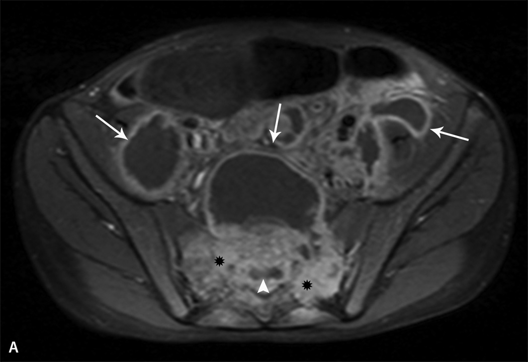
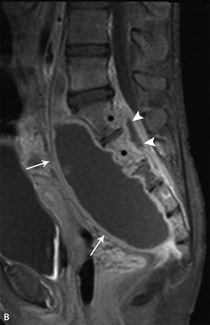
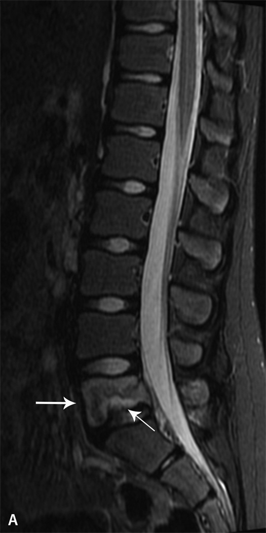
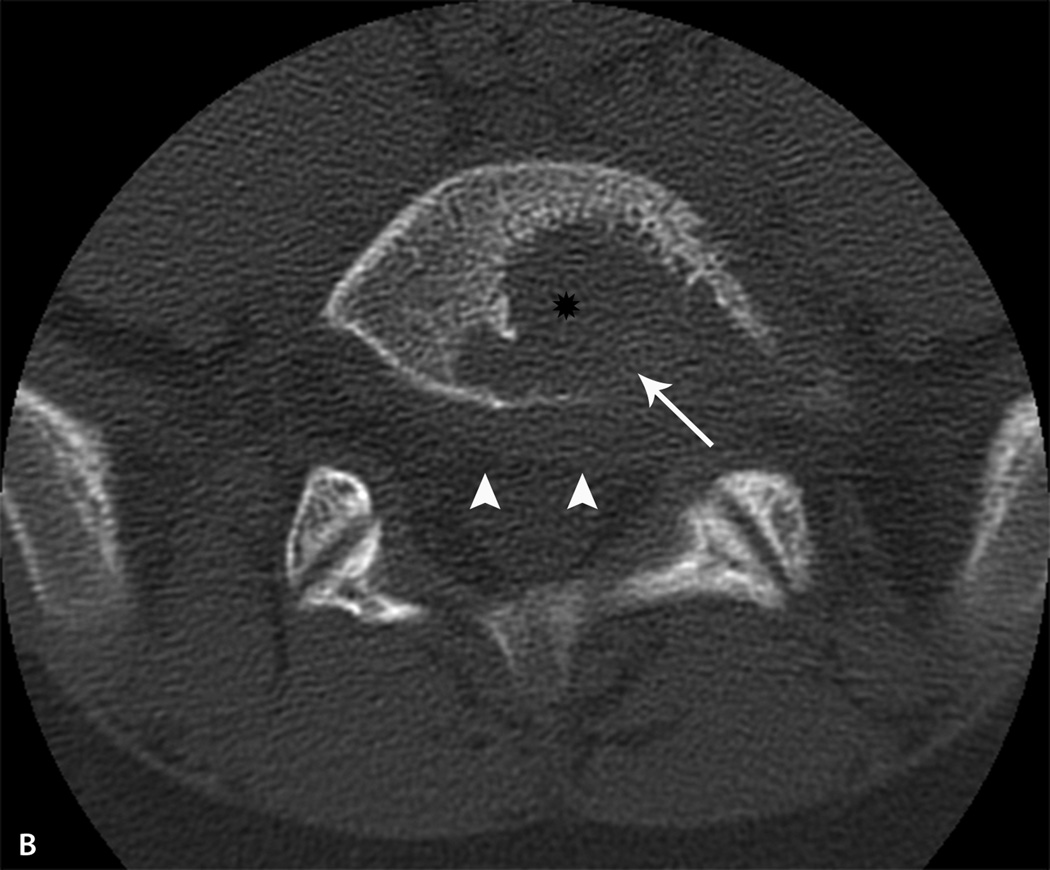
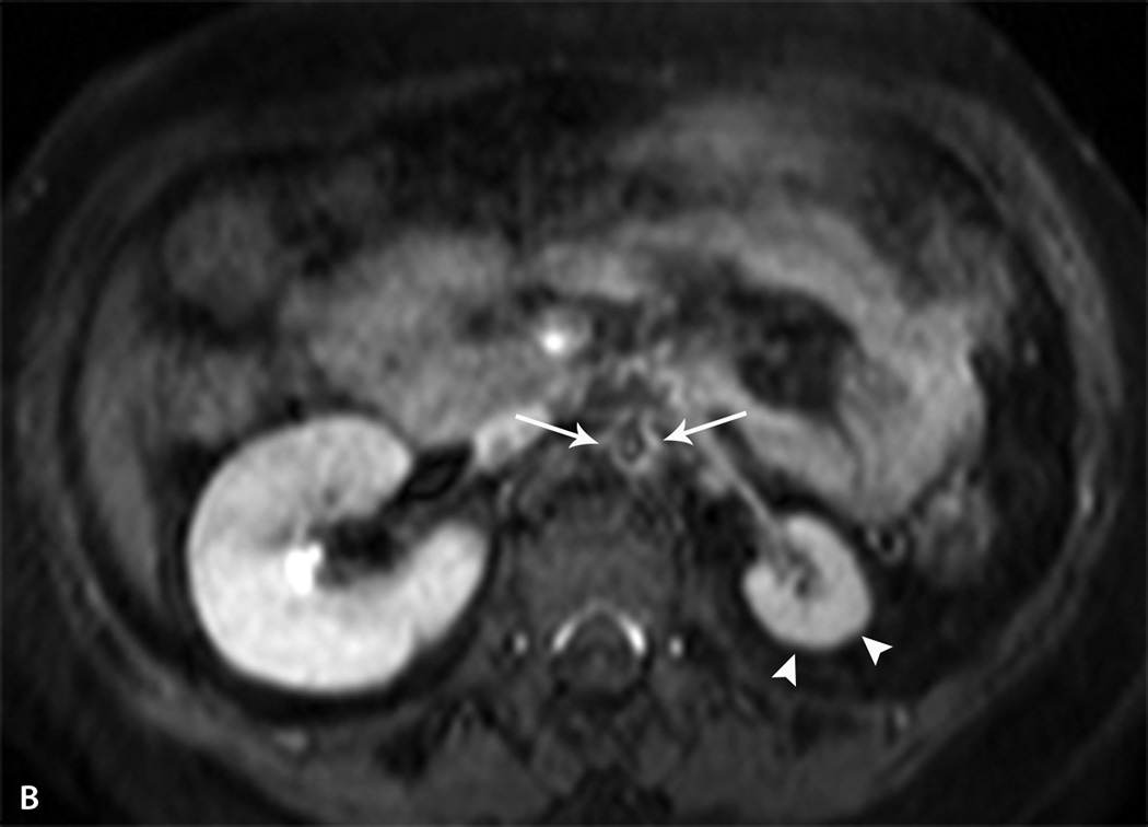
Back pain in the pediatric population is a common complaint presenting to sports medicine clinics. There is a wide differential that should be considered, including mechanical, infectious, neoplastic, inflammatory, and amplified musculoskeletal pain. The history, pain quality, and examination are key components to help distinguish the etiologies of the pain and direct further evaluation. Laboratory investigations, including blood counts and inflammatory markers, can provide insight into the diagnosis. The HLA-B27 antigen can be helpful if a spondyloarthropathy is suspected. Imaging as clinically indicated typically begins with radiographs, and the use of MRI, CT, or bone scan can provide additional information. Proper diagnosis of back pain is important because prognosis and treatments are significantly different. We review the pertinent evaluation, differential diagnoses, and treatment of low back pain in the pediatric population.


Figures

Figure 1
Figure 1
Tuberculosis. Axial (A) and sagittal (B) T1 fat-saturated post contrast images of the sacrum in a 10 year-old boy with lower back and abdominal pain demonstrate a large region of abnormal bone marrow enhancement (*) with bone destruction within the sacrum consistent with osteomyelitis. There is a large, multiseptated presacral abscess extending into the paraspinal and pelvic muscles (arrows) as well as an associated epidural abscess (arrowheads).
Figure 1
Figure 1
Tuberculosis. Axial (A) and sagittal (B) T1 fat-saturated post contrast images of the sacrum in a 10 year-old boy with lower back and abdominal pain demonstrate a large region of abnormal bone marrow enhancement (*) with bone destruction within the sacrum consistent with osteomyelitis. There is a large, multiseptated presacral abscess extending into the paraspinal and pelvic muscles (arrows) as well as an associated epidural abscess (arrowheads).
Fig. 2
Fig. 2
Langerhans Cell Histiocytosis. A. Sagittal fluid-sensitive MR image of the lumbar spine in a 12 yearold boy with back pain demonstrates edema within the L5 vertebral body (thick arrow) with bone destruction of the inferior aspect of the vertebra (thin arrow). B. Non-contrast CT axial image in bone algorithm shows a lytic lesion (*) with destruction of the cortex along the posterior aspect of the L5 vertebral body (arrow). There is abnormal soft tissue extending into the epidural space (arrowheads).
Fig. 2
Fig. 2
Langerhans Cell Histiocytosis. A. Sagittal fluid-sensitive MR image of the lumbar spine in a 12 yearold boy with back pain demonstrates edema within the L5 vertebral body (thick arrow) with bone destruction of the inferior aspect of the vertebra (thin arrow). B. Non-contrast CT axial image in bone algorithm shows a lytic lesion (*) with destruction of the cortex along the posterior aspect of the L5 vertebral body (arrow). There is abnormal soft tissue extending into the epidural space (arrowheads).
Fig. 3
Fig. 3
Chronic recurrent multifocal osteomyelitis. A. Sagittal fluid-sensitive MR image of the thoracolumbar spine in a 15 year-old girl with back pain demonstrates edema within the T10 and L4 vertebral bodies (thick arrows) with loss of vertebral body height. There is endplate irregularity of the T10 and L4 vertebral bodies with disc space narrowing above and below. Nuclear medicine bone scan was obtained to evaluate for additional lesions B. Tc -99m MDP 3- hour delayed whole body bone scan image in the anterior projection shows increased radiotracer activity within the T10 and L4 vertebral bodies. Abnormal radiotracer activity was also seen within the right greater trochanter (thin arrow) and within the right proximal tibia (circle) consistent with additional sites of inflammation. Normal, expected radiotracer activity is seen within the open physes.
Fig. 3
Fig. 3
Chronic recurrent multifocal osteomyelitis. A. Sagittal fluid-sensitive MR image of the thoracolumbar spine in a 15 year-old girl with back pain demonstrates edema within the T10 and L4 vertebral bodies (thick arrows) with loss of vertebral body height. There is endplate irregularity of the T10 and L4 vertebral bodies with disc space narrowing above and below. Nuclear medicine bone scan was obtained to evaluate for additional lesions B. Tc -99m MDP 3- hour delayed whole body bone scan image in the anterior projection shows increased radiotracer activity within the T10 and L4 vertebral bodies. Abnormal radiotracer activity was also seen within the right greater trochanter (thin arrow) and within the right proximal tibia (circle) consistent with additional sites of inflammation. Normal, expected radiotracer activity is seen within the open physes.
Fig. 4
Fig. 4
Enthesitis related arthritis. Axial fluid-sensitive MR image of a 16 year-old boy with lower back pain. Bone marrow edema is seen within the medial aspect of both iliac (thick arrows) and to a lesser extent, the left sacral ala (*). A small amount of fluid is seen within the right sacroiliac joint. There are erosive changes seen on the left (thin arrows). Follow-up laboratory studies revealed that the boy was HLA-B27 positive.
Fig. 5
Fig. 5
Takayasu arteritis. A. Coronal post contrast Maximum Intensity Projection (MIP) MR angiogram in the arterial phase of the chest, abdomen and pelvis in a 13 year-old girl demonstrates narrowing and irregularity of the mid to distal abdominal aorta (thin arrows). There is enlargement of the Arc of Rioland (thick arrow) due to severe stenosis of the celiac and superior mesenteric arteries (not shown). B. Axial post contrast T1 fat-saturated image shows severe narrowing of the abdominal aorta with a thick, enhancing wall (arrows). There is atrophy of the left kidney (arrowheads) due to severe stenosis of the left renal artery.
Fig. 5
Fig. 5
Takayasu arteritis. A. Coronal post contrast Maximum Intensity Projection (MIP) MR angiogram in the arterial phase of the chest, abdomen and pelvis in a 13 year-old girl demonstrates narrowing and irregularity of the mid to distal abdominal aorta (thin arrows). There is enlargement of the Arc of Rioland (thick arrow) due to severe stenosis of the celiac and superior mesenteric arteries (not shown). B. Axial post contrast T1 fat-saturated image shows severe narrowing of the abdominal aorta with a thick, enhancing wall (arrows). There is atrophy of the left kidney (arrowheads) due to severe stenosis of the left renal artery.

References

    1. Calvo-Munoz I, Gomez-Conesa A, Sanchez-Meca J. Prevalence of low back pain in children and adolescents: a meta-analysis. BMC Pediatr. 2013;13:14. - PMC - PubMed
    1. Anderson JA. Problems of classification of low-back pain. Rheumatol Rehabil. 1977 Feb;16(1):34–36. - PubMed
    1. Anderson L. Educational approaches to management of low back pain. Orthop Nurs. 1989 Jan-Feb;8(1):43–46. - PubMed
    1. d'Hemecourt PA, Gerbino PG, 2nd, Micheli LJ. Back injuries in the young athlete. Clin Sports Med. 2000 Oct;19(4):663–679. - PubMed
    1. Stracciolini A, Casciano R, Levey Friedman H, Meehan WP, 3rd, Micheli LJ. Pediatric sports injuries: an age comparison of children versus adolescents. Am J Sports Med. 2013 Aug;41(8):1922–1929. - PubMed

LinkOut - more resources