. 2014 May 1;123(18):2843-53.
doi: 10.1182/blood-2013-05-502435. Epub 2014 Feb 24.

Molecular mechanisms of neutrophil dysfunction in glycogen storage disease type Ib

Affiliations

Molecular mechanisms of neutrophil dysfunction in glycogen storage disease type Ib

Hyun Sik Jun et al. Blood. .

Abstract

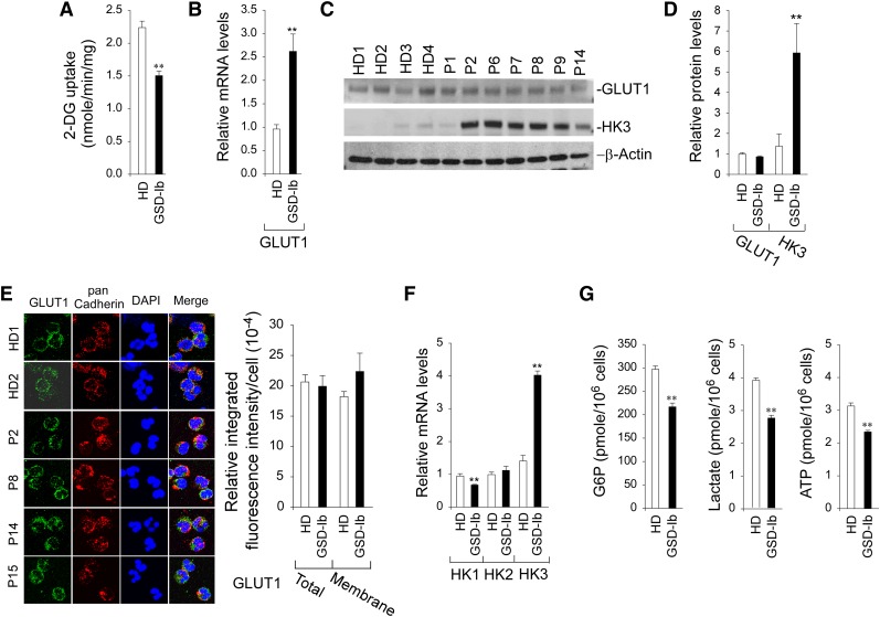
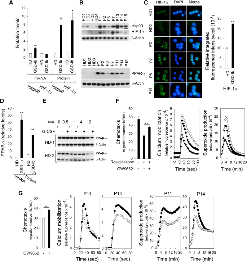
Glycogen storage disease type Ib (GSD-Ib) is an autosomal-recessive syndrome characterized by neutropenia and impaired glucose homeostasis resulting from a deficiency in the glucose-6-phosphate (G6P) transporter (G6PT). The underlying cause of GSD-Ib neutropenia is an enhanced neutrophil apoptosis, but patients also manifest neutrophil dysfunction of unknown etiology. Previously, we showed G6PT interacts with the enzyme glucose-6-phosphatase-β (G6Pase-β) to regulate the availability of G6P/glucose in neutrophils. A deficiency in G6Pase-β activity in neutrophils impairs both their energy homeostasis and function. We now show that G6PT-deficient neutrophils from GSD-Ib patients are similarly impaired. Their energy impairment is characterized by decreased glucose uptake and reduced levels of intracellular G6P, lactate, adenosine triphosphate, and reduced NAD phosphate, whereas functional impairment is reflected in reduced neutrophil respiratory burst, chemotaxis, and calcium mobilization. We further show that the mechanism of neutrophil dysfunction in GSD-Ib arises from activation of the hypoxia-inducible factor-1α/peroxisome-proliferators-activated receptor-γ pathway.


Figures

Figure 1
Figure 1
G6PT-deficient neutrophils of GSD-Ib patients with varying maturation states were dysfunctional. (A) Analysis of the levels of total (CD66b+) and immature (CD66b+CD16−/lo) neutrophils in the fixed, erythrocyte-depleted blood leukocytes by flow cytometry using CD66b and CD16 antibodies. Representative profiles from HDs and GSD-Ib and GSD-Ia patients are shown. (B) Hema-3–stained cytospins of isolated peripheral blood neutrophils. (C) Viability of freshly isolated and annexin V–depleted neutrophils. Annexin V–depleted blood neutrophils were used for respiratory burst, chemotaxis, and calcium mobilization analyses. (D) Neutrophil respiratory burst activity in response to PMA. Representative experiments are shown. (E) Neutrophil concentration-dependent chemotaxis in response to fMLP. Data represent the mean ± standard error of the mean (SEM) of 5 patients (P1, P8, P10, P12, and P14) examined in separate experiments. **P < .005. (F) Calcium mobilization in response to 10−7 M of fMLP. Representative experiments are shown. ○, HDs; ●, GSD-Ib patients; ▲, GSD-Ia patients.
Figure 2
Figure 2
Analysis of 2-DG uptake, the expression of GLUT1 and HK, and levels of intracellular G6P, lactate, and ATP in G6PT-deficient neutrophils of GSD-Ib patients. Annexin V–depleted peripheral blood neutrophils isolated from HDs and GSD-Ib patients were used in the study. For quantitative RT-PCR, data represent the mean ± SEM for HDs (n = 10) and GSD-Ib patients (n = 12). (A) Uptake of 2-DG. Data represent the mean ± SEM for HDs (n = 3) and GSD-Ib patients (n = 3). (B) Quantification of mRNA of GLUT1 by real-time RT-PCR. (C) Western blot analysis of protein extracts using antibodies against GLUT1 and HK3. Each lane contains 50 μg of protein. (D) Quantification of GLUT1 and HK3 protein levels by densitometry. Data represent the mean ± SEM for HDs (n = 8) and GSD-Ib patients (n = 7). (E) Confocal analysis of GLUT1 (green fluorescence), pan Cadherin membrane staining (red fluorescence), and DAPI nuclei staining (blue fluorescence) at original magnification ×630. (F) Quantification of HK1, HK2, and HK3 mRNA by real-time RT-PCR. (G) Quantification of G6P, lactate, and ATP. Data represent the mean ± SEM for HDs (n = 13) and GSD-Ib patients (n = 12). **P < .005.
Figure 3
Figure 3
Analysis of NADPH and the expression of NADPH oxidase in G6PT-deficient neutrophils of GSD-Ib patients. Annexin V–depleted peripheral blood neutrophils isolated from HDs and GSD-Ib patients were used in the study. (A) Levels of neutrophil NADPH. Data represent the mean ± SEM for HDs (n = 7) and GSD-Ib patients (n = 6). (B) Quantification of gp91phox, p22phox, and p47phox mRNA by real-time RT-PCR. Data represent the mean ± SEM for HDs (n = 10) and GSD-Ib patients (n = 12). (C) Western blot analysis of protein extracts using antibodies against gp91phox, p22phox, p47phox, or β-actin. Each lane contains 50 μg of protein. (D) The relative protein levels of gp91phox, p22phox, and p47phox were quantified by densitometry. Data represent the mean ± SEM for HDs (n = 6) and GSD-Ib patients (n = 6). (E) Confocal analysis of p47phox (green fluorescence), pan Cadherin membrane staining (red fluorescence), and DAPI nuclei staining (blue fluorescence) at original magnification ×630 and quantification of the relative integrated fluorescence intensity by ImageJ. The p47phox translocation from the cytoplasm to the plasma membrane was demonstrated by colocalization of p47phox with the plasma membrane marker pan Cadherin. **P < .005.
Figure 4
Figure 4
Analysis of levels of Hsp90, HIF-1α, and PPAR-γ in G6PT-deficient neutrophils and the effects of PPAR-γ antagonist/agonist on neutrophil function. Annexin V–depleted peripheral blood neutrophils isolated from HDs and GSD-Ib patients were used in the study. (A) Quantification of Hsp90 and HIF-1α mRNA levels by real-time RT-PCR and protein levels by densitometry. Data for RT-PCR represent the mean ± SEM for HDs (n = 10) and GSD-Ib patients (n = 12), and data for protein levels represent mean ± SEM for HDs (n = 11) and GSD-Ib patients (n = 7). (B) Western blot analysis of protein extracts using antibodies against Hsp90, HIF-1α, PPAR-γ, or β-actin. Data represent the mean ± SEM for HDs (n = 11) and GSD-Ib patients (n = 7). (C) Immunofluorescence of HIF-1α (green fluorescence) and DAPI nuclei staining (blue fluorescence) at original magnification ×400 and quantification of the relative integrated fluorescence intensity by ImageJ. (D) Quantification of PPAR-γ mRNA levels by real-time RT-PCR and protein levels by densitometry. Data for RT-PCR represent the mean ± SEM for HDs (n = 10) and GSD-Ib patients (n = 12), and data for PPAR-γ protein represent the mean ± SEM for HDs (n = 11) and GSD-Ib patients (n = 7). (E) Western blot analysis of the effects of G-CSF on PPAR-γ expression in HD neutrophils after in vitro culturing. (F) Effects of PPAR-γ agonist rosiglitazone and antagonist GW9662 on function of neutrophils isolated from HDs. Three independent experiments were conducted with similar results. Chemotaxis was examined in response to 10−7 M fMLP. Data represent the mean ± SEM. Calcium mobilization was examined in response to 10−7 M fMLP. Representative profiles are shown. Respiratory burst was examined in response to PMA. Representative profiles are shown. ○, control; ▲, PPAR-γ agonist rosiglitazone; ●, PPAR-γ agonist rosiglitazone followed by antagonist GW9662. (G) Effects of PPAR-γ antagonist GW9662 on function of G6PT-deficient neutrophils isolated from 2 GSD-Ib patients. Two independent experiments using neutrophils isolated from P11 and P14 were conducted. Chemotaxis was examined in response to 10−7 M fMLP. Data represent the mean ± SEM of both patients. Calcium mobilization was examined in response to 10−7 M fMLP. Respiratory burst was examined in response to PMA. ○, control; ●, PPAR-γ antagonist GW9662. **P < .005, *P < .05.
Figure 5
Figure 5
Inhibition of neutrophil function by PPAR-γ is mediated via HIF-1α signaling. Annexin V–depleted peripheral blood neutrophils isolated from HDs were used in the study. (A) Effects of the PPAR-γ antagonist GW9662 on chemotaxis, calcium mobilization, and respiratory burst activities of neutrophils exposing to hypoxic conditions, and western blot analysis of protein extracts using antibodies against HIF-1α, PPAR-γ, or β-actin. Three independent experiments were conducted with similar results. ○, normoxia; ●, hypoxia; ▾, hypoxia and PPAR-γ antagonist GW9662. (B) Effects of the PPAR-γ antagonist GW9662 on chemotaxis, calcium mobilization, and respiratory burst activities of neutrophils exposed to the hypoxia mimetic CoCl2, and western blot analysis of protein extracts using antibodies against HIF-1α, PPAR-γ, or β-actin. Three independent experiments were conducted with similar results. ○, control; ●,CoCl2; ▾, CoCl2 and PPAR-γ antagonist GW9662. (C) Effects of HIF-α inhibitor 2-ME2 on chemotaxis, calcium mobilization, and respiratory burst activities of neutrophils exposed to hypoxic conditions, and western blot analysis of protein extracts using antibodies against HIF-1α, PPAR-γ or β-actin. Three independent experiments were conducted with similar results. ○, normoxia; ●, hypoxia; ▾, hypoxia and 2-ME2. Chemotaxis was examined in response to 10−7 M fMLP. Data represent the mean ± SEM. Calcium mobilization was examined in response to 10−7 M fMLP. Representative profiles are shown. Respiratory burst was examined in response to PMA. Representative profiles are shown. **P < .005, *P < .05.
Figure 6
Figure 6
Proposed mechanisms that underlie neutrophil dysfunction in GSD-Ib. Glucose transported into the cytoplasm via GLUT1 is metabolized by HK to G6P, which participates in 3 major pathways: glycolysis, the HMS, and ER cycling. In cycling, G6P enters the ER via G6PT, where it can accumulate until it is hydrolyzed to glucose by G6Pase-β and transported back into the cytoplasm. By limiting the cytoplasmic glucose/G6P availability, cycling regulates the other 2 cytoplasmic pathways for G6P metabolism. Disruption of ER cycling in G6PT-deficient neutrophils results in reduced glucose uptake and impaired energy homeostasis and functionality. The underlying cause of neutropenia in GSD-Ib is enhanced neutrophil ER stress and oxidative stress. The increases in Hsp90 and ROS in G6PT-deficient neutrophils stabilize HIF-1α, an upstream activator of PPAR-γ. The increase in PPAR-γ downregulates neutrophil respiratory burst, chemotaxis, and calcium mobilization activities. GLUT1, responsible for the transport of glucose in and out of the cell, is shown embedded in the plasma membrane. The G6PT, responsible for the transport of G6P into the ER, and G6Pase-β, responsible for hydrolyzing G6P to glucose and phosphate, are shown embedded in the ER membrane. Thick arrows indicate the changes caused by a defect in G6PT activity.

Comment in

References

    1. Chou JY, Jun HS, Mansfield BC. Glycogen storage disease type I and G6Pase-β deficiency: etiology and therapy. Nat Rev Endocrinol. 2010;6(12):676–688. - PMC - PubMed
    1. Chou JY, Jun HS, Mansfield BC. Neutropenia in type Ib glycogen storage disease. Curr Opin Hematol. 2010;17(1):36–42. - PMC - PubMed
    1. Pan C-J, Lin B, Chou JY. Transmembrane topology of human glucose 6-phosphate transporter. J Biol Chem. 1999;274(20):13865–13869. - PubMed
    1. Lei K-J, Shelly LL, Pan C-J, Sidbury JB, Chou JY. Mutations in the glucose-6-phosphatase gene that cause glycogen storage disease type 1a. Science. 1993;262(5133):580–583. - PubMed
    1. Shieh J-J, Pan C-J, Mansfield BC, Chou JY. A glucose-6-phosphate hydrolase, widely expressed outside the liver, can explain age-dependent resolution of hypoglycemia in glycogen storage disease type Ia. J Biol Chem. 2003;278(47):47098–47103. - PubMed

Publication types

MeSH terms

Supplementary concepts