Review
. 2014 Apr;35(4):144-52.
doi: 10.1016/j.it.2014.01.002. Epub 2014 Feb 21.

Re-examining class-I presentation and the DRiP hypothesis

Affiliations
Review

Re-examining class-I presentation and the DRiP hypothesis

Kenneth L Rock et al. Trends Immunol. 2014 Apr.

Abstract

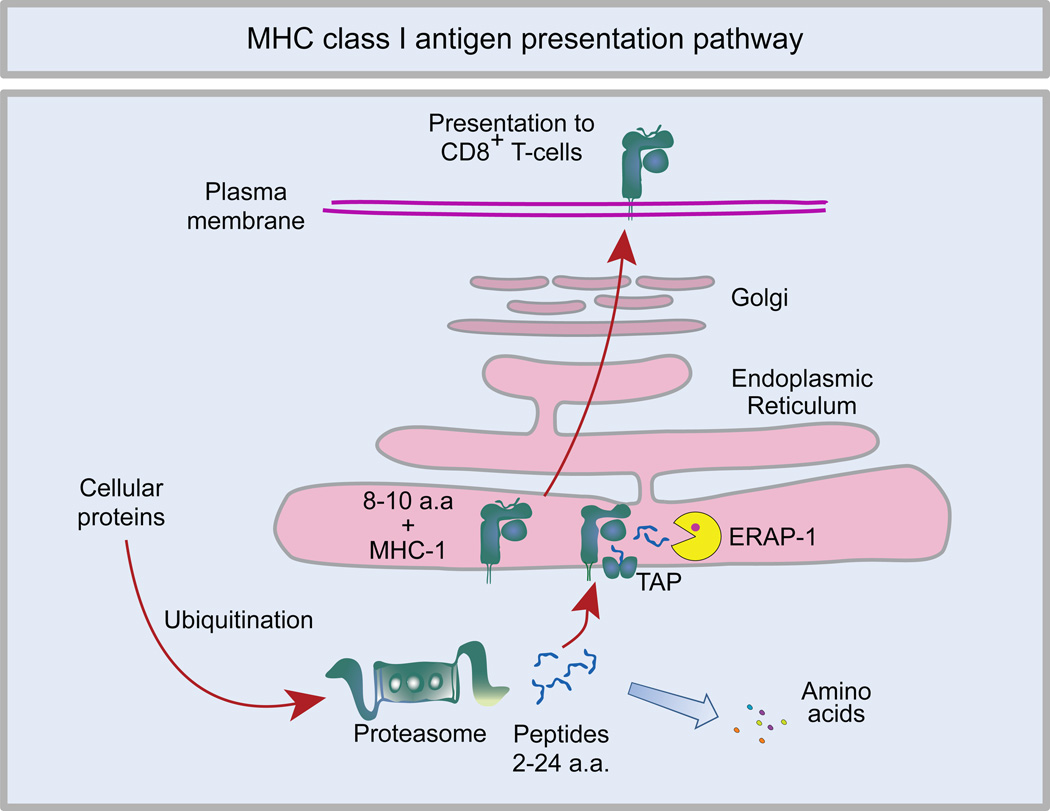
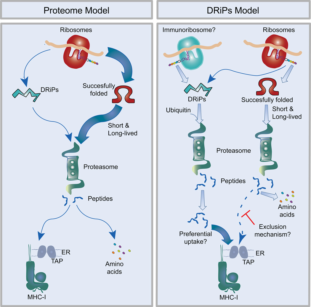
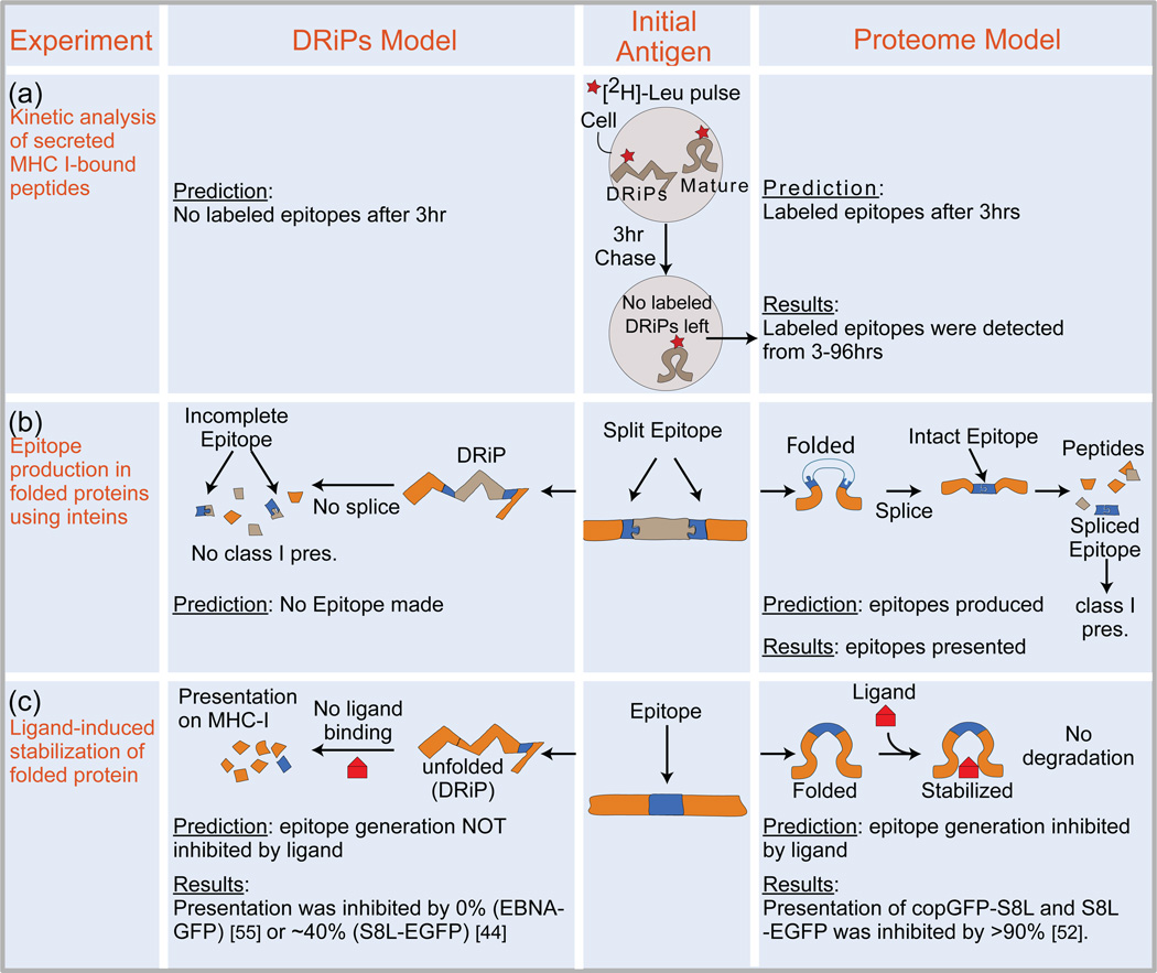
MHC class I molecules present peptides derived from intracellular proteins, enabling immune surveillance by CD8(+) T cells and the elimination of virus-infected and cancerous cells. It has been argued that the dominant source of MHC class I-presented peptides is through proteasomal degradation of newly synthesized defective proteins, termed defective ribosomal products (DRiPs). Here, we critically examine the DRiP hypothesis and discuss recent studies indicating that antigenic peptides are generated from the entire proteome and not just from failures in protein synthesis or folding.

Keywords: MHC class I; antigen presentation; defective ribosomal product; proteasome.


Figures

Figure 1
Figure 1. MHC class I antigen presentation pathway
Cellular proteins are degraded into oligopeptides by the ubiquitin-proteasome pathway. Most of these peptides are further degraded by peptidases into amino acids, but a small fraction are transferred into the lumen of the ER by the TAP-transporter, where they can be trimmed further into 8–10 amino acid fragments by the endoplasmic reticulum aminopeptidase-1 (ERAP-1). These peptides can then be bound by newly synthesized MHC class I and transported to the plasma membrane for display to CD8+ T cells.
Figure 2
Figure 2. Models for the source of MHC class I peptides
Proteome Model-Proteins are synthesized by ribosomes and subsequently fold into their mature conformation. The degradation of folded or unfolded proteins begins immediately after synthesis, following first order rate kinetics. Most of these cellular proteins are degraded by the ubiquitin-proteasome pathway into oligopeptides. A fraction of these peptides escape further hydrolysis into amino acids when they are transported into the ER. This process provides the MHC class I antigen presentation pathway with a sampling of peptides from all cellular proteins, including mature folded proteins and defective ones (the proteome). DRiPs Model- Ribosomes, or the hypothetical error-prone immunoribosomes, synthesizes a cohort of functional and defective proteins. The defective proteins (DRiPs) are ubiquitinated and rapidly degraded by the proteasome. By an unknown mechanism (possibly preferential uptake into the ER and/or exclusion of peptides derived from mature proteins) the DRiP-derived peptides may be preferentially loaded on MHC class I molecules. Consequently, newly synthesized defective proteins constitute the dominant source of presented peptides, either at early time points (e.g. post viral infection) and/or at all time points [35].
Figure 3
Figure 3. Experimental evidence supporting the DRiPs Model and the Proteome Model
(a) Cells were labeled for SILAC using heavy leucine (red star) for 7 days. Cells were then transferred to light leucine and secreted MHC:peptide complexes were collected every 3hrs for up to 96 hours. Peptides bound to the soluble MHC class I complexes were eluted and characterized by Mass Spectrometry. Since the labeled DRiPs would be very rapidly degraded, the DRiPs model predicts that peptides containing the heavy isotope would only be detected at early time points; in contrast the proteome model predicts that labeled peptides would continue to be generated over time. The actual data showed that peptides containing heavy leucine continued to be detected for ≥ 6 hours (with a half life of the label ≥3hrs), consistent with many presented peptides coming from the proteome [61] (b) Proteins were expressed with T cell epitopes (blue) split by an intervening sequence (inteins) so that the intact epitope would only be generated post-translationally from mature protein by intein catalysis and protein ligation. Since the epitope would not exist in an unfolded protein, the DRiP model predicted that no presented epitopes would be generated. The actual data showed the epitopes were presented with the same efficiency as ones that did not require splicing demonstrating that mature proteins were the dominant source of the presented peptides [64] (c) Antigens were expressed as protein fusions to a destabilizing domain causing the protein to rapidly degrade (left panel). Addition of a ligand (red) specific for the folded form of the protein prevented its turnover (right panel). In two studies, presentation of SIINFEKL fused to EGFP (S8L-EGFP) and the EBV protein EBNA1 fused to EGFP (EBNA-EGFP) were inhibited by 40% and 0%, respectively, suggesting that a majority of presented peptides came from unfolded protein species [44,55]. However, a later study using one of the same antigens (S8L-EGFP) and a new antigen (copepod GFP fused to SIINFEKL, copGFP-S8L), showed near complete inhibition (80–95%) of presentation in the presence of stabilizing ligand, indicating that the presented peptides came from mature protein [52].

References

    1. Rock KL, et al. Inhibitors of the proteasome block the degradation of most cell proteins and the generation of peptides presented on MHC class I molecules. Cell. 1994;78:761–771. - PubMed
    1. Zhao J, et al. FoxO3 coordinately activates protein degradation by the autophagic/lysosomal and proteasomal pathways in atrophying muscle cells. Cell Metab. 2007;6:472–483. - PubMed
    1. Glickman MH, Ciechanover A. The ubiquitin-proteasome proteolytic pathway: destruction for the sake of construction. Physiol. Rev. 2002;82:373–428. - PubMed
    1. Goldberg AL. Protein degradation and protection against misfolded or damaged proteins. Nature. 2003;426:895–899. - PubMed
    1. Pickart CM, Cohen RE. Proteasomes and their kin: proteases in the machine age. Nat. Rev. Mol. Cell Biol. 2004;5:177–187. - PubMed

Publication types

MeSH terms