. 2014 Feb 25:4:4163.
doi: 10.1038/srep04163.

Functional transformations of bile acid transporters induced by high-affinity macromolecules

Affiliations

Functional transformations of bile acid transporters induced by high-affinity macromolecules

Taslim A Al-Hilal et al. Sci Rep. .

Erratum in

Abstract

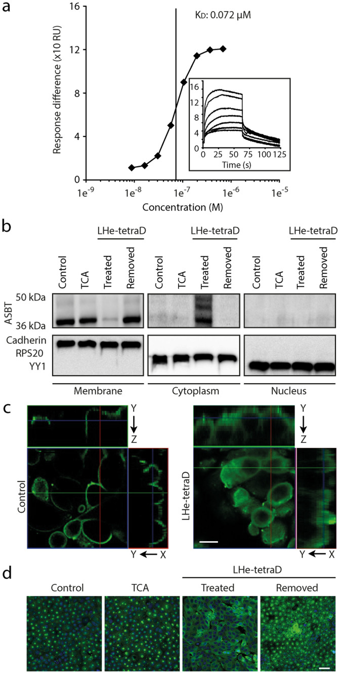
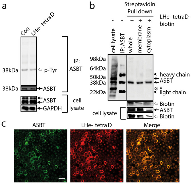
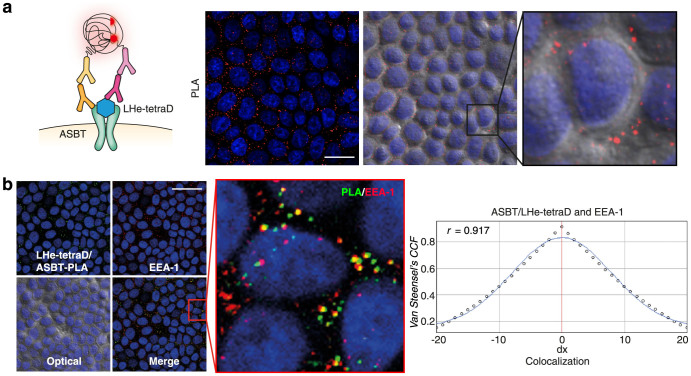
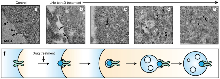
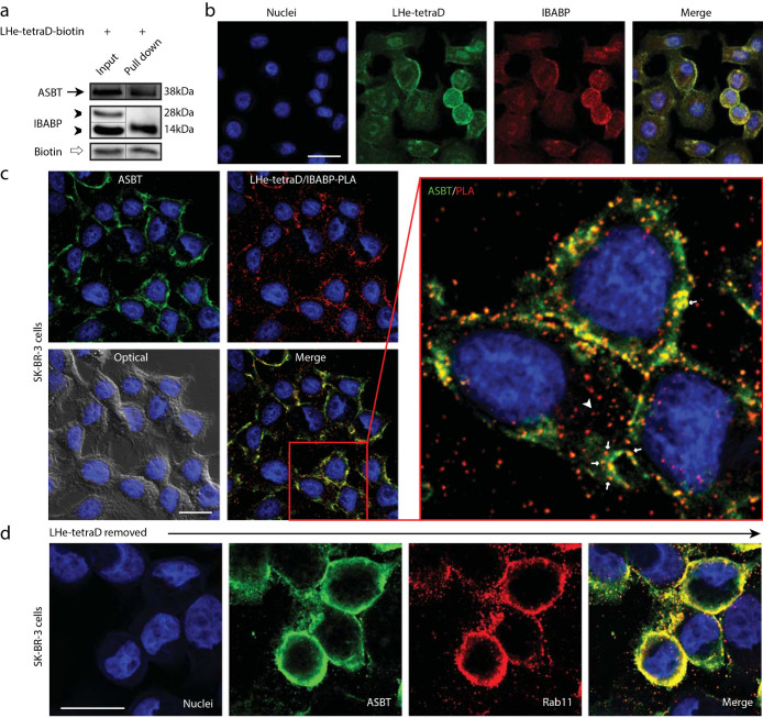
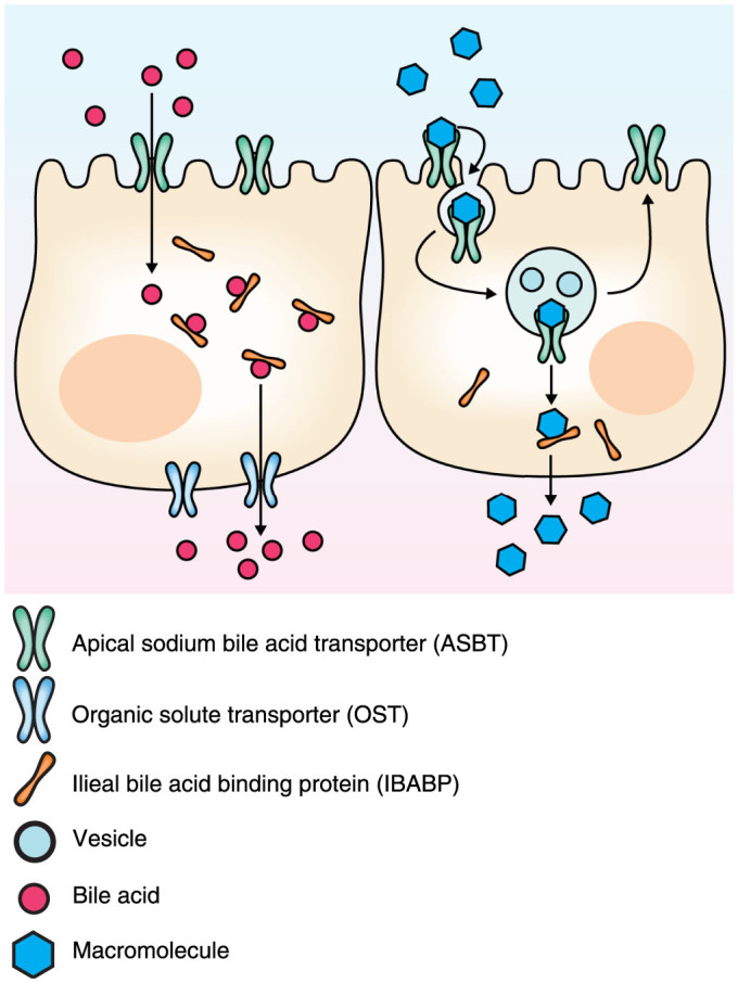
Apical sodium-dependent bile acid transporters (ASBT) are the intestinal transporters that form intermediate complexes with substrates and its conformational change drives the movement of substrates across the cell membrane. However, membrane-based intestinal transporters are confined to the transport of only small molecular substrates. Here, we propose a new strategy that uses high-affinity binding macromolecular substrates to functionally transform the membrane transporters so that they behave like receptors, ultimately allowing the apical-basal transport of bound macromolecules. Bile acid based macromolecular substrates were synthesized and allowed to interact with ASBT. ASBT/macromolecular substrate complexes were rapidly internalized in vesicles, localized in early endosomes, dissociated and escaped the vesicular transport while binding of cytoplasmic ileal bile acid binding proteins cause exocytosis of macromolecules and prevented entry into lysosomes. This newly found transformation process of ASBT suggests a new transport mechanism that could aid in further utilization of ASBT to mediate oral macromolecular drug delivery.


Figures

Figure 1
Figure 1. Distribution of ASBT due to the treatment of high-affinity binding LHe-tetraD in cells.
(a) Binding affinity of tetrameric deoxycholic acid (tetraDOCA) conjugated LMWH derivative (LHe-tetraD) with ASBT. The dose-response curve of LHe-tetraD binding to immobilized human ASBT protein. (b) Changes in the ASBT membrane expression after LHe-tetraD and sodium taurocholate (TCA) treatment in the MDCK-ASBT cells. To facilitate representation, the blots are cropped and the full-length blots are presented in the supplementary Figure S7. The translocation of ASBT (green) from membrane to cytoplasm was observed in both (c) Caco-2 cells; scale bar, 10 μm, and (d) MDCK-ASBT cells; scale bar, 20 μm.
Figure 2
Figure 2. Physical interactions of ASBT and LHe-tetraD in cells.
(a) Immunoprecipitation-immunoblot analysis for p-ASBT (IP-ASBT, IB-pTyr) and the total ASBT (IP-ASBT, IB-ASBT) from the non-treated and LHe-tetraD-treated MDCK-ASBT cells. (b) Biotin-labeled LHe-tetraD was incubated with MDCK-ASBT cells and pulled-down by streptavidin beads from membrane and cytoplasmic fraction as well as from the whole cell lysates, followed by immunoblotting for bound ASBT protein and biotin. The control included the ASBT band from lysates and the immunoprecipitates of the non-treated MDCK-ASBT cells. Asterisk indicates non-specific band; (c) The co-localization of LHe-tetraD (red) and ASBT (green) were also visualized in MDCK-ASBT cells. Scale bar indicates 20 μm.
Figure 3
Figure 3. Visualization of ASBT and LHe-tetraD complex internalization in cells.
(a) Proxitmity ligation assay (PLA; red dot) between ASBT and biotin-labeled LHe-tetraD in MDCK-ASBT treated cells. Scale bar indicates 20 μm. (b) The co-localization of PLA-labeled ASBT/LHe-tetraD complexes (green) with early endosome marker EEA-1 (red) in MDCK-ASBT cells. Scale bar indicates 20 μm. The co-localization analysis performed on original images using Van Steensel's cross-correlation coefficient (CCF) between PLA labeled ASBT/LHe-tetraD and EEA-1 with JACop software. The perfect bell-shaped curves and Pearson coefficient (r) ranging from 0.8 to 1 were observed for the ASBT/LHe-tetraD complexes with EEA-1 (right panel).
Figure 4
Figure 4. LHe-tetraD stimulates ASBT internalization in vesicles.
The vesicular transport of ASBT (a–e) as illustrated by the schematic representation (f) was observed by TEM. ASBT was detected (arrow) in the membrane of MDCK-ASBT cells as gold-marks (a). Following the LHe-tetraD treatment, the gold-marks appeared in distinctively concave-shaped membrane curvatures (b), in vesicles near the membrane (c), and in the multi-vesicular bodies (asterisks) in the cytoplasm (d and e). Scale bar indicates 200 nm.
Figure 5
Figure 5. Interaction of LHe-tetraD with IBABP and retro-translocation process of ASBT.
(a) The interaction between LHe-tetraD and IBABP along with ASBT in SK-BR-3 cells. To facilitate representation, the blots are cropped and the full-length blots are presented in the Supplementary Figure S7. (b) The colocalization of individually labeled LHe-tetraD (green) and IBABP (red) in SK-BR-3 cells (nucleus, blue). Scale bar indicates 10 μm. (c) PLA between IBABP and the biotin-labeled LHe-tetraD in the SK-BR-3-treated cells. Scale bar indicates 10 μm ASBT is stained as green. Arrowheads indicate the co-localization of LHe-tetraD/IBABP-PLA and ASBT in the membrane. Arrow indicates that the complexes of LHe-tetraD/IBABP (red dots) are being dissociated from ASBT in the cytoplasm. (d) SK-BR-3 cells were treated with LHe-tetraD for 30 min, washed and incubated for additional 15 min at 37°C in HBSS before staining. The extensive co-localization of individually labeled ASBT (green) and Rab11 (red), a marker of recycling endosome, indicated that free ASBTs were located in the recycling endosomes for membrane retro-translocation. Scale bar indicates 10 μm.
Figure 6
Figure 6. Redistribution of ASBT in vivo after LHe-tetraD treatment.
(a) The internalization of ASBT from the apical membrane (arrow) to the sub-apical mucosa (arrowhead) of rat ileum following treatment with LHe-tetraD. At 24 h after LHe-tetraD administration, ASBT were recycled back to the apical region as that of the control group. (b) Note that the total ileum ASBT protein expression was not changed. IBABP staining and expression in the sub-apical mucosa (arrowhead) of rat ileum following treatment with LHe-tetraD as shown by immunohistochemistry (c) and western blot analysis (d) from the whole-tissue lysates. Scale bars indicate 50 μm. To make a better representation, the blots are cropped and the full-length blots are presented in the supplementary Figure S7.
Figure 7
Figure 7. The schematic of the new model for the ASBT transport mechanism.
In the conventional transport mechanism, ASBT moves only small molecule substrates into the cytoplasm through its transmembrane domain. In the newly proposed vesicular transport mechanism, the transformation of ASBT can be induced by high-affinity binding macromolecular substrates that lend the transporter to carry its substrates to the cytoplasm in vesicles.

References

    1. Meier P. J. & Stieger B. Bile salt transporters. Annu. Rev. Physiol. 64, 635–661 (2002). - PubMed
    1. Lazaridis K. N. et al. Rat cholangiocytes absorb bile acids at their apical domain via the ileal sodium-dependent bile acid transporter. J. Clin. Invest. 100, 2714–2721 (1997). - PMC - PubMed
    1. Claro da Silva T., Polli J. E. & Swaan P. W. The solute carrier family 10 (SLC10): beyond bile acid transport. Mol. Aspects Med. 34, 252–269 (2013). - PMC - PubMed
    1. Hu N. J., Iwata S., Cameron A. D. & Drew D. Crystal structure of a bacterial homologue of the bile acid sodium symporter ASBT. Nature 478, 408–411 (2011). - PMC - PubMed
    1. van der Velden L. M. et al. Monitoring bile acid transport in single living cells using a genetically encoded Forster resonance energy transfer sensor. Hepatology 57, 740–752 (2013). - PubMed

Publication types

MeSH terms

Substances