. 2014 May;82(5):1872-9.
doi: 10.1128/IAI.01591-14. Epub 2014 Feb 24.

Protection of human podocytes from shiga toxin 2-induced phosphorylation of mitogen-activated protein kinases and apoptosis by human serum amyloid P component

Affiliations

Protection of human podocytes from shiga toxin 2-induced phosphorylation of mitogen-activated protein kinases and apoptosis by human serum amyloid P component

Anne K Dettmar et al. Infect Immun. 2014 May.

Abstract

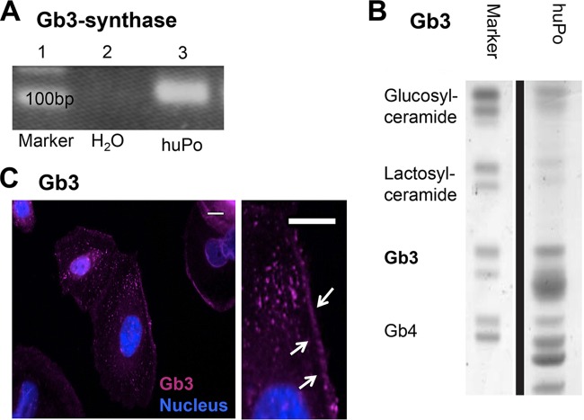
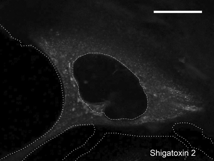
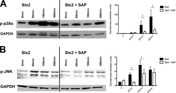
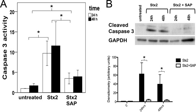
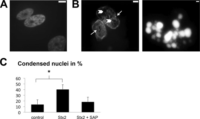
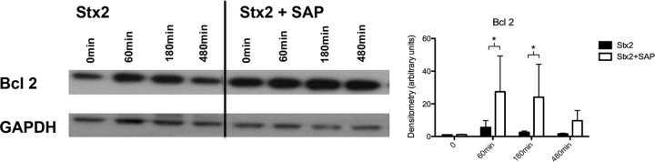
Hemolytic uremic syndrome (HUS) is mainly induced by Shiga toxin 2 (Stx2)-producing Escherichia coli. Proteinuria can occur in the early phase of the disease, and its persistence determines the renal prognosis. Stx2 may injure podocytes and induce proteinuria. Human serum amyloid P component (SAP), a member of the pentraxin family, has been shown to protect against Stx2-induced lethality in mice in vivo, presumably by specific binding to the toxin. We therefore tested the hypothesis that SAP can protect against Stx2-induced injury of human podocytes. To elucidate the mechanisms underlying podocyte injury in HUS-associated proteinuria, we assessed Stx2-induced activation of mitogen-activated protein kinases (MAPKs) and apoptosis in immortalized human podocytes and evaluated the impact of SAP on Stx2-induced damage. Human podocytes express Stx2-binding globotriaosylceramide 3. Stx2 applied to cultured podocytes was internalized and then activated p38α MAPK and c-Jun N-terminal kinase (JNK), important signaling steps in cell differentiation and apoptosis. Stx2 also activated caspase 3, resulting in an increased level of apoptosis. Coincubation of podocytes with SAP and Stx2 mitigated the effects of Stx2 and induced upregulation of antiapoptotic Bcl2. These data suggest that podocytes are a target of Stx2 and that SAP protects podocytes against Stx2-induced injury. SAP may therefore be a useful therapeutic option.


Figures

FIG 1
FIG 1
Expression of Gb3 synthase and Gb3 in human podocytes. (A) Expression of Gb3 synthase in untreated human podocytes (huPo) by conventional PCR (lane 3). (B) Lipid analysis for the presence of Gb3 in untreated human podocytes. (C) Localization of Gb3 in differentiated untreated podocytes along the membrane (arrows) and within the cytoplasm by immunofluorescence (pink) and in cell nuclei (blue). All experiments were repeated at least four times in independent experiments (scale bars = 15 μm).
FIG 2
FIG 2
Internalization of labeled Stx2 (white) in podocytes after 40 min of incubation. Distribution is predominantly within the cytoplasm. No Stx2 staining was found in the cell nucleus. The cell outline (dotted lines) and nucleus (dashed line) are marked (scale bar = 15 μm). The experiment was repeated at least three times independently.
FIG 3
FIG 3
Western blot and densitometry of phosphorylation of MAPK p38α and JNK. P-p38α (A) and p-JNK (B) after treatment with 15 ng/ml Stx2 and in combination with SAP (3 mg/liter) after 60, 180, or 480 min. Below are shown loading controls with GAPDH. *, P < 0.05. Means and standard errors of the means of five independent experiments, performed in duplicate, are shown.
FIG 4
FIG 4
Caspase 3 activity and cleaved caspase 3. (A) Caspase 3 activity (in AU) after Stx2 incubation (1.5 ng/ml) for 24 or 48 h compared to untreated cells and after coincubation with SAP (3 mg/liter) as evaluated by ELISA. For statistical analysis, the values at 24 and 48 h were compared. (B) Amounts of cleaved caspase 3 after Stx2 incubation with or without addition of SAP after 24 and 48 h. Below is shown loading control with GAPDH. *, P < 0.05. Means and standard errors of the means of four independent experiments, performed in duplicate, are shown.
FIG 5
FIG 5
Apoptosis, measured via the amount of condensed or apoptotic nuclei after Stx2 incubation. (A) Untreated control. (B) Nuclei of podocytes after treatment with 1.5 ng/ml Stx2. (Left) Typical enclosures/cysts (arrowheads) and indentations (arrows). (Right) An apoptotic body. (C) Content of condensed nuclei after Stx2 treatment compared to control or coincubation of Stx2 and SAP (3 mg/liter). *, P < 0.05. Means and standard deviations of three independent experiments are shown.
FIG 6
FIG 6
Induction of Bcl2 after Stx2 treatment (15 ng/ml) with or without coincubation with SAP (3 mg/liter) after 60, 180, and 480 min. Below is shown loading control with GAPDH. *, P < 0.05. Means and standard errors of the means of five independent experiments, performed in duplicate, are shown.

References

    1. Tarr PI. 2009. Shiga toxin-associated hemolytic uremic syndrome and thrombotic thrombocytopenic purpura: distinct mechanisms of pathogenesis. Kidney Int. Suppl. 112:S29–S32. 10.1038/ki.2008.615 - DOI - PubMed
    1. Orth D, Grif K, Khan AB, Naim A, Dierich MP, Wurzner R. 2007. The Shiga toxin genotype rather than the amount of Shiga toxin or the cytotoxicity of Shiga toxin in vitro correlates with the appearance of the hemolytic uremic syndrome. Diagn. Microbiol. Infect. Dis. 59:235–242. 10.1016/j.diagmicrobio.2007.04.013 - DOI - PubMed
    1. Tesh VL. 2012. Activation of cell stress response pathways by Shiga toxins. Cell. Microbiol. 14:1–9. 10.1111/j.1462-5822.2011.01684.x - DOI - PMC - PubMed
    1. Tesh VL. 2012. The induction of apoptosis by Shiga toxins and ricin. Curr. Top. Microbiol. Immunol. 357:137–178. 10.1007/82_2011_155 - DOI - PubMed
    1. Lingwood CA. 1999. Glycolipid receptors for verotoxin and Helicobacter pylori: role in pathology. Biochim. Biophys. Acta 1455:375–386. 10.1016/S0925-4439(99)00062-9 - DOI - PubMed

Publication types

Substances

LinkOut - more resources