. 2014 May;82(5):1968-81.
doi: 10.1128/IAI.00087-14. Epub 2014 Feb 24.

Symbiotic relationship between Streptococcus mutans and Candida albicans synergizes virulence of plaque biofilms in vivo

Affiliations

Symbiotic relationship between Streptococcus mutans and Candida albicans synergizes virulence of plaque biofilms in vivo

Megan L Falsetta et al. Infect Immun. 2014 May.

Abstract

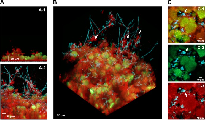
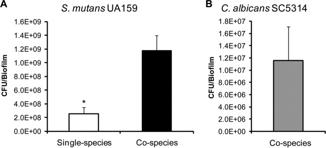
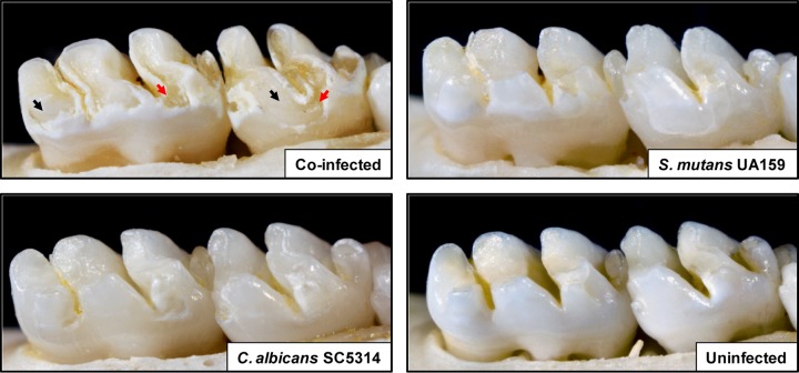
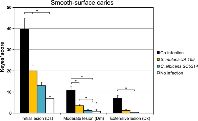
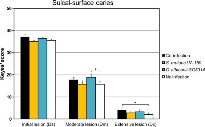
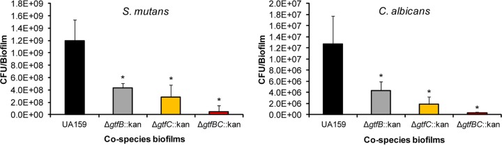
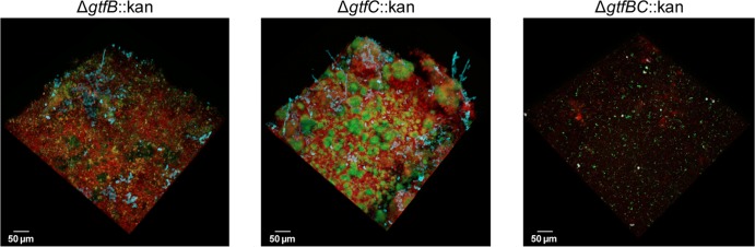
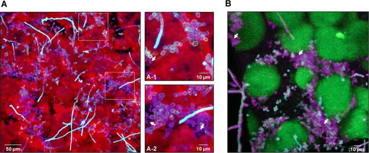
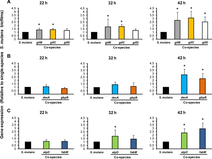
Streptococcus mutans is often cited as the main bacterial pathogen in dental caries, particularly in early-childhood caries (ECC). S. mutans may not act alone; Candida albicans cells are frequently detected along with heavy infection by S. mutans in plaque biofilms from ECC-affected children. It remains to be elucidated whether this association is involved in the enhancement of biofilm virulence. We showed that the ability of these organisms together to form biofilms is enhanced in vitro and in vivo. The presence of C. albicans augments the production of exopolysaccharides (EPS), such that cospecies biofilms accrue more biomass and harbor more viable S. mutans cells than single-species biofilms. The resulting 3-dimensional biofilm architecture displays sizeable S. mutans microcolonies surrounded by fungal cells, which are enmeshed in a dense EPS-rich matrix. Using a rodent model, we explored the implications of this cross-kingdom interaction for the pathogenesis of dental caries. Coinfected animals displayed higher levels of infection and microbial carriage within plaque biofilms than animals infected with either species alone. Furthermore, coinfection synergistically enhanced biofilm virulence, leading to aggressive onset of the disease with rampant carious lesions. Our in vitro data also revealed that glucosyltransferase-derived EPS is a key mediator of cospecies biofilm development and that coexistence with C. albicans induces the expression of virulence genes in S. mutans (e.g., gtfB, fabM). We also found that Candida-derived β1,3-glucans contribute to the EPS matrix structure, while fungal mannan and β-glucan provide sites for GtfB binding and activity. Altogether, we demonstrate a novel mutualistic bacterium-fungus relationship that occurs at a clinically relevant site to amplify the severity of a ubiquitous infectious disease.


Figures

FIG 1
FIG 1
Three-dimensional architecture of the cospecies biofilm. Representative images of single-species and cospecies biofilms grown for 42 h are shown. Bacterial microcolonies expressing GFP appear green, while fungal cells labeled with ConA-tetramethylrhodamine appear blue. EPS labeled with Alexa Fluor 647-dextran appear red. (A) Orthogonal views of the biofilms, illustrating the overall differences in the accumulation of biofilms between cospecies and S. mutans single-species biofilms. (B) Three-dimensional rendering of the cospecies biofilm, illustrating the complexity of its architecture. Bacterial microcolonies and yeast forms of C. albicans are enmeshed and surrounded by an EPS-rich matrix, while hyphae (indicated by white arrows) extend from the biofilm into the fluid phase and are coated with EPS. (C) Projection images of the first 20 μm (from the surface of attachment) of a cospecies biofilm, illustrating the spatial relationship between C. albicans, S. mutans, and EPS. (C-1) Merged image of all three components; (C-2) merged image of C. albicans and S. mutans; (C-3) C. albicans and EPS. The arrows indicate that there is little to no direct association between S. mutans and C. albicans (C-1 and C-2). In contrast, it is readily apparent that the fungal cells are associated with EPS (C-3), which then contacts the microcolonies (C-1).
FIG 2
FIG 2
Microbial populations in the cospecies biofilm. Shown are the total viable counts (CFU) of S. mutans in single-species and cospecies biofilms (A) and of C. albicans in cospecies biofilms (B), grown for 42 h. C. albicans alone lacked the capacity to form biofilms under our experimental conditions. The data are mean values ± standard deviations (n = 16). (A) The asterisk indicates that the values for single-species and cospecies biofilms are significantly different from each other (P, <0.05). There is a dramatic increase in the number of S. mutans CFU in cospecies biofilms (∼6-fold increase). (B) There is a large number of viable C. albicans cells in cospecies biofilms (∼107 CFU/biofilm).
FIG 3
FIG 3
Images of teeth from rats infected with S. mutans UA159 and/or C. albicans SC5314, or left uninfected, after 2 weeks. Photographs of lower molars in the rodent jaws are shown; jaws representing the average result have been selected. For the coinfected animal, black arrows indicate moderate to severe carious lesions where areas of the enamel are missing, exposing the underlying dentin. In some areas, the dentin is eroded or missing (red arrows), indicating the most severe carious lesions. In the S. mutans-infected animal, large areas of initial lesions were detected, although they were visibly less severe than those of coinfected animals. In the C. albicans-infected animal, small areas of demineralization and initial lesions were observed. In the uninfected animal, overt carious lesions are absent, while “white spots” (very early lesions) begin to appear in some localized areas.
FIG 4
FIG 4
Smooth-surface carious lesions in singly infected, coinfected, or uninfected animals. Smooth-surface caries scores are presented as mean values ± standard deviations (n = 11). Scores are recorded as stages of carious lesion severity according to Larson's modification of Keyes' scoring system: Ds, initial lesion (surface enamel white, broken, and/or dry); Dm, moderate lesion (dentin exposed); Dx, extensive lesion (dentin soft or missing). Asterisks indicate that the values for different experimental groups are significantly different from each other (P, <0.05).
FIG 5
FIG 5
Sulcal-surface carious lesions in singly infected, coinfected, or uninfected animals. Sulcal-surface caries scores are presented as mean values ± standard deviations (n = 11). Scores are recorded as stages of carious lesion severity according to Larson's modification of Keyes' scoring system: Ds, initial lesion (surface enamel white, broken and/or dry); Dm, moderate lesion (dentin exposed); Dx, extensive lesion (dentin soft or missing). Asterisks indicate that the values for different experimental groups are significantly different from each other (P, <0.05).
FIG 6
FIG 6
Viable counts in cospecies biofilms formed with Δgtf::kan mutant strains of S. mutans UA159. Shown are the total viable counts of S. mutans and C. albicans in 42-h cospecies biofilms formed with C. albicans SC5314 and one of the following S. mutans strains: the parental strain, UA159 (black bars), the ΔgtfB::kan mutant (gray bars), the ΔgtfC::kan mutant (orange bars), or the ΔgtfBC::kan mutant (red bars). The data are mean values ± standard deviations (n, ≥22). All cospecies biofilms formed with any of the three mutant strains contained significantly fewer viable counts of S. mutans and C. albicans than those formed in the presence of UA159 (*, P < 0.05).
FIG 7
FIG 7
Architecture of cospecies biofilms formed with Δgtf mutants. Shown are representative images of the architectures of cospecies biofilms formed by each of the Δgtf::kan mutant strains (at 42 h). Cospecies biofilms formed by the parental strain, UA159 (image not displayed; refer to Fig. 1), were always included (as a control) for comparison. Overall, biofilms formed with the ΔgtfB::kan mutant were thin and flat; they were devoid of microcolony structures and contained few yeast cells and almost no hyphae. The presence of the ΔgtfC::kan mutant strain also altered the overall architecture of the cospecies biofilms, which contained small microcolonies, few fungal cells, and largely defective EPS-rich matrix production. The ΔgtfBC::kan mutant strain was virtually incapable of forming cospecies biofilms with C. albicans.
FIG 8
FIG 8
Visualization and spatial distribution of β-glucan within cospecies biofilms. (A) Projection image of 42-h cospecies biofilms labeled with an anti-β-glucan antibody (purple), Alexa Fluor 647-dextran (EPS) (red), and ConA-tetramethylrhodamine (C. albicans cells) (blue). The image shows the presence of β-glucan (purple) within the biofilm, while the arrows in the closeup images of selected areas indicate punctate accumulations of β-glucan (A-1) that appear to be localized extracellularly (A-2). (B) Three-dimensional projection of a separate 42-h cospecies biofilm labeled with the anti-β-glucan antibody (purple), GFP (S. mutans cells) (green), and ConA-tetramethylrhodamine (C. albicans cells) (blue). The arrows indicate extracellular accumulations of β-glucan that appear to enmesh the C. albicans cells. Clearly, β-glucan can be found intercalated between C. albicans cells and S. mutans microcolonies, potentially having a structural role.
FIG 9
FIG 9
Expression profiles of S. mutans UA159 genes during the development of cospecies biofilms. The expression of selected S. mutans genes associated with EPS synthesis (A), EPS degradation and binding (B), and acid stress survival (C) is shown. The data (gene expression in cospecies biofilms relative to that in single-species S. mutans biofilms, represented by the black bars) are depicted as the mean ± standard deviation (n = 8). An asterisk indicates that the expression level of a specific S. mutans gene is significantly different for single-species and cospecies biofilms (P, <0.05).

References

    1. Hall-Stoodley L, Stoodley P. 2009. Evolving concepts in biofilm infections. Cell. Microbiol. 11:1034–1043. 10.1111/j.1462-5822.2009.01323.x - DOI - PubMed
    1. Dye BA, Tan S, Smith V, Lewis BG, Barker LK, Thornton-Evans G, Eke PI, Beltran-Aguilar ED, Horowitz AM, Li CH. 2007. Trends in oral health status: United States, 1988–1994 and 1999–2004. Vital Health Stat. 11:1–92 - PubMed
    1. Berkowitz RJ, Turner J, Hughes C. 1984. Microbial characteristics of the human dental caries associated with prolonged bottle-feeding. Arch. Oral Biol. 29:949–951. 10.1016/0003-9969(84)90097-9 - DOI - PubMed
    1. Milnes AR, Bowden GH. 1985. The microflora associated with developing lesions of nursing caries. Caries Res. 19:289–297. 10.1159/000260858 - DOI - PubMed
    1. Hallett KB, O'Rourke PK. 2002. Early childhood caries and infant feeding practice. Community Dent. Health 19:237–242 - PubMed

Publication types