Review
. 2014 Oct 7;43(19):6750-64.
doi: 10.1039/c3cs60459d.

Disordered amyloidogenic peptides may insert into the membrane and assemble into common cyclic structural motifs

Affiliations
Review

Disordered amyloidogenic peptides may insert into the membrane and assemble into common cyclic structural motifs

Hyunbum Jang et al. Chem Soc Rev. .

Abstract

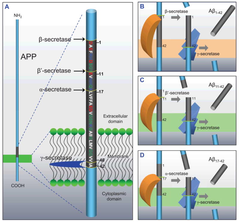
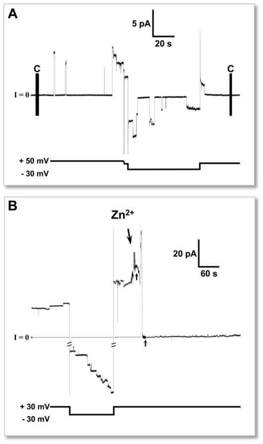
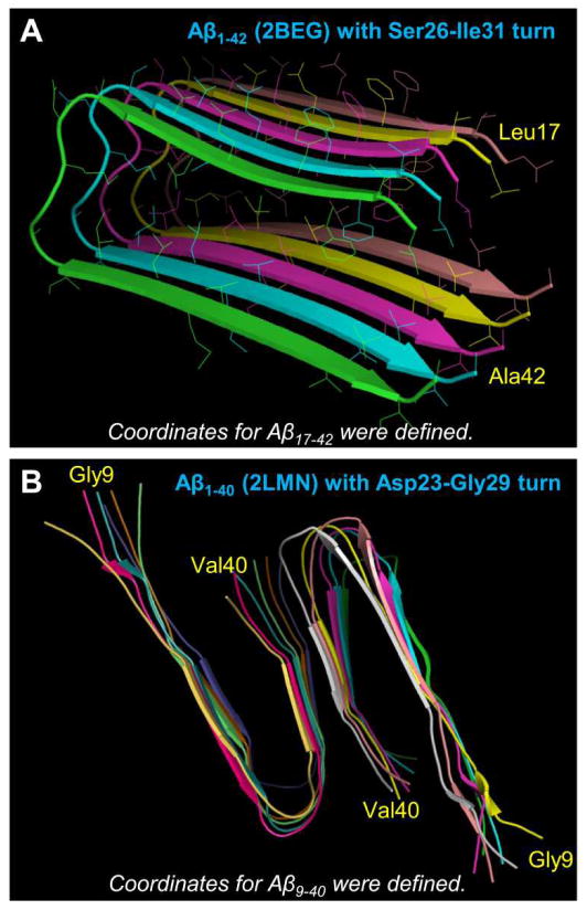
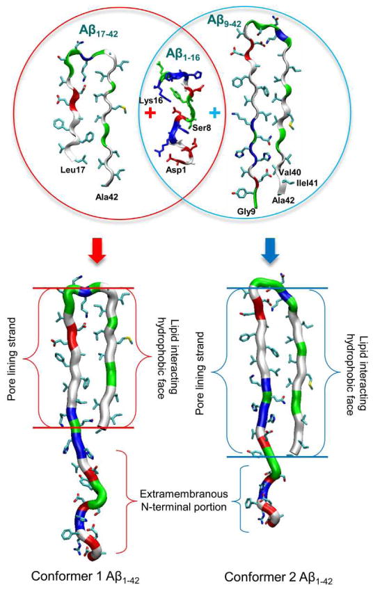
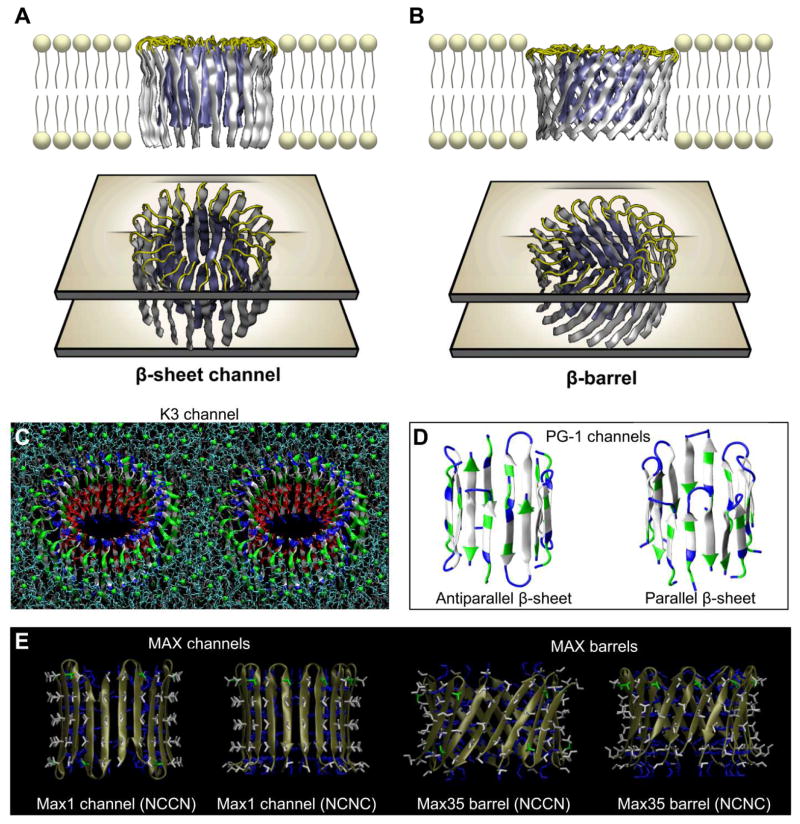
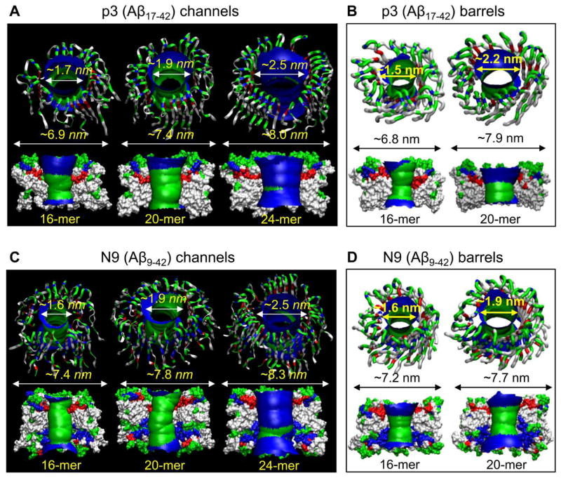
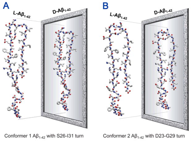
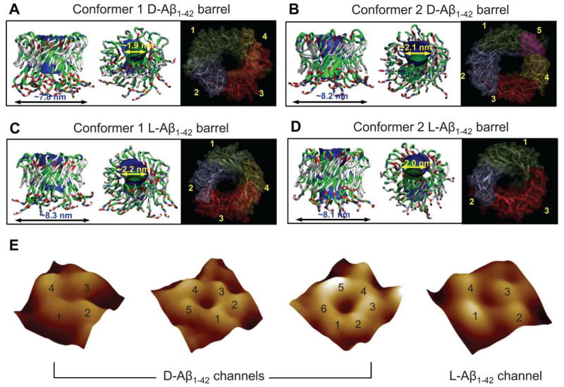
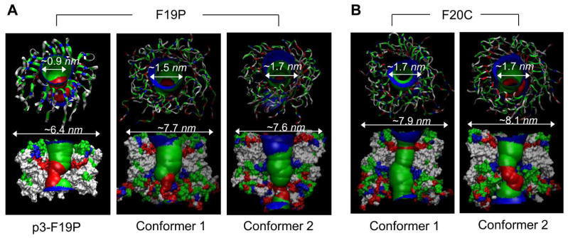
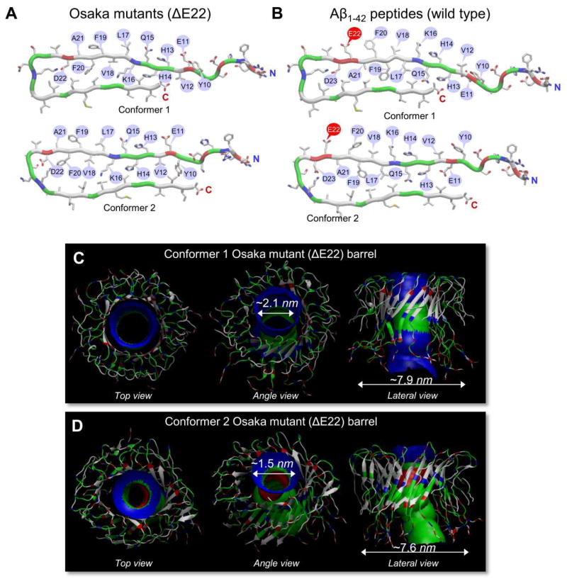
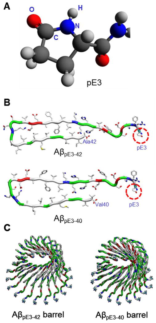
Aggregation of disordered amyloidogenic peptides into oligomers is the causative agent of amyloid-related diseases. In solution, disordered protein states are characterized by heterogeneous ensembles. Among these, β-rich conformers self-assemble via a conformational selection mechanism to form energetically-favored cross-β structures, regardless of their precise sequences. These disordered peptides can also penetrate the membrane, and electrophysiological data indicate that they form ion-conducting channels. Based on these and additional data, including imaging and molecular dynamics simulations of a range of amyloid peptides, Alzheimer's amyloid-β (Aβ) peptide, its disease-related variants with point mutations and N-terminal truncated species, other amyloidogenic peptides, as well as a cytolytic peptide and a synthetic gel-forming peptide, we suggest that disordered amyloidogenic peptides can also present a common motif in the membrane. The motif consists of curved, moon-like β-rich oligomers associated into annular organizations. The motif is favored in the lipid bilayer since it permits hydrophobic side chains to face and interact with the membrane and the charged/polar residues to face the solvated channel pores. Such channels are toxic since their pores allow uncontrolled leakage of ions into/out of the cell, destabilizing cellular ionic homeostasis. Here we detail Aβ, whose aggregation is associated with Alzheimer's disease (AD) and for which there are the most abundant data. AD is a protein misfolding disease characterized by a build-up of Aβ peptide as senile plaques, neurodegeneration, and memory loss. Excessively produced Aβ peptides may directly induce cellular toxicity, even without the involvement of membrane receptors through Aβ peptide-plasma membrane interactions.


Figures

Figure 1
Figure 1
Productions of β-amyloid (Aβ) via various cleavages. (A) A cartoon representing the cleavage process by α-, β-, β′- and γ-secretases of amyloid precursor protein (APP). The Aβ1-42 domain is shown in gray with the sequence and numbering of the amino acids. In single letter amino acid codes, hydrophobic, polar/Gly, positively charged, and negatively charged residues are colored white, green, blue, and red, respectively. Various Aβ fragments are processed by different secretase combinations. (B) Amyloidogenic fragment of Aβ1-40/42 by β- and γ-secretases, and non-amyloidogenic fragments of (C) Aβ11-40/42 by β′- and γ-secretases and (D) Aβ17-40/42 by α- and γ-secretases. These cartoons were inspired by previous publication. (B and D from Jang et al., 2010, are reprinted with permission).
Figure 2
Figure 2
An example of stepwise current feature across planar lipid bilayer (PLB, or BLM representing “black lipid membrane”) membrane produced by amyloid channels. (A) The electrophysiological activity of Aβ1-42 ion channels embedded in PLB. (B) Inhibition of channel activity by Zn2+ addition. Time of Zn2+ addition (2 mM) is marked by arrow. (From Capone et al., 2012, reprinted with permission).
Figure 3
Figure 3
Three-dimensional structures of Alzheimer’s amyloids. (A) The pentameric Aβ1-42 fibril structure obtained from a combination of hydrogen/deuterium-exchange nuclear magnetic resonance (NMR) data, side-chain packing constraints from pairwise mutagenesis, solid state NMR (ssNMR) and electron microscopy (EM) (pdb id: 2BEG). The coordinates for residues 1–16 were missing due to disorder. (B) The ssNMR model for small Aβ1-40 protofibrils (pdb ids: 2LMN). The coordinates for residues 1–8 were missing due to disorder. Residues at both termini are marked.
Figure 4
Figure 4
A cartoon representing the constructions of the full-length Aβ1-42 peptides. The N-terminal truncated Aβ monomers are U-shape with the β-strand-turn-β-strand motif. The missing N-terminal portions of these NMR-derived Aβ peptides are recovered with the coordinates from the solution structure of Aβ1-16 (pdb id: 1ZE7). By covalently connecting the N-terminal to the truncated Aβ peptides, two Aβ1-42 conformers (conformer 1 and 2) with different turns can be generated.
Figure 5
Figure 5
Conventional β-sheet channel vs. β-barrel designs. (A) The conventional β-sheet channel has the β-strands that orient parallel to the membrane normal, (B) while the β-strands that orient obliquely to the membrane normal generate β-barrel structure. Above examples are shown for the p3 (Aβ17-42) channel and barrel. Other examples of the β-sheet channel and barrel formed by the U-shaped K3 (a fragment of β2-microglobulin) peptide and by the PG-1 and MAX β-hairpins. (C) The 24-mer channel embedded in the DOPC bilayer in stereo view. (D) The 8-mer PG-1 channels with the antiparallel and parallel β-strand arrangements. (E) The 10-mer antiparallel MAX channels and barrels in the NCCN and NCNC packing modes (from Gupta et al., 2013, reprinted with permission).
Figure 6
Figure 6
Truncated Aβ channel/barrel conformations in the lipid bilayer. Averaged pore structures calculated by the HOLE program embedded in the averaged channel/barrel conformations during the simulations for the p3 (Aβ17-42) (A) channels and (B) barrels, and the N9 (Aβ9-42) (C) channels and (D) barrels. (A from Jang et al., 2010, B and D from Jang et al., 2010, and C from from Jang et al., 2009, reprinted with permission).
Figure 7
Figure 7
Schematic diagrams representing the all L- and all-D-amino acids Aβ1-42 peptides with different conformers. The coordinates of all D-amino acids Aβ1-42 are mirror-imaged coordinates of all L-amino acids Aβ1-42 for (A) the conformer 1 and (B) the conformer 2. The standard CHARMM force can be directly used for D-amino acids. However, the parameters include the dihedral angle cross-term map (CMAP), which, for D-amino acids, needs to be corrected since the map was constructed for L-amino acids. To simulate D-amino acids, corrected CMAP data for D-amino acids should be applied via reflecting the phi-psi CMAP matrix for L-amino acids.,,
Figure 8
Figure 8
Full-length Aβ1-42 barrel conformations in the lipid bilayer. Simulated barrel structure with an embedded pore structure and highlighted subunits for (A) the conformer 1 and (B) the conformer 2 D-Aβ1-42 barrels, and (C) the conformer 1 and (D) the conformer 2 L-Aβ1-42 barrels. (E) High resolution atomic force microscopy (AFM) images of D- and L-Aβ1-42 reconstituted in the lipid bilayer. The number of subunits is resolved and indicated for each channel. (From Connelly et al., 2012, reprinted with permission).
Figure 9
Figure 9
Aβ mutant channels/barrels in the lipid bilayers. (A) Simulated channel/barrel structures with an embedded pore structure for the p3-F19P (from the truncated Aβ17-42) mutant channel (left panel, from Jang et al., 2010, reprinted with permission) and for the conformer 1 and the conformer 2 F19P (from the full-length Aβ1-42) mutant barrels (middle and right panels, from Connelly et al., 2012, reprinted with permission). (B) Simulated barrel structures with an embedded pore structure for the conformer 1 and the conformer 2 F20C (from the full-length Aβ1-42) mutant barrels (from Connelly et al., 2012, reprinted with permission).
Figure 10
Figure 10
Osaka mutant (ΔE22) barrels in the lipid bilayers. (A) Monomer conformations of the Osaka mutant with different turns at Ser25-Ile30 (conformer 1) and Asp22-Gly28 (conformer 2). (B) The U-shaped monomer conformations are similar to the wild type Aβ1-42 peptides with different turns at Ser26-Ile31 (conformer 1) and Asp23-Gly29 (conformer 2). Simulated barrel structures with an embedded pore structure for (C) the conformer 1 and (D) the conformer 2 Osaka mutant (ΔE22) barrels. (From Jang et al., 2013, reprinted with permission).
Figure 11
Figure 11
Computational modelling of pyroglutamate (pE) modified Aβ barrels. (A) Molecular structure of pyroglutamate (pE) at position 3. (B) Monomer conformations of AβpE3-42 (upper) and AβpE3-40 (lower) peptides. (C) Modelled structures of 18-mer AβpE3-42 (left) and AβpE3-40 (right) barrels.

References

    1. Cohen FE, Kelly JW. Nature. 2003;426:905–909. - PubMed
    1. Temussi PA, Masino L, Pastore A. Embo J. 2003;22:355–361. - PMC - PubMed
    1. Dobson CM. Nature. 2003;426:884–890. - PubMed
    1. Chiti F, Dobson CM. Annu Rev Biochem. 2006;75:333–366. - PubMed
    1. DeToma AS, Salamekh S, Ramamoorthy A, Lim MH. Chem Soc Rev. 2012;41:608–621. - PMC - PubMed

Publication types

MeSH terms