Review
. 2014 Mar;14(3):195-208.
doi: 10.1038/nri3622.

Regulation of immune responses by extracellular vesicles

Affiliations
Review

Regulation of immune responses by extracellular vesicles

Paul D Robbins et al. Nat Rev Immunol. 2014 Mar.

Abstract

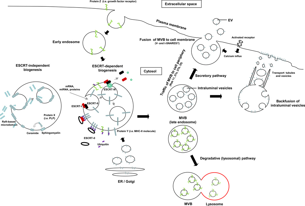
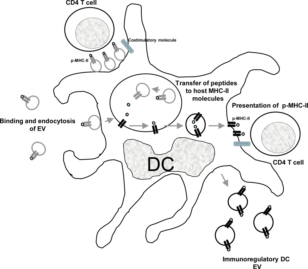
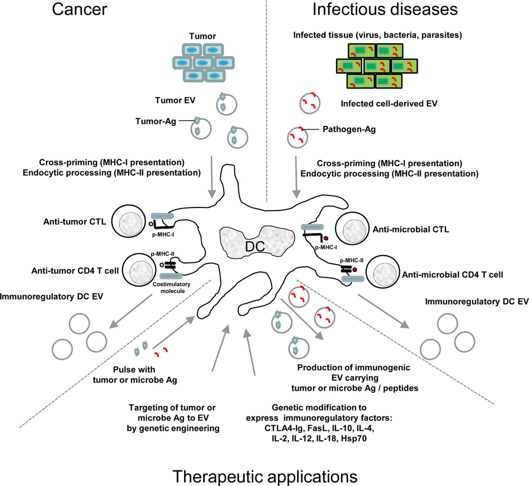
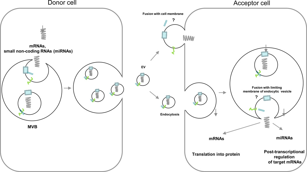
Extracellular vesicles, including exosomes, are small membrane vesicles derived from multivesicular bodies or from the plasma membrane. Most, if not all, cell types release extracellular vesicles, which then enter the bodily fluids. These vesicles contain a subset of proteins, lipids and nucleic acids that are derived from the parent cell. It is thought that extracellular vesicles have important roles in intercellular communication, both locally and systemically, as they transfer their contents, including proteins, lipids and RNAs, between cells. Extracellular vesicles are involved in numerous physiological processes, and vesicles from both non-immune and immune cells have important roles in immune regulation. Moreover, extracellular vesicle-based therapeutics are being developed and clinically tested for the treatment of inflammatory diseases, autoimmune disorders and cancer. Given the tremendous therapeutic potential of extracellular vesicles, this Review focuses on their role in modulating immune responses, as well as their potential therapeutic applications.


Figures

Figure 1
Figure 1. Biogenesis of extracellular vesicles
EVs are generated as ILVs in MVBs by ESCRT-dependent or –independent mechanisms. Proteins transported from the Golgi (i.e. MHC Class-II molecules), or internalized from the cell surface (i.e. activated growth factor receptors) are ubiquitylated on their cytosolic domains. However, not all proteins such as MHC Class-II required ubiquitinylation for targeting to vesicles. The ESCRT-0 complex recognizes the ubiquitylated proteins on the cytosolic side of the endosome / MVB membrane, segregates the proteins into microdomains, and binds the ESCRT-I complex, which in turn recruits ESCRT–II subunits. ESCRT-I and –II initiate the reverse budding of the nascent ILVs within MVBs. At this time, cytosolic RNAs and proteins have direct access into the interior of the forming vesicles. Next, the ESCRT-II complex recruits ESCRT-III subunits inside the neck of the nascent ILVs, which results in their cleavage into free vesicles. The free ubiquitin molecules and ESCRT subunits are released into the cytosol for recycling. Certain proteins (i.e. the proteolipid protein, PLP) are sorted into ILVs independently of the ESCRT machinery through raft-based microdomains rich in sphingolipids, from which ceramides are formed by sphingomyelinases. Ceramide induces coalescence of the microdomains and triggers ILV formation. The dashed line indicates that the role of ceramide in ILV formation is still controversial. The MVBs then follow either the secretory or degradative pathway. In the former, MVBs traffic to the cell periphery and fuse with the cell membrane, releasing the ILVs (now termed EV) constitutively, or following activation of surface receptors that trigger calcium influx. In the degradative route, MVBs released the ILVs into lysosomes. The lysosomal pathway is critical for limiting signaling of activated growth factor receptors. It is likely that differences in the MVBs confer the route of traffic.
Figure 2
Figure 2. Role of extracellular vesicles in Ag-presentation for cellular immunity
Professional APCs (i.e. DCs) present p-MHC complexes derived from captured exosomes. EVs retained on the APC surface present their p-MHC complexes directly to T cells, although the costimulatory molecules are provided by the APC. Alternatively, internalized EVs transfer their Ag-peptides to MHC molecules of the host APCs. The host MHC molecules loaded with the exosome-derived Ag-peptide are then transported to the APC surface for presentation to T cells. The APCs also release EVs able to regulate Ag-specific immune responses. Although for only MHC Class-II complexes are shown in fig. 2, a similar process occurs for exosomal MHC Class-I for regulation of CD8+ cells.
Figure 3
Figure 3. Role of extracellular vesicles in regulating tumor and microbe immunity that can be modified for therapeutic applications
Professional APCs (i.e. DCs) process tumor- or microbe-derived Ags for presentation to CTLs or CD4 T cells. A similar approach can be employed in vitro for generation of immunogenic EVs for therapeutic applications, by pulsing the APCs with tumor- or pathogen-derived Ags, or by genetically engineered the APCs to target the desired-Ags to the exosome membrane. Similarly, the APC can be modified to express immunosuppressive or immunostimulatory cytokines or ligands, rendering the EVs released from the APCs able to suppress or stimulate Ag-specific immune responses.
Figure 4
Figure 4. Mechanism of transfer of exosomal shuttle RNAs between cells
mRNAs and small non-coding RNAs, including miRNAs are transported inside the lumen of secreted EVs. Once released, the EVs are trapped by acceptor cells. Release of the vesicular RNAs into the cytosol of the target cell requires fusion of the exosome membrane with the plasma membrane or more likely with the limiting membrane of endocytic vesicles, once the EVs are internalized.

References

    1. Thery C, Zitvogel L, Amigorena S. Exosomes: composition, biogenesis and function. Nat Rev Immunol. 2002;2:569–579. - PubMed
    1. S ELA, Mager I, Breakefield XO, Wood MJ. Extracellular vesicles: biology and emerging therapeutic opportunities. Nat Rev Drug Discov. 2013;12:347–357. - PubMed
    1. Thery C, Ostrowski M, Segura E. Membrane vesicles as conveyors of immune responses. Nat Rev Immunol. 2009;9:581–593. - PubMed
    1. Yang C, Robbins PD. Immunosuppressive exosomes: a new approach for treating arthritis. Int J Rheumatol. 2012;2012:573528. - PMC - PubMed
    1. Booth AM, et al. Exosomes and HIV Gag bud from endosome-like domains of the T cell plasma membrane. J Cell Biol. 2006;172:923–935. - PMC - PubMed

Publication types