Comparative Study
. 2014 Mar 11;111(10):3793-8.
doi: 10.1073/pnas.1324266111. Epub 2014 Feb 24.

Toll-like receptor 10 is involved in induction of innate immune responses to influenza virus infection

Affiliations
Comparative Study

Toll-like receptor 10 is involved in induction of innate immune responses to influenza virus infection

Suki M Y Lee et al. Proc Natl Acad Sci U S A. .

Abstract

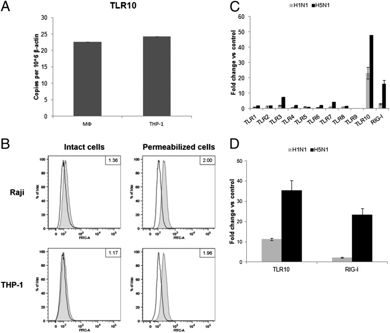
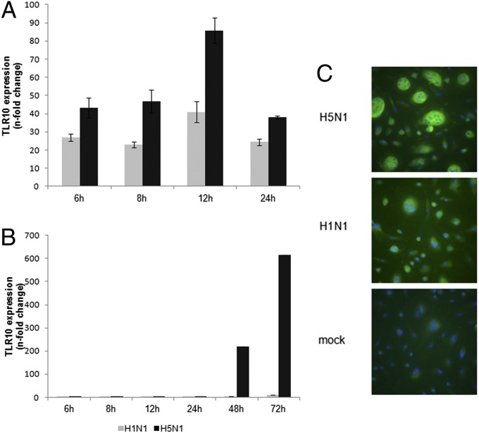
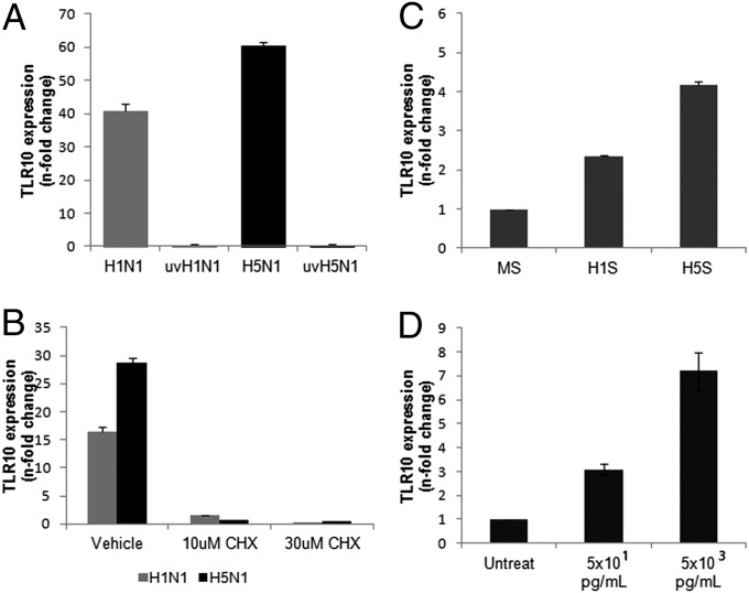
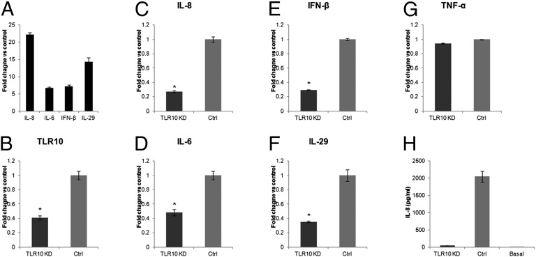
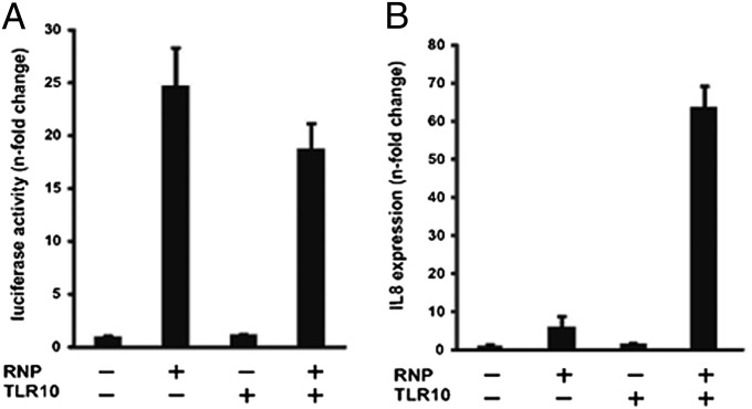
Toll-like receptors (TLRs) play key roles in innate immune recognition of pathogen-associated molecular patterns of invading microbes. Among the 10 TLR family members identified in humans, TLR10 remains an orphan receptor without known agonist or function. TLR10 is a pseudogene in mice and mouse models are noninformative in this regard. Using influenza virus infection in primary human peripheral blood monocyte-derived macrophages and a human monocytic cell line, we now provide previously unidentified evidence that TLR10 plays a role in innate immune responses following viral infection. Influenza virus infection increased TLR10 expression and TLR10 contributed to innate immune sensing of viral infection leading to cytokine induction, including proinflammatory cytokines and interferons. TLR10 induction is more pronounced following infection with highly pathogenic avian influenza H5N1 virus compared with a low pathogenic H1N1 virus. Induction of TLR10 by virus infection requires active virus replication and de novo protein synthesis. Culture supernatants of virus-infected cells modestly up-regulate TLR10 expression in nonvirus-infected cells. Signaling via TLR10 was activated by the functional RNA-protein complex of influenza virus leading to robust induction of cytokine expression. Taken together, our findings identify TLR10 as an important innate immune sensor of viral infection and its role in innate immune defense and immunopathology following viral and bacterial pathogens deserves attention.


Conflict of interest statement

The authors declare no conflict of interest.

Figures

Fig. 1.
Fig. 1.
Expression of TLR10 in primary human macrophages and THP-1 cells. (A) Basal mRNA expression of TLR10 in primary human macrophages (MФ) and THP-1 cells determined by RT-PCR. (B) Determination of TLR10 protein by flow cytometry in intact or permeabilized human Raji and THP-1 cell lines. Cells were stained with either FITC-conjugated isotype control antibody (open histogram) or FITC-conjugated anti-human TLR10 antibody (gray tinted histogram). Data shown are the ratio value of a signal-to-isotype control based on the median of FITC fluorescence intensity. Changes in expression level of TLR10 in response to infection by H1N1 and H5N1 influenza A viruses with MOI of 2 at 6 h after infection compared with mock infection in (C) primary human macrophages and (D) THP-1 cells. Results shown are representative of biological replicates performed in three independent experiments and error bars indicate SD of technical triplicates.
Fig. 2.
Fig. 2.
Kinetics of influenza A virus-induced TLR10 expression in human macrophages. Expression of TLR10 in human macrophages infected by H1N1 or H5N1 influenza A viruses at (A) MOI of 2 or (B) 0.001 compared with mock infection at different postinfection time were assessed by RT-PCR. The result of one representative experiment from three independent experiments with three donors is shown. Error bars indicate SD of three technical triplicates. (C) TLR10 protein expression (FITC-green) was detected using immunofluorescent staining. Cells were counterstained with DAPI (blue) and viewed in a fluorescent microscope (magnification 400×). Multinucleate giant cells are seen, especially in H5N1 virus infected cells.
Fig. 3.
Fig. 3.
Mechanism of TLR10 induction by influenza A virus infection. (A) TLR10 expression in cells infected by UV-irradiated H1N1 (uvH1N1) and H5N1 (uvH5N1) influenza A viruses at MOI of 2 were compared with nonirradiated virus infection at 6 h postinfection. (B) Suppression of TLR10 expression in H1N1 and H5N1 virus infected cells (MOI of 2) by treatment with protein synthesis inhibitor, cycloheximide (CHX). (C) Induction of TLR10 expression in uninfected human macrophages after challenged with H1N1 supernatant (H1S) and H5N1 supernatant (H5S) compared with mock supernatant (MS) at 3 h after stimulation. (D) Induction of TLR10 by TNF-α in a dose-dependent manner. Data shown are representative of biological replicates performed in three independent experiments and error bars indicate SD of technical triplicates.
Fig. 4.
Fig. 4.
Cytokines induced by influenza A virus are regulated via TLR10. (A) Induction of proinflammatory cytokines and IFNs in H1N1 virus infected THP-1 cells. (B) The knockdown efficiencies of TLR10 shRNA knockdown (TLR10 KD) in THP-1 cells assessed by RT-PCR. Relative expression of virus induced proinflammatory cytokine genes, (C) IL-8 and (D) IL-6 and type I and III IFNs, (E) IFN-β and (F) IL-29 in TLR10 shRNA knockdown cells compared with control at 6 h after H1N1 virus infection. (G) TNF-α gene expression was not affected by TLR10 knockdown. (H) Secretory IL-8 protein level in culture supernatant collected from TLR10 knockdown and control cells after virus infection determined using ELISA. Basal IL-8 protein level in culture supernatant before infection was included for comparison. Data shown are average of two independent experiments and error bars indicate SD of six measurements obtained from the two independent experiments. (*P < 0.05).
Fig. 5.
Fig. 5.
Expression of proinflammatory cytokine IL-8 was mediated via TLR10 during influenza A virus replication. (A) Dual-luciferase assay: 293T cells were transfected with pPOLI-NS-Luc reporter and pRL-CMV control plasmid, together with the plasmids including the expression plasmids of PB2, PB1, PA and NP (RNP) and human TLR10 expression plasmid (TLR10). Cells were harvested at 20 h after transfection. Luciferase activity shown was the Firefly luciferase activity normalized to Renilla luciferase activity. (B) RT-PCR analysis of proinflammatory cytokines IL-8 mRNA expression. Similar to A, 293T cells were harvested at 20 h after transfection. Data shown are representative of two independent experiments and error bars indicate SD of technical triplicates.

Similar articles

Cited by

References

    1. Kawai T, Akira S. Toll-like receptors and their crosstalk with other innate receptors in infection and immunity. Immunity. 2011;34(5):637–650. - PubMed
    1. Medzhitov R. Recognition of microorganisms and activation of the immune response. Nature. 2007;449(7164):819–826. - PubMed
    1. Diebold SS, Kaisho T, Hemmi H, Akira S, Reis e Sousa C. Innate antiviral responses by means of TLR7-mediated recognition of single-stranded RNA. Science. 2004;303(5663):1529–1531. - PubMed
    1. Lund JM, et al. Recognition of single-stranded RNA viruses by Toll-like receptor 7. Proc Natl Acad Sci USA. 2004;101(15):5598–5603. - PMC - PubMed
    1. Kawai T, Akira S. The role of pattern-recognition receptors in innate immunity: Update on Toll-like receptors. Nat Immunol. 2010;11(5):373–384. - PubMed

Publication types

MeSH terms

LinkOut - more resources