. 2014 Mar 11;111(10):3787-92.
doi: 10.1073/pnas.1401105111. Epub 2014 Feb 24.

Autoantibodies to IgG/HLA class II complexes are associated with rheumatoid arthritis susceptibility

Affiliations

Autoantibodies to IgG/HLA class II complexes are associated with rheumatoid arthritis susceptibility

Hui Jin et al. Proc Natl Acad Sci U S A. .

Abstract

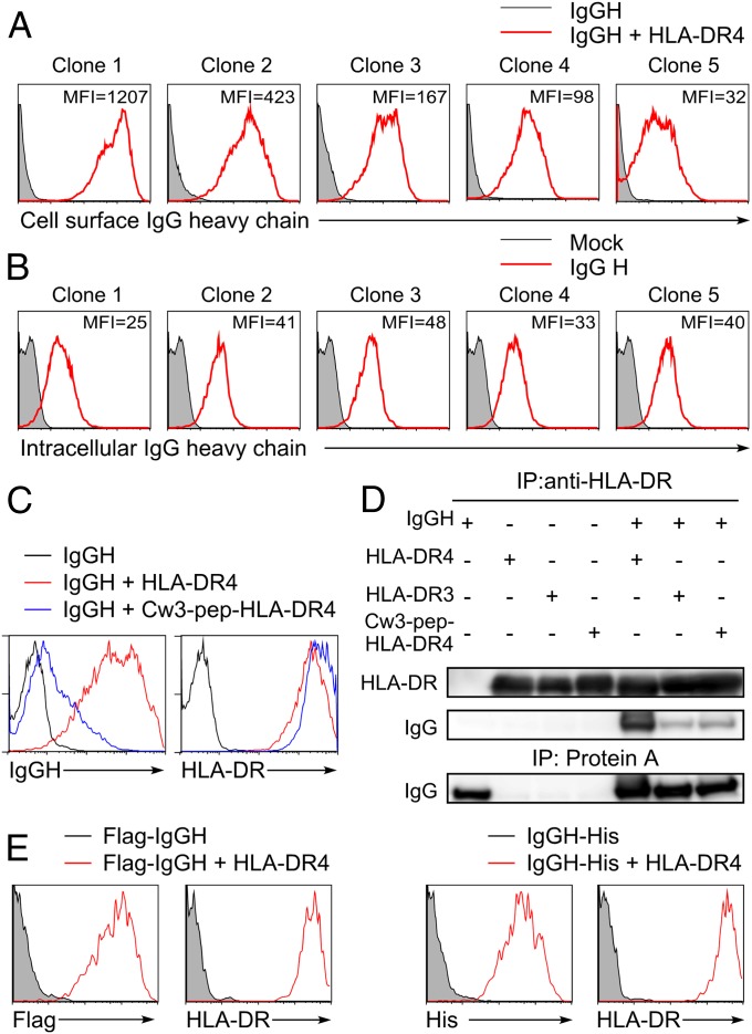
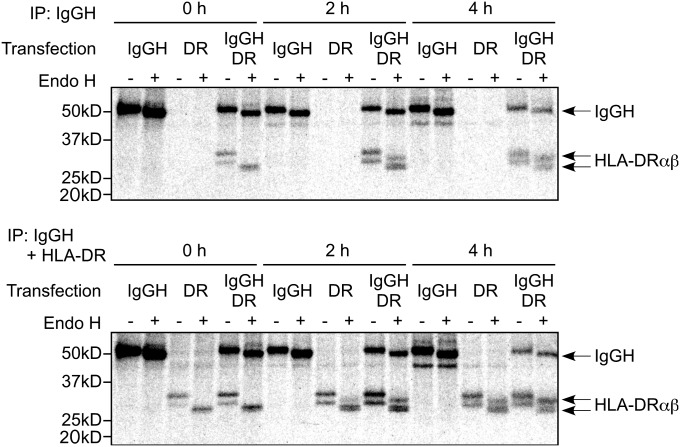
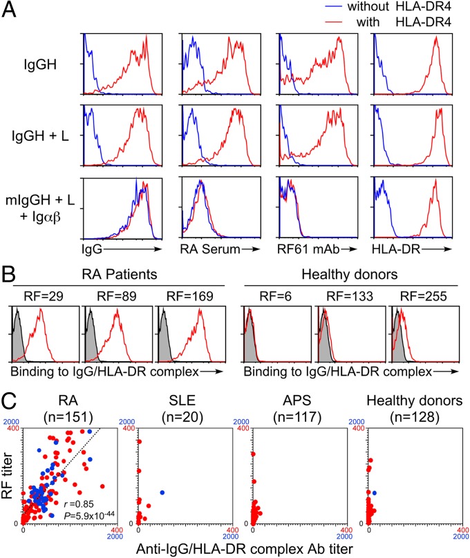
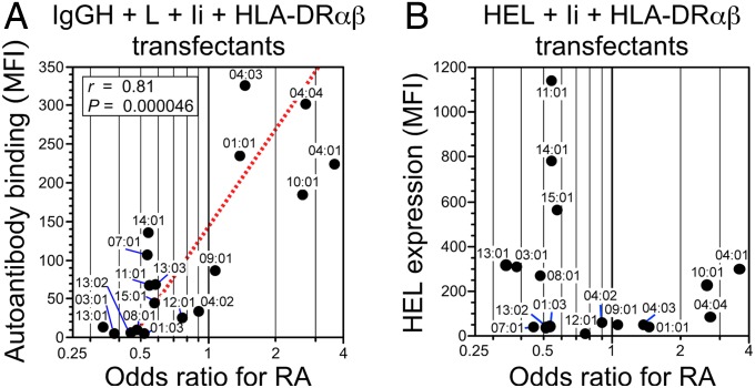
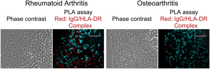
Specific HLA class II alleles are strongly associated with susceptibility to rheumatoid arthritis (RA); however, how HLA class II regulates susceptibility to RA has remained unclear. Recently, we found a unique function of HLA class II molecules: their ability to aberrantly transport cellular misfolded proteins to the cell surface without processing to peptides. Rheumatoid factor (RF) is an autoantibody that binds to denatured IgG or Fc fragments of IgG and is detected in 70-80% of RA patients but also in patients with other diseases. Here, we report that intact IgG heavy chain (IgGH) is transported to the cell surface by HLA class II via association with the peptide-binding groove and that IgGH/HLA class II complexes are specifically recognized by autoantibodies in RF-positive sera from RA patients. In contrast, autoantibodies in RF-positive sera from non-RA individuals did not bind to IgGH/HLA class II complexes. Of note, a strong correlation between autoantibody binding to IgG complexed with certain HLA-DR alleles and the odds ratio for that allele's association with RA was observed (r = 0.81; P = 4.6 × 10(-5)). Our findings suggest that IgGH complexed with certain HLA class II alleles is a target for autoantibodies in RA, which might explain why these HLA class II alleles confer susceptibility to RA.

Keywords: autoimmune disease; major histocompatibility complex.


Conflict of interest statement

The authors declare no conflict of interest.

Figures

Fig. 1.
Fig. 1.
IgGH is transported to the cell surface by HLA-DR. (A) The secreted forms of IgGHs cloned from human PBMCs that have different V regions were cotransfected with HLA-DRα, DRB1*04:04 (HLA-DR4), and GFP. Cell surface IgG on GFP-expressing cells was detected with anti-human IgG Fc-specific Ab (red lines). Cells transfected with IgGH alone were stained as control (shaded histograms). (B) The secreted form of IgGH clones was cotransfected with GFP, and intracellular IgGH was detected by intracellular staining with anti-human IgG Fc-specific Ab (red lines). Cells transfected with GFP alone were stained as control (shaded histograms). MFIs of IgG complexed with HLA-DR or intracellular IgG are shown in the figure (B). Data are representative of three independent experiments. (C) HLA-DR4 (red lines), Cw3-pep-HLA-DR4 (blue lines), or control plasmids (black lines) were cotransfected with IgGH clone 1 and GFP. Expression of IgG and HLA-DR on GFP-positive cells is shown. (D) HLA-DR4, DR3, Cw3-pep-DR4, and IgGH clone 1 (IgGH) were transfected, HLA-DR was immunoprecipitated from cell lysates, and IgGH and HLA-DR in precipitates and whole-cell lysates were immunoblotted. (E) N-terminal Flag-tagged or C-terminal His-tagged IgGH was transfected with GFP in the presence (red lines) or absence (shaded histograms) of HLA-DR4. Cell surface IgG on GFP-positive cells was detected by anti-Flag or anti-His mAb, respectively. Data are representative of at least three independent experiments.
Fig. 2.
Fig. 2.
Association of IgGH with HLA-DR in the ER. IgGH and HLA-DR were transfected into 293T cells and were labeled with [35S]methionine and cysteine for 15 min and chased for the indicated times in the absence of radioactivity. IgGH was precipitated with protein A (Upper) and both IgGH and HLA-DR were precipitated with protein A plus anti–HLA-DR mAb (Lower). The precipitates were boiled, treated with Endo H or mock, and resolved by SDS/PAGE. HLA-DRαβ and IgGH are indicated. Data are representative of three independent experiments.
Fig. 3.
Fig. 3.
IgGH complexed with HLA-DR is specifically recognized by autoantibodies in RA patients. (A) The IgGH, light chain (L), and GFP were cotransfected with (red) or without (blue) HLA-DR4. The membrane form of IgGH containing the same V region as IgGH clone 1 (mIgGH) was transfected with the light chain, Igα, Igβ (Igαβ), and GFP with (red) or without (blue) HLA-DR. Cell surface expression of HLA-DR or IgG and binding of autoantibodies in the RF standard serum and RF61 mAb by GFP-positive cells are shown. (B) IgGH, HLA-DR4, and GFP were cotransfected. Binding of IgM autoantibody in sera from RA patients and healthy controls to GFP-positive cells expressing IgGH/HLA-DR complexes is shown (red lines). The same cells were stained by APC-conjugated anti-human IgM Ab only as control (shaded histograms). RF activities (units per milliliter) of serum samples are shown in the figure. (C) RF titers of sera from 151 RA patients, 20 SLE patients, 117 APS patients, and 128 healthy controls were plotted against IgM autoantibody titers of each serum to IgGH complexed with HLA-DR4 (anti-IgGH/HLA-DR Ab titer). Samples with autoantibody titers under 400 were plotted in red and those between 400 and 2,000 were plotted in blue. Linear regression line (dashed line), the correlation coefficient (r), and P value of the regression line are shown in the plots of RA patients.
Fig. 4.
Fig. 4.
Autoantibody binding to IgGH complexed with HLA-DR molecules is different between RA-susceptible and RA-resistant alleles. (A and C) The IgGH and GFP were cotransfected together with HLA-DR into 293T cells in the presence (red lines) or absence (blue lines) of Ii (A) or HLA-DM (C), and surface expression of HLA-DR and IgG and binding of autoantibodies in the RF standard serum on GFP-positive cells were analyzed. Cells transfected with the IgGH alone were stained as a control (shaded histograms). Expression of Ii (A) or HLA-DM (C) was analyzed by intracellular staining. (B) Flag-tagged cysteine mutant HEL and HLA-DR were cotransfected with GFP in the presence (red lines) or absence (blue lines) of Ii. The transfectants were stained with anti-Flag and anti–HLA-DR mAbs, and expression on GFP-positive cells was determined. Cells transfected with mutHEL alone were stained as a control (shaded histograms). Data are representative of at least three independent experiments.
Fig. 5.
Fig. 5.
A strong correlation between autoantibody binding to IgG complexed with each HLA-DR allele and the odds ratio for that allele’s association with RA. (A) The IgGH was cotransfected with Ig light chain (L), Ii, and HLA-DRα in combination with each HLA-DRB1 allele indicated, and the MFIs of binding of autoantibodies in the RF standard serum to IgG complexed with HLA-DR were plotted against the odds ratios for RA susceptibility for each HLA-DRB1 allele. The odds ratios for the association between different HLA-DRB1 alleles and rheumatoid arthritis, which were obtained from a recently reported large-scale genetic study (6), were log-transformed to normalize the distribution. Linear regression line (dashed line), the correlation coefficient (r), and P value of the regression line are shown. (B) Cysteine mutant HEL was cotransfected with Ii and HLA-DRα in combination with each HLA-DRB1 allele, and the MFIs of HEL staining of the transfectants were plotted against the odds ratios for RA susceptibility for each HLA-DRB1 allele. Data are representative of four independent experiments.
Fig. 6.
Fig. 6.
In situ association of IgGH with HLA-DR in synovial membranes of RA patients. In situ association of IgGH with HLA-DR in tissue sections from synovial membranes of RA patients (Left) and osteoarthritis (Right) patients were analyzed by PLA. PLA signals and nuclei are shown as red and light blue, respectively. (Scale bars, 50 μm.) Representative data of tissue sections from three different patients are shown.

References

    1. Dörner T, Egerer K, Feist E, Burmester GR. Rheumatoid factor revisited. Curr Opin Rheumatol. 2004;16(3):246–253. - PubMed
    1. Jones EY, Fugger L, Strominger JL, Siebold C. MHC class II proteins and disease: A structural perspective. Nat Rev Immunol. 2006;6(4):271–282. - PubMed
    1. Neefjes J, Jongsma ML, Paul P, Bakke O. Towards a systems understanding of MHC class I and MHC class II antigen presentation. Nat Rev Immunol. 2011;11(12):823–836. - PubMed
    1. Chicz RM, et al. Specificity and promiscuity among naturally processed peptides bound to HLA-DR alleles. J Exp Med. 1993;178(1):27–47. - PMC - PubMed
    1. Nicholson MJ, Hahn M, Wucherpfennig KW. Unusual features of self-peptide/MHC binding by autoimmune T cell receptors. Immunity. 2005;23(4):351–360. - PMC - PubMed

Publication types