. 2014 Apr 1;111(13):4940-5.
doi: 10.1073/pnas.1316013111. Epub 2014 Feb 24.

Oral glycotoxins are a modifiable cause of dementia and the metabolic syndrome in mice and humans

Affiliations

Oral glycotoxins are a modifiable cause of dementia and the metabolic syndrome in mice and humans

Weijing Cai et al. Proc Natl Acad Sci U S A. .

Abstract

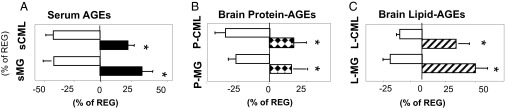
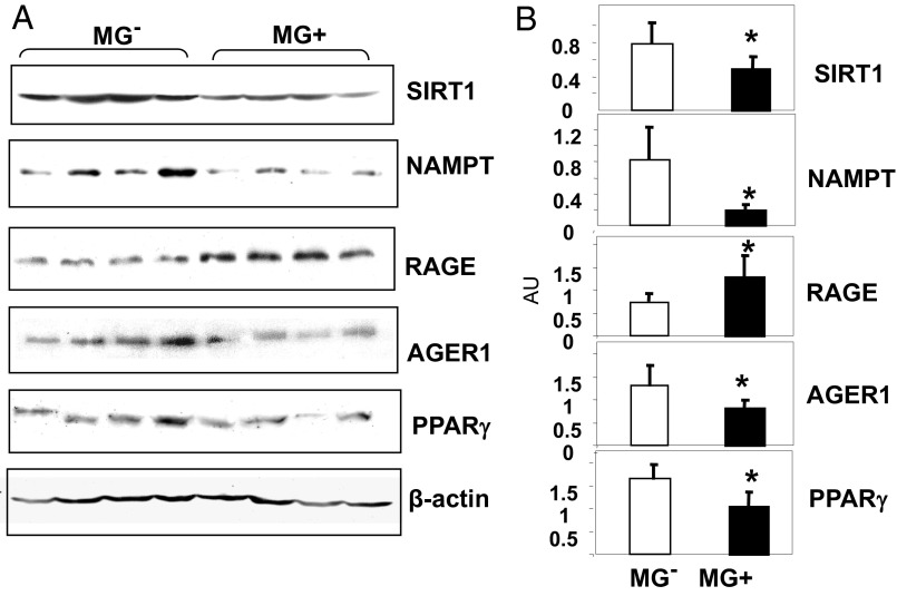
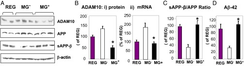
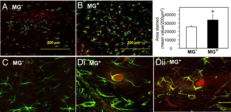
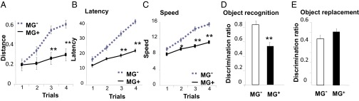
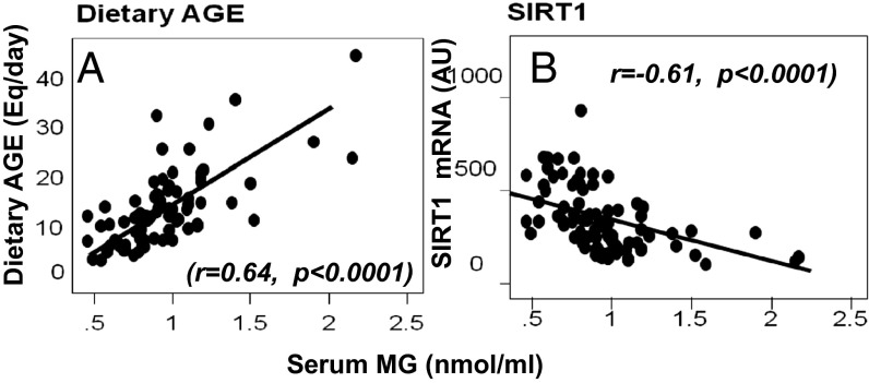
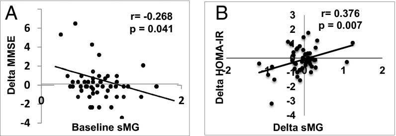
Age-associated dementia and Alzheimer's disease (AD) are currently epidemic. Neither their cause nor connection to the metabolic syndrome (MS) is clear. Suppression of deacetylase survival factor sirtuin 1 (SIRT1), a key host defense, is a central feature of AD. Age-related MS and diabetes are also causally associated with suppressed SIRT1 partly due to oxidant glycotoxins [advanced glycation end products (AGEs)]. Changes in the modern diet include excessive nutrient-bound AGEs, such as neurotoxic methyl-glyoxal derivatives (MG). To determine whether dietary AGEs promote AD, we evaluated WT mice pair-fed three diets throughout life: low-AGE (MG(-)), MG-supplemented low-AGE (MG(+)), and regular (Reg) chow. Older MG(+)-fed mice, similar to old Reg controls, developed MS, increased brain amyloid-β42, deposits of AGEs, gliosis, and cognitive deficits, accompanied by suppressed SIRT1, nicotinamide phosphoribosyltransferase, AGE receptor 1, and PPARγ. These changes were not due to aging or caloric intake, as neither these changes nor the MS were present in age-matched, pair-fed MG(-) mice. The mouse data were enhanced by significant temporal correlations between high circulating AGEs and impaired cognition, as well as insulin sensitivity in older humans, in whom dietary and serum MG levels strongly and inversely associated with SIRT1 gene expression. The data identify a specific AGE (MG) as a modifiable risk factor for AD and MS, possibly acting via suppressed SIRT1 and other host defenses, to promote chronic oxidant stress and inflammation. Because SIRT1 deficiency in humans is both preventable and reversible by AGE reduction, a therapeutic strategy that includes AGE reduction may offer a new strategy to combat the epidemics of AD and MS.

Keywords: caloric restriction; insulin resistance; neural; nutrition; obesity.


Conflict of interest statement

The authors declare no conflict of interest.

Figures

Fig. 1.
Fig. 1.
Oral MG+ leads to increased systemic and brain protein and lipid AGEs. Data are from 18-mo WT C57BL6 mice pair-fed MG+ or MG diet and control (Reg) mice (24–26 mo, n = 8/group). (A) Serum CML and MG levels. (B) Brain protein CML and protein MG. (C) Brain lipid CML and lipid MG. Data are percent (mean ± SEM) above or below Reg controls (as shown in Table 1). *P < 0.05, MG+ vs. MG mice.
Fig. 2.
Fig. 2.
Oral MG+ alters brain SIRT1, NAMPT, AGER1, RAGE, and PPARγ protein expression in MG+-fed mice brains. (A) Representative Western blots from brain extracts of 18-mo C57BL6 mice fed an MG+ or MG diet (n = 3–5) and (B) densitometry of A, shown as ratio (mean ± SEM) of target protein to β-actin. *P < 0.05 vs. MG mice.
Fig. 3.
Fig. 3.
Oral MG+ suppresses ADAM10 expression and increases Aβ in brain. Data are from 18-mo C57BL6 mice fed an MG+ or MG diet and Reg mice (24–26 mo, n = 8/group). (A) Western blots of brain extracts for ADAM10, APP, and soluble or sAPPβ (n = 3–5). β-actin is used as control. (B, i) Densitometry of ADAM10 protein data shown in A and (B, ii) of ADAM10 mRNA levels, by RT-PCR, shown as AU (n = 5/group). (C) Densitometry of sAPPβ and APP shown in A and expressed as sAPPβ/APP ratio. (D) Aβ1–42 levels. Data in B–D are shown as percent (mean ± SEM) of Reg. *P < 0.05 MG+ vs. MG mice.
Fig. 4.
Fig. 4.
Oral MG+ promotes brain gliosis and AGE deposits. Confocal microscopy of coronal hippocampal sections from (A) MG+ and (B) MG mice (n = 8/group) immunostained for glia cells (Magnification, 20×), and (C) MG and (D, i and ii) MG+ for both glia and AGEs, using anti-GFAP and anti-AGE, as primary antibodies and Alexa Fluor-488 (green) and Fluor-594 (red), respectively, as secondary antibodies (magnification, 200×). Bar graph shows the quantification of GFAP staining. (Scale bar, 200 µm.) *P < 0.05, MG+ vs. MG mice.
Fig. 5.
Fig. 5.
Oral MG+ impairs learning and memory capacities. (A–C) Accelerating rotarod test indicating that MG+ mice (straight line) can travel a shorter distance (A), have reduced latency until fall (B), and a slower speed than MG mice (broken line) (C). Data (mean ± SEM) were analyzed by two-way repeated-measures ANOVA. n = 10/group. *P < 0.05, **P < 0.01 vs. MG mice. (D) Object recognition and (E) object replacement tests show that MG+ mice have a deficient memory compared with MG mice. Data are shown as mean ± SEM (n = 10/group, **P < 0.01 vs. MG mice).
Fig. 6.
Fig. 6.
Serum MG levels correlate directly with dietary AGE intake (A) and inversely with MNC SIRT1 mRNA (B). Baseline fasting sMG levels, shown as mean ± SEM (nmol/mL), are plotted against daily dietary AGE intake, shown as Eq/d (A) or against MNC SIRT1 mRNA of healthy older adults (B). Fitted regression lines are as shown.
Fig. 7.
Fig. 7.
(A) Human serum MG (sMG) levels predict changes in cognition over time. Levels of fasting sMG at baseline are plotted against changes (Δ) in MMSE of healthy older adults, over 9 mo. (B) Changes (Δ) in sMG levels over time correlate with changes (Δ) in HOMA-IR over the same period. All tests were performed in fasting sera; fitted regression lines are as shown.

Comment in

References

    1. Janson J, et al. Increased risk of type 2 diabetes in Alzheimer disease. Diabetes. 2004;53(2):474–481. - PubMed
    1. Guarente L, Franklin H. Franklin H. Epstein Lecture: Sirtuins, aging, and medicine. N Engl J Med. 2011;364(23):2235–2244. - PubMed
    1. Talbot K, et al. Demonstrated brain insulin resistance in Alzheimer’s disease patients is associated with IGF-1 resistance, IRS-1 dysregulation, and cognitive decline. J Clin Invest. 2012;122(4):1316–1338. - PMC - PubMed
    1. Reddy VP, Zhu X, Perry G, Smith MA. Oxidative stress in diabetes and Alzheimer’s disease. J Alzheimers Dis. 2009;16(4):763–774. - PMC - PubMed
    1. Loy CT, Twigg SM. Growth factors, AGEing, and the diabetes link in Alzheimer’s disease. J Alzheimers Dis. 2009;16(4):823–831. - PubMed

Publication types

MeSH terms