. 2014 Mar 11;111(10):E943-52.
doi: 10.1073/pnas.1321390111. Epub 2014 Feb 24.

Mycobacterium abscessus cording prevents phagocytosis and promotes abscess formation

Affiliations

Mycobacterium abscessus cording prevents phagocytosis and promotes abscess formation

Audrey Bernut et al. Proc Natl Acad Sci U S A. .

Abstract

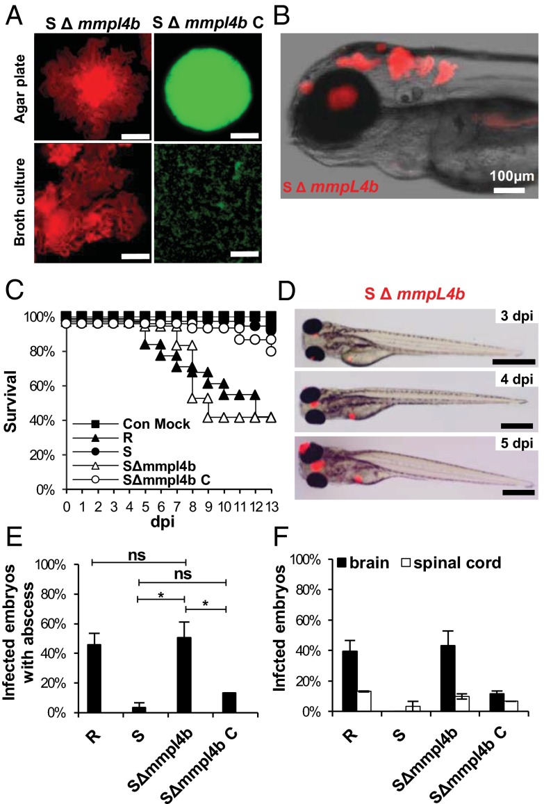
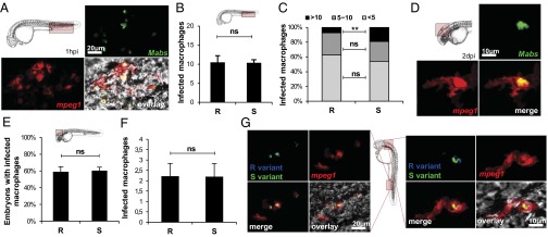
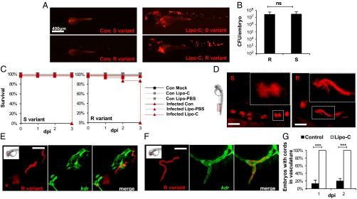
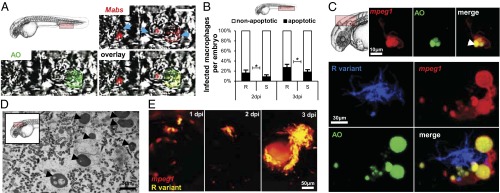
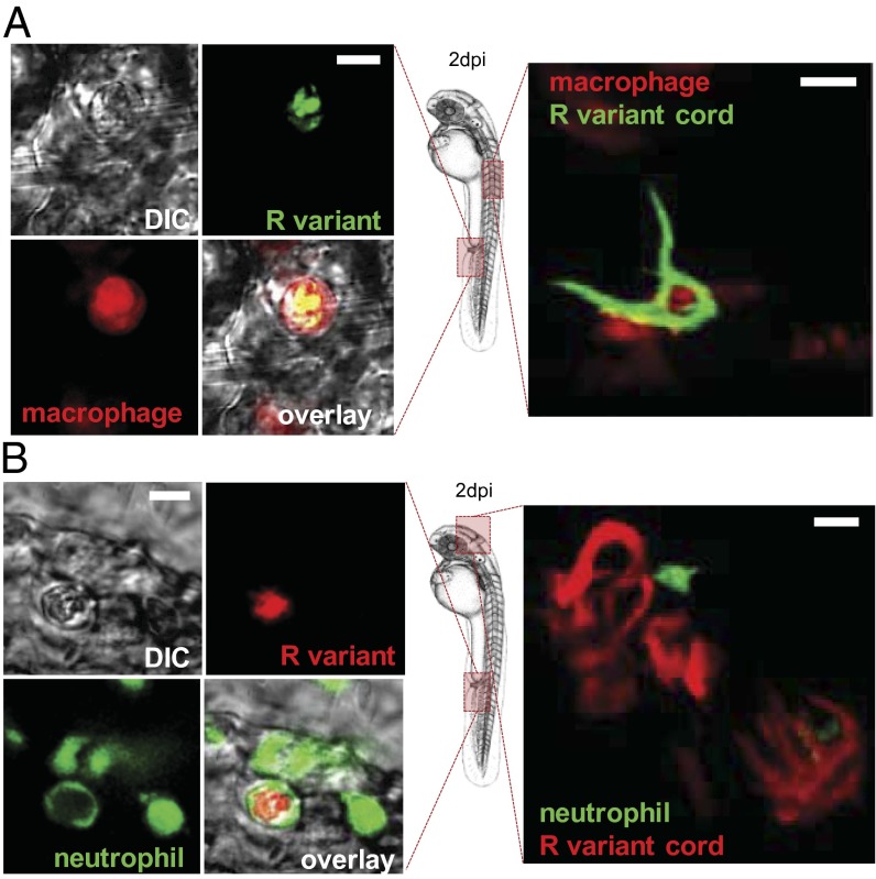
Mycobacterium abscessus is a rapidly growing Mycobacterium causing a wide spectrum of clinical syndromes. It now is recognized as a pulmonary pathogen to which cystic fibrosis patients have a particular susceptibility. The M. abscessus rough (R) variant, devoid of cell-surface glycopeptidolipids (GPLs), causes more severe clinical disease than the smooth (S) variant, but the underlying mechanisms of R-variant virulence remain obscure. Exploiting the optical transparency of zebrafish embryos, we observed that the increased virulence of the M. abscessus R variant compared with the S variant correlated with the loss of GPL production. The virulence of the R variant involved the massive production of serpentine cords, absent during S-variant infection, and the cords initiated abscess formation leading to rapid larval death. Cording occurred within the vasculature and was highly pronounced in the central nervous system (CNS). It appears that M. abscessus is transported to the CNS within macrophages. The release of M. abscessus from apoptotic macrophages initiated the formation of cords that grew too large to be phagocytized by macrophages or neutrophils. This study is a description of the crucial role of cording in the in vivo physiopathology of M. abscessus infection and emphasizes cording as a mechanism of immune evasion.

Keywords: granuloma; innate immunity; morphotype; pathogenesis.


Conflict of interest statement

The authors declare no conflict of interest.

Figures

Fig. 1.
Fig. 1.
Increased virulence and specific neurotropism of rough M. abscessus in ZF embryos. (A) Survival of embryos infected with ∼300 colony forming units (cfu) of the M. abscessus R or S variant compared with mock-infected controls (Con) (n = 20). Shown are representative results of three independent experiments. Embryos are significantly more susceptible to R-variant infection (P < 0.0001, log-rank test) than S-variant infection (statistically not different from mock-infected control). (B) Survival curves of embryos infected at various doses of M. abscessus R or S variant. Data shown are representative of two independent experiments; n = 20. Survival of embryos infected with the R variant depends on the dose (P < 0.0001, log-rank test). No statistically significant difference is seen between the control group and the embryos injected with the S variant, regardless of the dose. (C) Spatiotemporal visualization of the infection by either the R or the S variant expressing mCherry (∼300 cfu): representative fluorescence and transmission overlay of whole embryos and close-up of the brain. The yolk is auto-fluorescent. (Scale bars, 400 μm.) (D) Frequency of R- and S-variant abscesses in whole embryos over 13 dpi (∼300 cfu; n = 40, three independent experiments). (E) Average localization of abscesses of the infected embryos in D. Embryos infected with the R variant developed more abscesses within the brain or the spinal cord than embryos infected with the S variant (P < 0.001). (F) Bacterial loads of embryos infected with the R or S variant (∼200 cfu). Embryos infected with the R variant were separated into two groups, with (R+) or without (R) abscesses. Results are expressed as mean cfu per embryo from three independent experiments. Significant expansion of bacterial loads was observed up to 5 dpi within embryos infected with the R or the S variant (P = 0.001). Except for the bacterial load at 3 dpi in embryos infected with the R variant without abscess and embryos infected with the S variant, the bacterial burdens were significantly different between each group and at each time point. (GJ) DIC and fluorescent overlays of 3-dpi (boxed in G) and 7-dpi (boxed in H) embryos used for the TEM analysis of brain infection in embryos infected with the R variant (Scale bars, 200 μm.) (G, H, I, and J). Representative electron micrographs show a transverse section through the brain of embryos infected with the R variant at 3 dpi (G) and 7 dpi (HJ). (G) Two macrophages with intracellular bacteria internalized into multiple phagosomes present in the boxed infection area are shown. n, nucleus. Close-up view of one phagosome showing the membrane of phagosomal compartment (arrowheads). (H) TEM image of a typical brain lesion (boxed) with extensive extracellular bacterial replication and cellular debris. (I) Bacterial division. Arrowheads indicate division septum. (J) Healthy brain tissue. Significance was assessed by a generalized mixed-effects model fitted with a binomial (D and E) or Poisson distribution (F), comparing the proportion of both strains in each category. **P < 0.01; ***P < 0.001. Error bars represent the SD. (K) Golden embryos i.v. infected by several mycobacterial species expressing mCherry. Representative fluorescence and DIC overlay of R variants of M. abscessus complex members (M. abscessus senso stricto, M. massiliense, and M. bolletii), M. chelonae, or M. marinum infections in whole infected embryos at 5 dpi. (Scale bars, 400 μm.)
Fig. 2.
Fig. 2.
In vivo cording of the R variant initiates abscess formation. (A) Fluorescent images of R or S morphotypes on 7H10 agar plates or 7H9/OADC/Tween broth. The S variant appears hemispherical and smooth, whereas the R variant displays serpentine cords. Only rough bacteria produce multicellular cords. (Scale bars, 1 mm.) (B and C) R-variant–infected embryos expressing mCherry (B) or GFP (C) at 2 dpi. (B) Fluorescence and DIC overlay of the head. (Scale bar, 150 μm.) The enlarged view at right shows a maximum intensity projection of confocal imaging of a serpentine cord. (C) Fluorescence and DIC overlay of part of the tail. (Scale bar, 100 μm.) The enlarged view at right shows a maximum intensity projection of confocal imaging of cords within the spinal cord. (D) Kinetic of cord formation in infected embryos (∼300 cfu of M. abscessus S or R variant; n = 40). Only embryos infected with the R variant develop serpentine cords (P < 0.001 at each time point). The number R-variant–infected embryos with cords increased significantly up to 2 dpi (P = 0.03). (E) Average number of R-variant cords per embryo in D. The number of cords within R-variant–infected embryos increased significantly up to 3 dpi (P < 0.002). (F) The average localization of R-variant cords in D. Overall, the number of embryos with cords within the CNS and eyes increased significantly up to 3 dpi (P < 0.001 and P = 0.008, respectively). There was a significant difference in the localization of cords within embryos (P = 0.0008). (G) Abscess formation in an embryo infected with the M. abscessus R variant. (Scale bar, 200 μm.) Statistics were calculated using a generalized mixed-effects model fitted with a Poisson distribution (D and E) or a binomial distribution (F), comparing the proportion in each category across both strains. All results are expressed as the average from three independent experiments; error bars represent the SD.
Fig. 3.
Fig. 3.
Loss of mmpL4b confers high pathogenicity to the S variant. (A) Fluorescent pictures showing the general aspect of the S variant M. abscessus ΔmmpL4b mutant (SΔmmpL4b) and its complemented strain (SΔmmpL4b_C) on 7H10 agar or in 7H9/OADC/Tween broth medium. Hemispherical, smooth colonies are characteristic of SΔmmpL4b_C, whereas the SΔmmpL4b strain forms serpentine cords. (Scale bars, 0.5 mm.) (B) Fluorescence and DIC overlay of the whole head of infected embryos with red fluorescent SΔmmpL4b showing large numbers of serpentine cord within the brain. (C) Survival of embryos infected with ∼200 cfu M. abscessus R variant, S variant, SΔmmpL4b, or SΔmmpL4b_C (n = 30–40 per group). Shown are representative results from two independent experiments. Embryos are significantly more susceptible to infection by the SΔmmpL4b mutant and the R variant than by the parental M. abscessus S variant (P < 0.0001, log-rank test). Complementation restored attenuation (no statistically significant differences were seen in the survival of embryos infected with SΔmmpL4b_C and the embryos injected with the M. abscessus S variant. (D) Evolution of the infection by SΔmmpL4b (∼200 cfus) expressing mCherry. (Scale bars, 400 μm.) (E) Frequency of abscesses at 13 dpi (∼200 cfus; n = 30–40 per group; data shown are the average of two independent experiments). *P = 0.03; ns, not significant. (F) Average localization of mycobacterial abscesses in embryos from E. Overall, SΔmmpL4b-infected embryos developed more abscesses within the brain or the spinal cord than embryos infected with the S variant or SΔmmpL4b_C (P < 0.005) but did not develop more abscesses than embryos infected with the R variant. A generalized mixed-effects model fitted with a binomial distribution was used for statistics in E and F. Error bars represent the SD.
Fig. 4.
Fig. 4.
Macrophages rapidly phagocytize bacteria with no preference for S or R variants. M. abscessus (∼100 cfu) expressing GFP (green; A and D) or tdTomato (B, C, E, and F) were injected in tg(mpeg1:mCherry) embryos. (A) Maximum intensity projection of macrophage-phagocytosed bacteria close to the injection site. (B) Number of individual infected macrophages at the injection site (2 hpi; n = 30) as counted in the displayed region (boxed area, Upper Left). (C) Average proportion of macrophages containing fewer than five, 5–10, or >10 bacteria within infected embryos in B. The number of individual infected macrophages with >10 bacteria is significantly greater in S-variant–infected embryos than in R-variant–infected embryos. (D) Confocal fluorescence of a maximum intensity projection of a M. abscessus-infected macrophage within the brain. (E and F) Infected macrophages found within the brain of tg(mpeg1:mCherry/kdr:egfp) embryos (red macrophages; green vasculature) infected with M. abscessus expressing tdTomato (∼100 cfu) (n = 30). (E) Frequency of either R-variant– or S-variant–infected macrophages at 1 dpi as counted in the displayed region (Inset). (F) Number of individual infected macrophages within the brain of infected embryos in E. (G) tg(mpeg1:mCherry) embryos injected with M. abscessus expressing GFP and tdTomato. DIC microscopy and confocal fluorescence of maximum intensity projection of R/S-coinfected macrophages at the infection site (4 hpi, Left) and the brain (1 dpi, Right). Significance was assessed by a generalized mixed-effects model fitted with a binomial distribution (C and E) or a Poisson distribution (B and F) comparing the proportion in each category across both strains. **P < 0.01; ns, not significant. All results are expressed as averages of three independent experiments; error bars represent the SD.
Fig. 5.
Fig. 5.
M. abscessus infection is exacerbated and is constrained within the vasculature in the absence of macrophages. (A) Whole control embryos and Lipo-C–injected embryos infected with either the S (Upper) or the R (Lower) variant (tdTomato, ∼300 cfu) at 2 dpi. (B) Bacterial loads within Lipo-C–injected embryos infected with either the R or the S variant (∼300 cdu) at 2 dpi. (C) Survival curves of S- (Left) or R- (Right) infected control (con) and Lipo-C–injected embryos (∼300 cfu, n = 20). Embryos treated with Lipo-C are hypersusceptible to both M. abscessus variants (P < 0.0001, log-rank test). Shown are representative results from two independent experiments. (D) Confocal fluorescence microscopy of embryos injected with Lipo-C and infected with the R or S variant at 2 dpi. (Insets) Morphology of extracellular replicating S- and R-variant bacteria within the vasculature. In the absence of macrophages, the S variant produces clumps that are clearly different from the serpentine cords induced by the R variant (tdTomato). (Scale bars, 400 μm.) (E and F) Control (E), or Lipo-C–injected (F) tg(kdr:egfp) embryos infected with the tdTomato-expressing R variant (2 dpi). Control embryos show bacteria within the brain that do not colocalize with green fluorescent vasculature, whereas cords colocalize perfectly with the vasculature in the Lipo-C–infected embryo. (Scale bars, 50 μm in E; 30 μm in F.) (G) Frequency of cord formation in the vasculature-infected control and Lipo-C–injected embryos (n = 30, three independent experiments). Significance was assessed by a generalized mixed-effects model fitted with a binomial distribution (G) or a Poisson distribution (B), comparing the proportion in each category across both strains. ***P < 0.001; ns, not significant.
Fig. 6.
Fig. 6.
Apoptosis of infected macrophages releases bacteria. (A and B) AO (green) staining of apoptosis cells in living (A and C) or fixed (B) infected embryos. (A) Representative single confocal planes fluorescence and DIC overlay of three individual macrophages (blue arrows) infected with tdTomato M. abscessus (red). (Inset) AO labeling (green) shows the presence of one M. abscessus-infected apoptotic cell in the vasculature of a 3-dpi embryo. (Scale bar, 10 μm.) (B) Proportion of apoptotic infected macrophages compared with the number of nonapoptotic infected macrophages at the infection site at 2 and 3 dpi [∼300 cfu of the M. abscessus R or S variant injected in tg(mpeg1:mCherry) embryos as counted in the region displayed in the Inset]. Results are expressed as the averages of three independent experiments; error bars represent the SD. Significance was assessed by one-way ANOVA. (C) (Upper) Maximum intensity projection of confocal images with a representative apoptotic noninfected macrophage with apoptotic bodies (arrowhead). (Lower) tg(mpeg1:mCherry) embryos infected with R variant M. abscessus (tdTomato). Maximum intensity projection of confocal images show a serpentine cord (blue) surrounded by several macrophages (red) with apoptotic bodies (green) at 3 dpi. (D) Representative electron micrograph shows extracellular bacteria surrounded by cellular debris and typical apoptotic nuclei (arrowheads) at 7 dpi. (E) Spatiotemporal visualization of cord formation emerging from a macrophage infected with the R variant expressing mCherry in tg(mpeg1:mCherry) embryos.
Fig. 7.
Fig. 7.
In vivo cording of the R variant prevents phagocytosis. (A) Tg(mpeg1:mCherry) embryos harboring red-fluorescent macrophages were infected with GFP-expressing R variants. (Left) Maximum intensity projection of confocal images of macrophages capable of phagocytosing the M. abscessus R variant in the vascular system. (Right) Macrophages are unable to internalize M. abscessus R cords. (Scale bars, 10 μm.) (B) Tg(mpx:egfp) embryos harboring green-fluorescent neutrophils were infected with mCherry-expressing R variants. (Left) Maximum intensity projection of confocal images of M. abscessus R-variant internalized neutrophils in the vascular system. (Scale bar, 10 μm). (Right) Neutrophils are unable to internalize M. abscessus R-variant cords. (Scale bars, 15 μm.)
Fig. 8.
Fig. 8.
Modeling the physiopathology of M. abscessus in ZF embryos. After injection in the blood flow (1), M. abscessus are rapidly engulfed by macrophages attracted by chemotaxis to the injection site (2 and 3). Infected macrophages migrate from the vasculature to the nervous tissues, become heavily infected (4), and eventually die (5). Apoptosis leads to the release of S variants (blue pathway) that subsequently are phagocytosed by newly recruited macrophages to form cellular aggregates (6). Bacterial multiplication is controlled, and a chronic infection is established. For the M. abscessus R variant (red pathway), apoptosis correlates with the appearance of extracellular serpentine cords (6), preventing phagocytosis by macrophages and neutrophils (7), leading to unrestricted cording, abscess formation with tissue destruction, and larval death (8).

References

    1. Brown-Elliott BA, Wallace RJ., Jr Clinical and taxonomic status of pathogenic nonpigmented or late-pigmenting rapidly growing mycobacteria. Clin Microbiol Rev. 2002;15(4):716–746. - PMC - PubMed
    1. Wallace RG, Jr, Silcox V, Brown BA. Taxonomy of rapidly growing mycobacteria. Clin Infect Dis. 1994;18(1):121–122. - PubMed
    1. Olivier KN, et al. Nontuberculous Mycobacteria in Cystic Fibrosis Study Group Nontuberculous mycobacteria. I: Multicenter prevalence study in cystic fibrosis. Am J Respir Crit Care Med. 2003;167(6):828–834. - PubMed
    1. Jönsson BE, et al. Molecular epidemiology of Mycobacterium abscessus, with focus on cystic fibrosis. J Clin Microbiol. 2007;45(5):1497–1504. - PMC - PubMed
    1. Roux AL, et al. Jean-Louis Herrmann for the OMA Group Multicenter study of prevalence of nontuberculous mycobacteria in patients with cystic fibrosis in france. J Clin Microbiol. 2009;47(12):4124–4128. - PMC - PubMed

Publication types

MeSH terms

LinkOut - more resources