. 2014 Mar 11;111(10):3745-50.
doi: 10.1073/pnas.1313886111. Epub 2014 Feb 24.

G-protein coupled receptor BAI3 promotes myoblast fusion in vertebrates

Affiliations

G-protein coupled receptor BAI3 promotes myoblast fusion in vertebrates

Noumeira Hamoud et al. Proc Natl Acad Sci U S A. .

Abstract

Muscle fibers form as a result of myoblast fusion, yet the cell surface receptors regulating this process are unknown in vertebrates. In Drosophila, myoblast fusion involves the activation of the Rac pathway by the guanine nucleotide exchange factor Myoblast City and its scaffolding protein ELMO, downstream of cell-surface cell-adhesion receptors. We previously showed that the mammalian ortholog of Myoblast City, DOCK1, functions in an evolutionarily conserved manner to promote myoblast fusion in mice. In search for regulators of myoblast fusion, we identified the G-protein coupled receptor brain-specific angiogenesis inhibitor (BAI3) as a cell surface protein that interacts with ELMO. In cultured cells, BAI3 or ELMO1/2 loss of function severely impaired myoblast fusion without affecting differentiation and cannot be rescued by reexpression of BAI3 mutants deficient in ELMO binding. The related BAI protein family member, BAI1, is functionally distinct from BAI3, because it cannot rescue the myoblast fusion defects caused by the loss of BAI3 function. Finally, embryonic muscle precursor expression of a BAI3 mutant unable to bind ELMO was sufficient to block myoblast fusion in vivo. Collectively, our findings provide a role for BAI3 in the relay of extracellular fusion signals to their intracellular effectors, identifying it as an essential transmembrane protein for embryonic vertebrate myoblast fusion.

Keywords: RhoGTP; ced-12; model system; myogenesis; myotube formation.


Conflict of interest statement

The authors declare no conflict of interest.

Figures
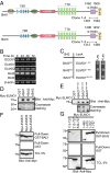
Fig. 1.
Fig. 1.
The GPCR BAI3 is expressed in myoblasts and interacts with the ELMO family of proteins. (A) Schematic representation of mBAI1 and mBAI3 receptors isolated as ELMO-binding partners in a yeast two-hybrid screen. Four identical BAI1 clones and three identical BAI3 clones were recovered from the screen, and the binding regions are denoted in the schematics. Important structural domains are shown [hormone-binding domain (HBD); transmembrane domains (TM); predicted α-helix (α-Helix) (Fig. S1A); binding sequence for PDZ domains (Gln-Thr-Glu-Val, QTEV)]. (B) Expression of DOCK1, ELMO1, ELMO2, BAI2, and BAI3 mRNAs in differentiating C2C12 myoblasts (0–72 h) was measured by semiquantitative RT-PCR. (C) The N terminus of ELMO1 is required for BAI3 binding. Yeasts were cotransformed with the indicated LexA fusion ELMO constructs and a B42 fusion of BAI3 construct ICD and were grown on selective (−leu) and nonselective plates (+leu) to assay for protein–protein interaction. (D) The C termini of BAI1, BAI2, and BAI3 interact with ELMO1. HEK293 cells were transfected with Myc–ELMO1, and cleared lysate was subjected to GST pull-downs with the indicated GST–BAI fusion proteins or GST alone. Bound Myc–ELMO1 was detected by immunoblotting. (E) Identification of the minimal BAI3 region sufficient for interaction with ELMO1. Cleared lysates as in D were subjected to pull-down assays with the indicated GST–BAI3 fragments. Bound Myc–ELMO1 was detected by immunoblotting. (F) The BAI3 C terminus binds ELMO1–3. HEK293 cells were transfected with Myc–ELMO1, Myc–ELMO2, or Myc–ELMO3, and clarified lysates were subjected to GST–BAI3 pull downs. Bound Myc–ELMO1–3 were detected by immunoblotting. (G) Mapping the BAI3-binding site on ELMO1. HEK293 cells were transfected with a panel of Myc–ELMO1 constructs, and clarified lysates were subjected to GST–BAI3 pull downs. Bound Myc–ELMO1 fragments were detected by immunoblotting. TCL, Total Cell Lysate.
Fig. 2.
Fig. 2.
BAI3, ELMO1, and ELMO2 are essential for myoblast fusion. (AL) C2C12 cells expressing GFP (no hairpin) or shRNAs targeting BAI1, BAI3, ELMO1, or ELMO2 were generated by retroinfections. (A) Down-regulation of BAI3 and ELMO2 impairs myoblast fusion after 48 h in differentiation conditions. (Right) Dotted white boxes are shown at a higher magnification. (BC) Real-time Q-RT-PCR amplifications against BAI3 or ELMO2 were performed to confirm specific knockdowns. (D) Quantification of experiments shown in A. (EL) Expression of hBAI3 in BAI3–shRNA, Myc–ELMO1 in ELMO2–shRNA, and Myc–ELMO2 in ELMO1–shRNA C2C12 cells restored myoblast fusion. (E, G, J) Representative images of myoblast fusion for the indicated conditions. (F, I, L) Quantification of experiments shown in E, G, and J. (H, K) Real-time Q-RT-PCR amplifications against ELMO1 or ELMO2 were performed to confirm specific knockdowns. (A, E, G, and J) Myofibers were stained for Myosin Heavy Chain [MHC, MF20 antibody (red)] and nuclei revealed by Hoechst (blue). Error bars indicate SD. One-way ANOVA followed by a Bonferroni test calculated the P values; ****P < 0.0001. (Scale bar, 100 μm.)
Fig. 3.
Fig. 3.
BAI3 coupling to ELMO2 is necessary for myoblast fusion. (A) Identification of BAI3 residues important for interaction to ELMO1. HEK293 cells were transfected with Myc–ELMO1 and clarified lysates were subjected to pull-downs with GST alone, GST–BAI3, or the indicated mutants of BAI3. Bound Myc–ELMO1 was detected by immunoblotting. (B) Expression of BAI3 mutants unable to engage ELMO fails to rescue myoblast fusion in C2C12 cells depleted of BAI3. Experiments were carried out as in Fig. 2. (C) Quantification of fibers with three nuclei and more and (D) quantification of fibers with one nucleus. (E) Overexpression of BAI3 mutants lacking ELMO-binding activity blocks myoblast fusion in parental C2C12 cells. Cells were transfected with the indicated BAI3 mutants and differentiated for 48 h before analyzing fusion. (F) Quantification of the experiment shown in E. Error bars indicate SD. One-way ANOVA followed by a Bonferroni test calculated the P values; ****P < 0.0001. (Scale bar, 100 μm.)
Fig. 4.
Fig. 4.
The interaction of BAI3 with ELMO is essential in vivo for myoblast fusion. (A) In situ hybridization of antisense DIG-labeled riboprobes demonstrates that BAI3 (Right) is coexpressed by muscle precursors that also express the myocyte differentiation marker MyoD (Left) in the developing muscles of E5 chicken embryos. BAI3 is also expressed in the spinal cord. SC, spinal cord; M, muscle. (B) Schematic of the strategy to express constructs in muscle progenitors. Somitocoeles of interlimb somites of embryos between 13 and 18 HH (E2.5) were microinjected with plasmids and electroporated as indicated. Seventy-two hours after electroporation, embryos expressing GFP were collected and analyzed for myoblast fusion. Identical muscle development takes place in nonelectroporated and electroporated (GFP plasmid) sides of the embryo as demonstrated by staining for MHC and GFP. (C) Differentiation of myoblasts is not impaired by expression of GFP, BAI3, or the indicated mutant of BAI3 lacking ELMO-binding activity. Cryosections were stained with anti-Desmin and anti-GFP antibodies. (D) Quantification of cells double positive for Desmin and GFP. (E) Expression of BAI3 lacking ELMO-binding activity, but not GFP or wild-type BAI3, blocks myoblast fusion in vivo. Cryosections as in C were stained with anti-MHC and anti-GFP antibodies, and nuclei were revealed with Hoechst. (F) Length of myofibers was quantified (µM) in the indicated conditions. Error bars indicate SD. One-way ANOVA followed by a Bonferroni test calculated the P values; ****P < 0.0001.

References

    1. Bentzinger CF, Wang YX, Rudnicki MA. Building muscle: Molecular regulation of myogenesis. Cold Spring Harb Perspect Biol. 2012;4(2):1–16. - PMC - PubMed
    1. Abmayr SM, Pavlath GK. Myoblast fusion: Lessons from flies and mice. Development. 2012;139(4):641–656. - PMC - PubMed
    1. Powell GT, Wright GJ. Do muscle founder cells exist in vertebrates? Trends Cell Biol. 2012;22(8):391–396. - PubMed
    1. Ruiz-Gómez M, Coutts N, Price A, Taylor MV, Bate M. Drosophila dumbfounded: A myoblast attractant essential for fusion. Cell. 2000;102(2):189–198. - PubMed
    1. Strünkelnberg M, et al. rst and its paralogue kirre act redundantly during embryonic muscle development in Drosophila. Development. 2001;128(21):4229–4239. - PubMed

Publication types