

Comment
doi: 10.1073/pnas.1401325111.
Epub 2014 Feb 24.
Examining how enzymes self-organize in a membrane
Affiliations
- PMID: 24567406
- PMCID: PMC3956151
- DOI: 10.1073/pnas.1401325111
Item in Clipboard
Comment
Examining how enzymes self-organize in a membrane
Proc Natl Acad Sci U S A.
.
No abstract available
Conflict of interest statement
The author declares no conflict of interest.
Figures
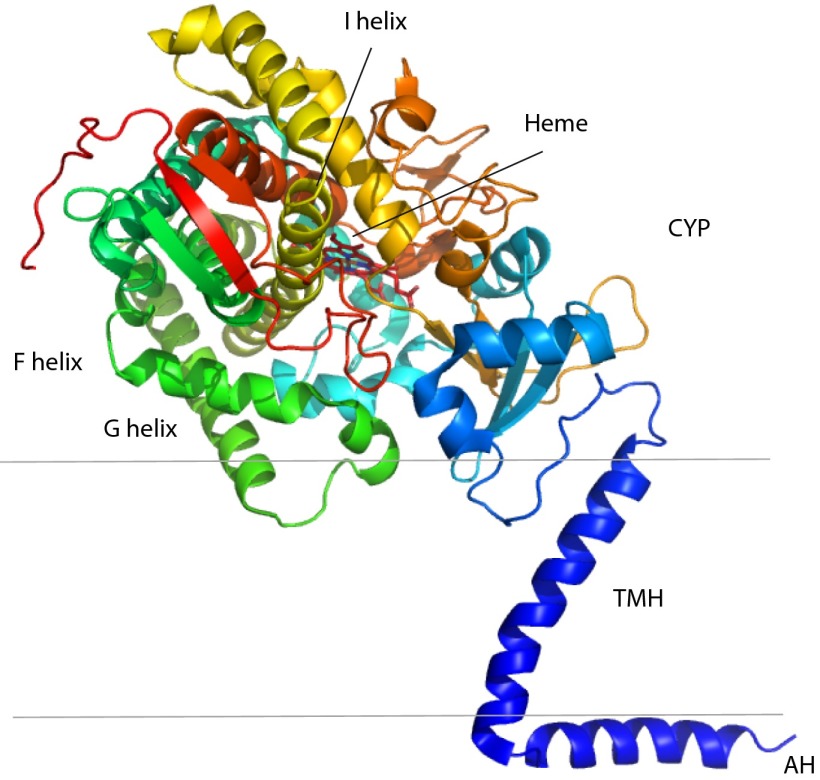
Three-domain structure of CYP51 from S. cerevisiae as described in ref. (PDB code 4K0F). The ribbon diagram is rainbow coded (N-terminal, blue; C-terminal, red). An N-terminal AH is predicted to bind to the inner surface of the ER membrane in vivo and is connected to the CYP on the outer membrane surface via a TMH. Positions of some secondary structural features in the CYP domain, including the active site heme and the F, G, and I helices, are noted. Parallel black lines indicate approximate position proposed for the lipid bilayer. Graphics were generated using PyMOL (11).

Residues involved in polar interactions between the TMH and CYP domains of lanosterol 14α demethylase (CYP51) from S. cerevisiae (PDB code 4K0F).

Alignment of membrane-interacting regions of two CYP51 structures: one from Monk et al. (4) (PDB code 4K0F) in magenta and that of the truncated T. cruzi enzyme (10) (PDB code 3K1O) in cyan. The region interacting with the TMH in 4K0F (near the A helix and β1 sheet) is shown for both enzymes. The loop connecting the F and G helices in 3K1O is disordered in the T. cruzi enzyme between Pro-216 and Leu-221, as is the loop following the G helix, between Lys-254 and Asn-257. The structure is ordered in both places in the 4K0F structure.
Comment on
-
Architecture of a single membrane spanning cytochrome P450 suggests constraints that orient the catalytic domain relative to a bilayer.Proc Natl Acad Sci U S A. 2014 Mar 11;111(10):3865-70. doi: 10.1073/pnas.1324245111. Epub 2014 Feb 3. Proc Natl Acad Sci U S A. 2014. PMID: 24613931 Free PMC article.
References
Publication types
MeSH terms
Substances
Grants and funding
LinkOut - more resources
Full Text Sources
Other Literature Sources
Molecular Biology Databases
