. 2014 Mar 11;111(10):3727-32.
doi: 10.1073/pnas.1322778111. Epub 2014 Feb 24.

Crystal structures of human soluble adenylyl cyclase reveal mechanisms of catalysis and of its activation through bicarbonate

Affiliations

Crystal structures of human soluble adenylyl cyclase reveal mechanisms of catalysis and of its activation through bicarbonate

Silke Kleinboelting et al. Proc Natl Acad Sci U S A. .

Abstract

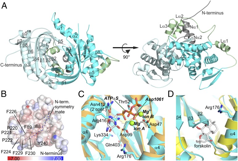
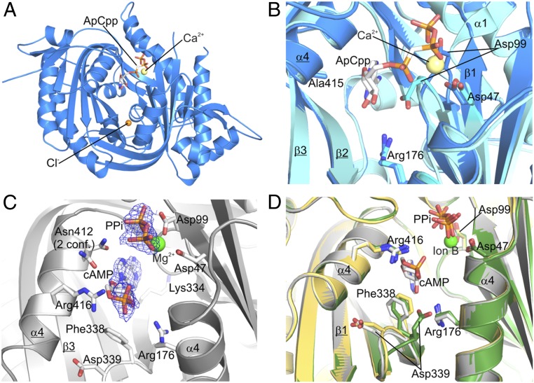
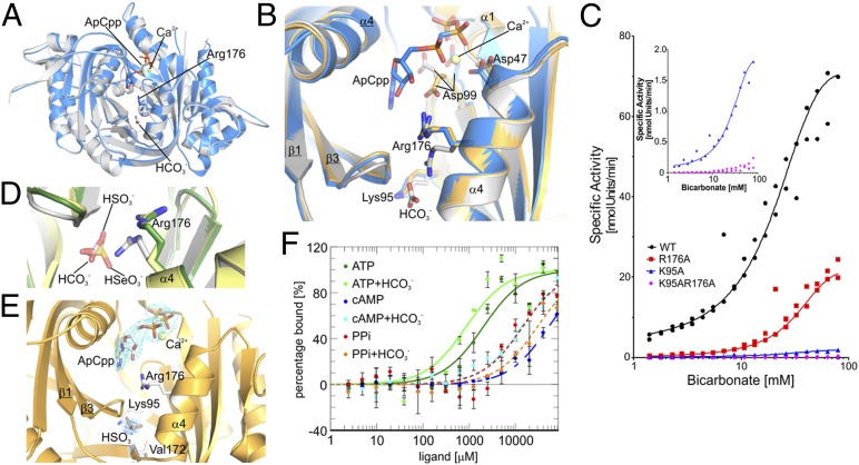
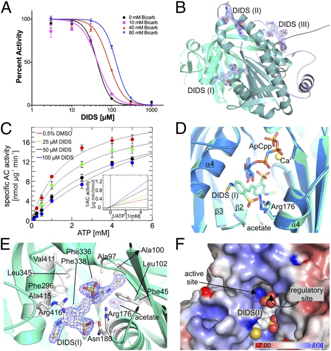
cAMP is an evolutionary conserved, prototypic second messenger regulating numerous cellular functions. In mammals, cAMP is synthesized by one of 10 homologous adenylyl cyclases (ACs): nine transmembrane enzymes and one soluble AC (sAC). Among these, only sAC is directly activated by bicarbonate (HCO3(-)); it thereby serves as a cellular sensor for HCO3(-), carbon dioxide (CO2), and pH in physiological functions, such as sperm activation, aqueous humor formation, and metabolic regulation. Here, we describe crystal structures of human sAC catalytic domains in the apo state and in complex with substrate analog, products, and regulators. The activator HCO3(-) binds adjacent to Arg176, which acts as a switch that enables formation of the catalytic cation sites. An anionic inhibitor, 4,4'-diisothiocyanatostilbene-2,2'-disulfonic acid, inhibits sAC through binding to the active site entrance, which blocks HCO3(-) activation through steric hindrance and trapping of the Arg176 side chain. Finally, product complexes reveal small, local rearrangements that facilitate catalysis. Our results provide a molecular mechanism for sAC catalysis and cellular HCO3(-) sensing and a basis for targeting this system with drugs.

Keywords: activation mechanism; bicarbonate signaling; catalytic mechanism; inhibition mechanism.


Conflict of interest statement

Conflict of interest statement: J.B. and L.R.L. own equity interest in CEP Biotech, which has licensed commercialization of a panel of monoclonal antibodies directed against sAC.

Figures

Fig. 1.
Fig. 1.
Crystal structure of human sAC-cat apo enzyme. (A) Overall sAC-cat structure, with C1 in cyan and C2 in pale cyan and secondary structures labeled (C2 labels underlined). (Right) This view highlights the N terminus (dark gray) and C1-C2 linker (green). (B) A sAC-cat apo surface region (colored according to electrostatic potential indicated in the scale bar) interacts with the N terminus of a symmetry mate (stick presentation). (C) An overlay of the active sites of sAC-cat apo (cyan; side chains colored according to atom type) and CyaC/ATPαS (orange; PDB ID code 1WC1) shows the overlap between human sAC Asp99 and ion site A (CyaC labels in italics). (D) Pseudosymmetrical sites of overlaid sAC-cat (cyan) and tmAC-C1C2 (yellow; PDB ID code 1AZS) structures, with the tmAC activator forskolin colored according to atom type.
Fig. 2.
Fig. 2.
Crystal structure of sAC-cat with bound ATP analog. (A) Overall structure of the sAC-cat/Ca2+/ApCpp complex, with Ca2+ in yellow and ApCpp colored according to atom type. The chlorine in the pseudosymmetrical site is shown as an orange sphere. (B) Active site overlay of sAC-cat apo (cyan) and the ApCpp complex (blue), with relevant residues shown in stick presentation. (C) sAC product complex (cAMP, PPi/Mg2+) colored according to atom type and overlaid with 2Fo-Fc density contoured at 1σ. (D) An overlay of the active sites of the sAC-cat complex with both products cAMP and PPi (gray), the sAC-cat/PPi complex resulting from soaking with PPi (green), and the sAC-cat/PPi complex resulting from soaking with ATP and substrate turnover (yellow) highlights the local rearrangement of the β2/β3 loop around Asp339.
Fig. 3.
Fig. 3.
sAC-cat activation by bicarbonate. (A) Overall structure of the sAC-cat bicarbonate complex (gray), overlaid with the sAC-cat/ApCpp complex (blue). Ligands are shown as sticks (ApCpp, bicarbonate) or as a sphere (Ca2+). (B) Overlay of active site regions of apo sAC (yellow), sAC/ApCpp (blue), and the sAC/bicarbonate complex (gray) shows the HCO3-triggered Arg176 movement. (C) HCO3 concentration–response for AC activity of WT human sAC-cat (black dots) and for the mutants R176A (red squares), K95A (blue triangles), and K95A/R176A (magenta diamonds). (Inset) Data for K95A and K95AR176A with an expanded y axis. (D) Overlay of the regulatory sites of sAC shows the different geometries and binding details of HCO3 (gray), HSO3 (green), and HSeO3 (yellow) and illustrates the Arg176 movement uniquely triggered by HCO3. Ligands are shown as sticks colored according to atom type. (E) sAC/ApCpp complex with bisulfite bound in the regulatory site, overlaid with 2Fo-Fc density (1σ; cyan) and anomalous scattering density (3σ; blue). (F) Binding affinity to sAC-cat for ATP (Kd = 2.0 ± 0.4 mM), cAMP (Kd = 54.2 ± 13.4 mM), and PPi (Kd = 14.2 ± 0.3 mM). Addition of 50 mM HCO3 changed the Kd for ATP (0.9 ± 0.1 mM), cAMP (18.6 ± 6.1 mM), and PPi (30.7 ± 12.1 mM). Error bars indicate SD (n = 2).
Fig. 4.
Fig. 4.
sAC-cat inhibition by DIDS. (A) WT human sAC-cat activity assayed at the indicated concentrations of DIDS and in the presence of increasing amounts of HCO3. DIDS IC50 determination in 0 mM HCO3 (IC50 43 μM), in 10 mM HCO3 (44 μM), in 40 mM HCO3 (85 μM), and in 80 mM HCO3 (130 μM) is shown. Error bars indicate SD (n = 3). (B) Overall structure of sAC (C1, teal; C2, cyan; linker, blue) with all three DIDS molecules (sticks) overlaid with 2Fo-Fc density (1σ). (C) sAC activity at various ATP and DIDS concentrations yields a constant Vmax (24.6 ± 3.7 nmol⋅μg−1⋅min−1) and increasing apparent Km values for ATP [0 μM DIDS (1.7 ± 0.3 mM), 25 μM DIDS (2.4 ± 0.4 mM), 50 μM DIDS (4.1 ± 0.5 mM), 100 μM DIDS (7.3 ± 2.7 mM)]. Error bars indicate SD (n = 2). (Inset) Lineweaver–Burk plot of the same data. (D) Overlay of the sAC-cat/DIDS structure (cyan) and the sAC/ApCpp complex (blue). (E) Details of the DIDS binding site in sAC show the interacting hydrophobic and positively charged residues (stick presentation) and the acetate trapped in the regulatory site. The 2Fo-Fc density around the ligands is contoured at 1σ. (F) Protein surface of sAC-cat/DIDS colored according to electrostatic potential (indicated in scale bar), with DIDS (shown as spheres) partially blocking the active site entrance. Access to the regulatory site, behind the indicated DIDS sulfonyl group, is fully blocked.

References

    1. Hall RA, et al. CO(2) acts as a signalling molecule in populations of the fungal pathogen Candida albicans. PLoS Pathog. 2010;6(11):e1001193. - PMC - PubMed
    1. Hanoune J, Defer N. Regulation and role of adenylyl cyclase isoforms. Annu Rev Pharmacol Toxicol. 2001;41:145–174. - PubMed
    1. Kamenetsky M, et al. Molecular details of cAMP generation in mammalian cells: A tale of two systems. J Mol Biol. 2006;362(4):623–639. - PMC - PubMed
    1. Sunahara RK, Taussig R. Isoforms of mammalian adenylyl cyclase: Multiplicities of signaling. Mol Interv. 2002;2(3):168–184. - PubMed
    1. Zippin JH, et al. Bicarbonate-responsive “soluble” adenylyl cyclase defines a nuclear cAMP microdomain. J Cell Biol. 2004;164(4):527–534. - PMC - PubMed

Publication types

LinkOut - more resources