Differences in neural connectivity between the substantia nigra and ventral tegmental area in the human brain
- PMID: 24567711
- PMCID: PMC3915097
- DOI: 10.3389/fnhum.2014.00041
Differences in neural connectivity between the substantia nigra and ventral tegmental area in the human brain
Abstract
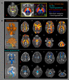
Objectives: Many animal and a few human studies have reported on the neural connectivity of the substantia nigra (SN) and the ventral tegmental area (VTA). However, it has not been clearly elucidated so far. We attempted to investigate any differences in neural connectivity of the SN/VTA in the human brain, using diffusion tensor imaging (DTI).
Methods: Sixty-three healthy subjects were recruited for this study. DTIs were acquired using a sensitivity-encoding head coil at 1. 5T. Connectivity was defined as the incidence of connection between the SN/VTA and each brain regions in the brain.
Results: The connectivity of SN was higher than that of the VTA. This included in the primary motor cortex, primary somatosensory cortex, premotor cortex, prefrontal cortex, caudate nucleus, globus pallidus, putamen, nucleus accumbens, temporal lobe, amygdala, pontine basis, occipital lobe, anterior and posterior lobe of cerebellum, corpus callosum, and external capsule (p < 0.05). However, no significant differences were observed in the red nucleus, thalamus, pontine tegmentum, and medial temporal lobe between the SN and VTA (p > 0.05).
Conclusions: We found the differences in neural connectivity of the SN/VTA in the human brain. The method and results of this study can provide useful information for clinicians and researchers in neuroscience, especially who work for Parkinson's disease and patients with brain injury.
Keywords: diffusion tensor imaging; dopamine; structural connectivity; substantia nigra; ventral tegmental area.
Figures

Similar articles
-
Internet gaming disorder: deficits in functional and structural connectivity in the ventral tegmental area-Accumbens pathway.Brain Imaging Behav. 2019 Aug;13(4):1172-1181. doi: 10.1007/s11682-018-9929-6. Brain Imaging Behav. 2019. PMID: 30054871
-
The neural connectivity of the inferior olivary nucleus in the human brain: a diffusion tensor tractography study.Neurosci Lett. 2012 Aug 8;523(1):67-70. doi: 10.1016/j.neulet.2012.06.043. Epub 2012 Jun 26. Neurosci Lett. 2012. PMID: 22743659
-
Novelty increases the mesolimbic functional connectivity of the substantia nigra/ventral tegmental area (SN/VTA) during reward anticipation: Evidence from high-resolution fMRI.Neuroimage. 2011 Sep 15;58(2):647-55. doi: 10.1016/j.neuroimage.2011.06.038. Epub 2011 Jun 24. Neuroimage. 2011. PMID: 21723396
-
The human substantia nigra and ventral tegmental area. A neuroanatomical study with notes on aging and aging diseases.Adv Anat Embryol Cell Biol. 1991;121:1-132. Adv Anat Embryol Cell Biol. 1991. PMID: 2053466 Review.
-
Reward and aversion in a heterogeneous midbrain dopamine system.Neuropharmacology. 2014 Jan;76 Pt B(0 0):351-9. doi: 10.1016/j.neuropharm.2013.03.019. Epub 2013 Apr 8. Neuropharmacology. 2014. PMID: 23578393 Free PMC article. Review.
Cited by
-
Utilising activity patterns of a complex biophysical network model to optimise intra-striatal deep brain stimulation.Sci Rep. 2024 Aug 14;14(1):18919. doi: 10.1038/s41598-024-69456-7. Sci Rep. 2024. PMID: 39143173 Free PMC article.
-
Dopamine Dysregulation in Reward and Autism Spectrum Disorder.Brain Sci. 2024 Jul 22;14(7):733. doi: 10.3390/brainsci14070733. Brain Sci. 2024. PMID: 39061473 Free PMC article. Review.
-
Cerebellum in levodopa-induced dyskinesias: the unusual suspect in the motor network.Front Neurol. 2014 Aug 18;5:157. doi: 10.3389/fneur.2014.00157. eCollection 2014. Front Neurol. 2014. PMID: 25183959 Free PMC article. Review.
-
Evidence for direct dopaminergic connections between substantia nigra pars compacta and thalamus in young healthy humans.Front Neural Circuits. 2025 Jan 9;18:1522421. doi: 10.3389/fncir.2024.1522421. eCollection 2024. Front Neural Circuits. 2025. PMID: 39850841 Free PMC article.
-
Neuroprotective Effects of Bacterial Melanin in a Rotenone-Induced Parkinson's Disease Rat Model: Electrophysiological Evidence from Cortical Stimulation of Substantia Nigra Neurons.Biomedicines. 2025 May 28;13(6):1317. doi: 10.3390/biomedicines13061317. Biomedicines. 2025. PMID: 40564036 Free PMC article.
References
LinkOut - more resources
Full Text Sources
Other Literature Sources

