Case Reports
. 2014 Jan 31;7(1):6-10.
doi: 10.4066/AMJ.2014.1875. eCollection 2014.

An uncommon cause of ascites: spontaneous rupture of biliary cystadenoma

Affiliations
Case Reports

An uncommon cause of ascites: spontaneous rupture of biliary cystadenoma

Sasidharan Abhishek et al. Australas Med J. .

Abstract

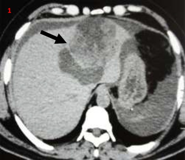
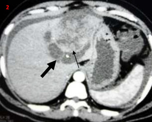
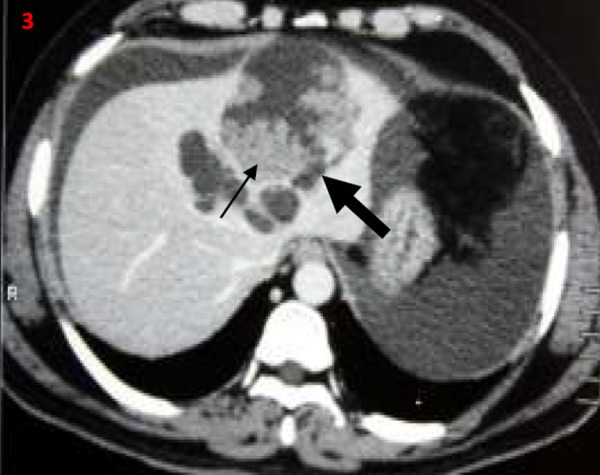
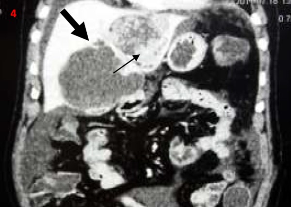
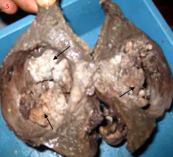
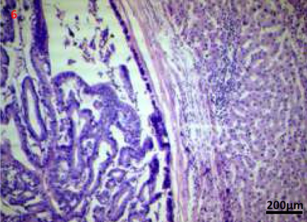
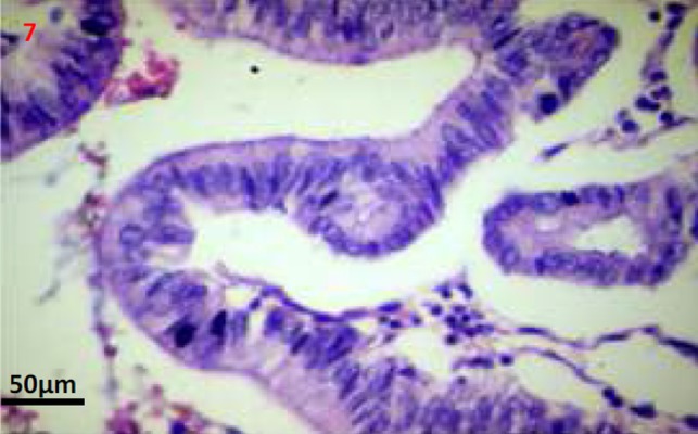
Biliary cystadenomas are cystic hepatic tumours of biliary origin. Cystadenomas are often slow-growing benign tumours, but always harbour the risk of malignant transformation. Cystadenomas are often asymptomatic, but may present with abdominal pain and distension. Though suspected with cross-sectional abdominal imaging, definitive diagnosis almost always requires histology. Spontaneous rupture of cystadenoma had been reported three times in the medical literature to date, all presenting with peritonitis. Here we report a case of spontaneous intraperitoneal rupture of biliary cystadenoma presenting as ascites without peritonitis.

Keywords: Nonparasitic hepatic cyst; abdominal pain; cystic neoplasm; leaking cyst.


Conflict of interest statement

CONFLICTS OF INTEREST

The authors declare that they have no competing interests.

Figures

Figures 1–4
Figures 1–4. Contrast-enhanced computerised tomography abdomen transverse (1-3) and coronal images (4); showing predominantly cystic left hepatic lobe lesions (thick arrow) with mural nodules/papillary projections (thin arrow).
Figures 1–4
Figures 1–4. Contrast-enhanced computerised tomography abdomen transverse (1-3) and coronal images (4); showing predominantly cystic left hepatic lobe lesions (thick arrow) with mural nodules/papillary projections (thin arrow).
Figures 1–4
Figures 1–4. Contrast-enhanced computerised tomography abdomen transverse (1-3) and coronal images (4); showing predominantly cystic left hepatic lobe lesions (thick arrow) with mural nodules/papillary projections (thin arrow).
Figures 1–4
Figures 1–4. Contrast-enhanced computerised tomography abdomen transverse (1-3) and coronal images (4); showing predominantly cystic left hepatic lobe lesions (thick arrow) with mural nodules/papillary projections (thin arrow).
Figure 5
Figure 5. Postoperative gross specimen showing cut section through the cyst with papillary projections/mural nodules (thin arrows)
Figures 6, 7
Figures 6, 7. Photomicrographs (Haematoxylin and eosin stain; 50x and 200x images) shows a well encapsulated cystic neoplasm with papillary projections lined by single layered epithelium with basement membrane and mesenchymal stroma. There is no invasion into the underlying stroma, no nuclear atypia and only scant mitosis.

References

    1. Mortelé KJ, Ros PR. Cystic focal liver lesions in the adult: differential CT and MR imaging features. Radiographics. 2001;21:895–910. - PubMed
    1. Palacios E, Shannon M, Solomon C, Guzman M. Biliary cystadenoma: ultrasound, CT, and MRI. Gastrointest Radiol. 1990;15:313–6. - PubMed
    1. Buetow PC, Midkiff RB. Primary malignant neoplasms in the adult. Magn Reson Imaging Clin N Am. 1997;5:289–318. - PubMed
    1. Ahanatha Pillai S, Velayutham V, Perumal S, Ulagendra Perumal S, Lakshmanan A, Ramaswami S, Ramasamy R, Sathyanesan J, Palaniappan R, Rajagopal S. Biliary cystadenomas: a case for complete resection. HPB Surg. 2012;501705 doi: 10.1155/2012/501705. - PMC - PubMed
    1. Preetha M, Chung AY, Lim-Tan SK, Lim DT, Thng CH. Intrahepatic biliary cystadenoma presenting with obstructive jaundice. Asian J Surg. 2004;27:243–5. - PubMed

Publication types

LinkOut - more resources