. 2014 May;13(5):1323-33.
doi: 10.1158/1535-7163.MCT-13-0904. Epub 2014 Feb 25.

The G protein-coupled receptor GALR2 promotes angiogenesis in head and neck cancer

Affiliations

The G protein-coupled receptor GALR2 promotes angiogenesis in head and neck cancer

Rajat Banerjee et al. Mol Cancer Ther. 2014 May.

Abstract

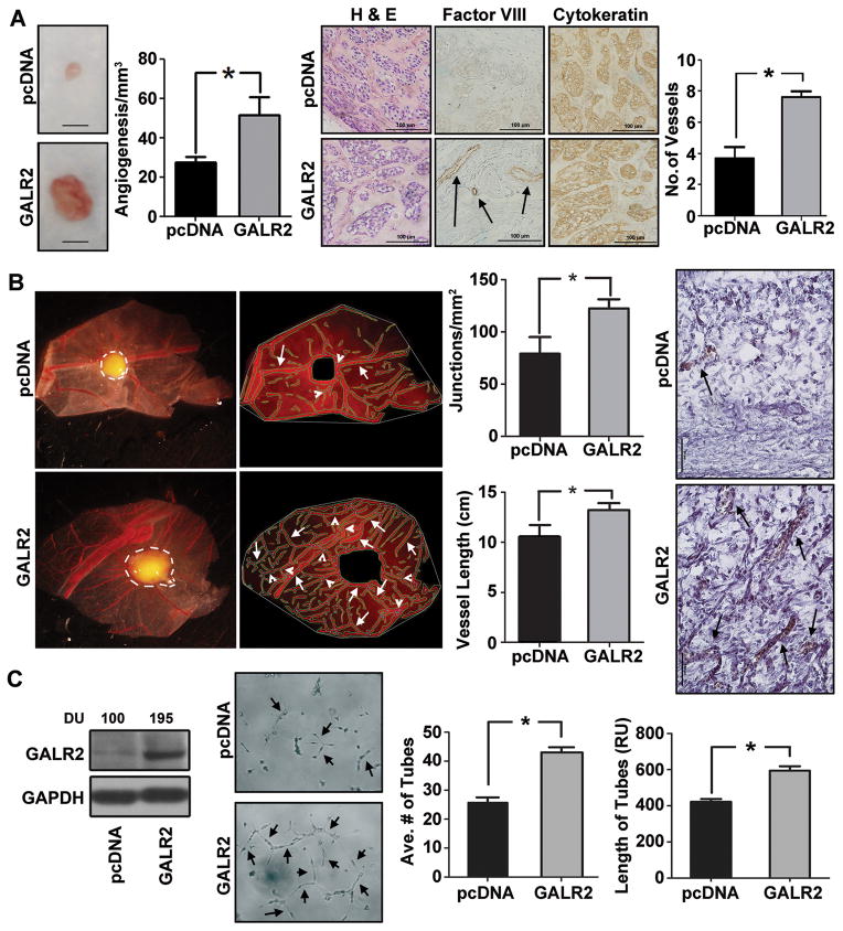
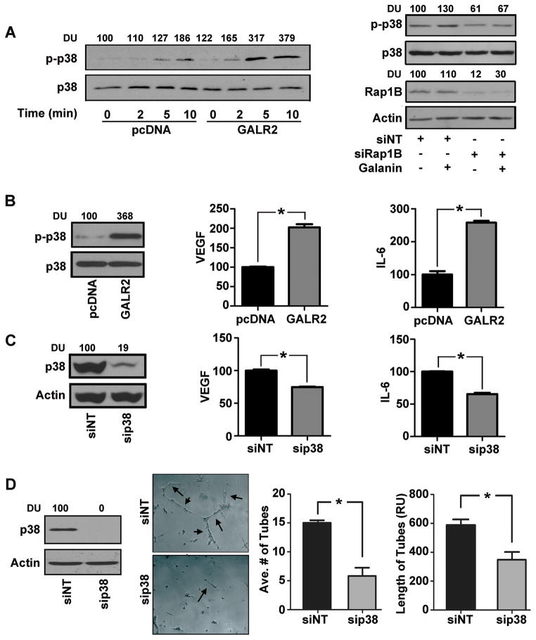
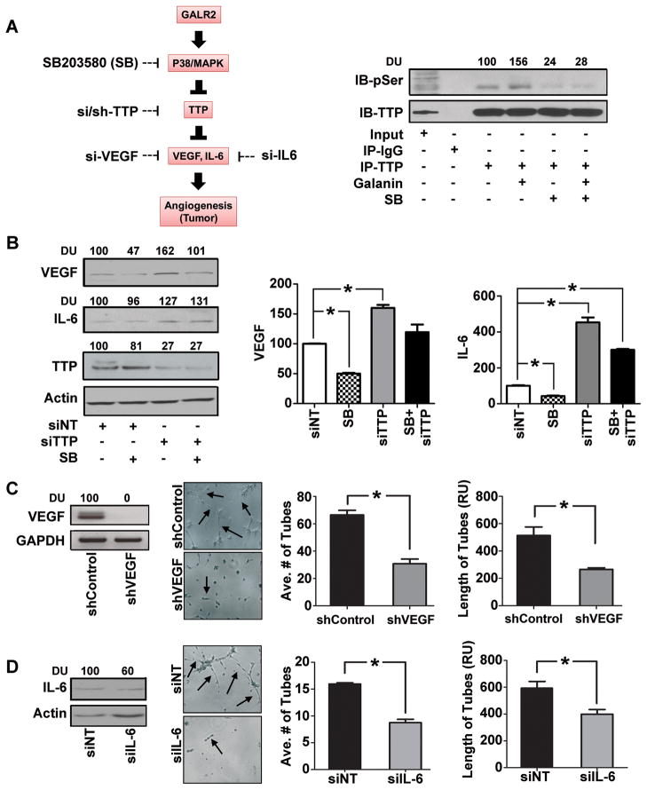
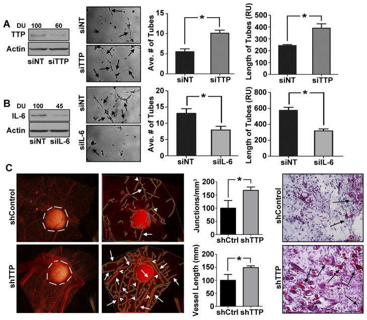
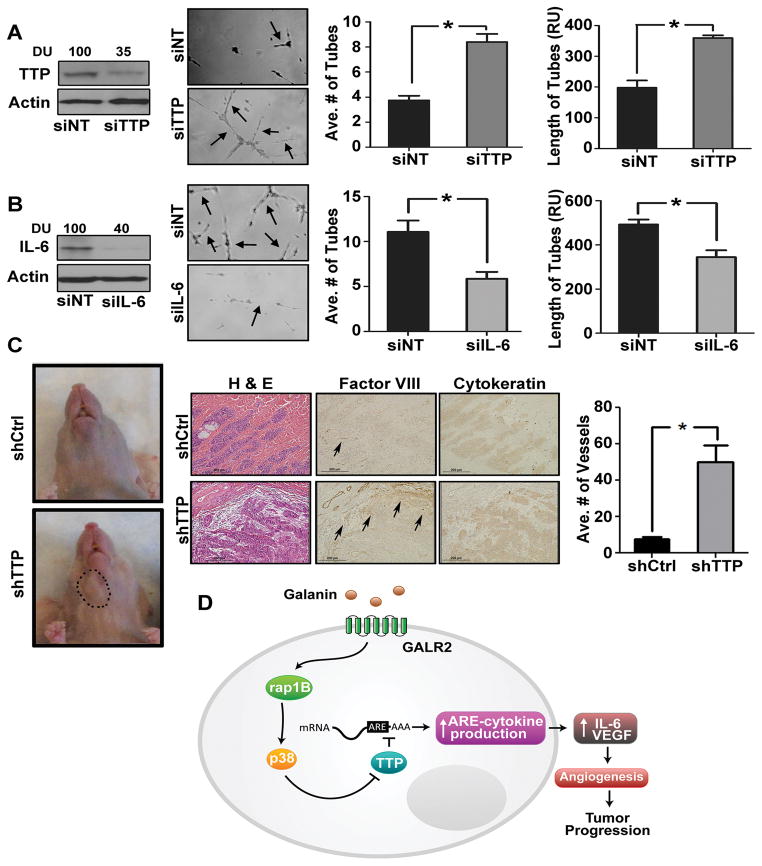
Squamous cell carcinoma of the head and neck (SCCHN) is an aggressive disease with poor patient survival. Galanin receptor 2 (GALR2) is a G protein-coupled receptor that induces aggressive tumor growth in SCCHN. The objective of this study was to investigate the mechanism by which GALR2 promotes angiogenesis, a critical oncogenic phenotype required for tumor growth. The impact of GALR2 expression on secretion of proangiogenic cytokines in multiple SCCHN cell lines was investigated by ELISA and in vitro angiogenesis assays. Chemical inhibitor and genetic knockdown strategies were used to understand the key regulators. The in vivo impact of GALR2 on angiogenesis was investigated in mouse xenograft, chick chorioallantoic membrane, and the clinically relevant mouse orthotopic floor-of-mouth models. GALR2 induced angiogenesis via p38-MAPK-mediated secretion of proangiogenic cytokines, VEGF, and interleukin-6 (IL-6). Moreover, GALR2 activated small-GTP-protein, RAP1B, thereby inducing p38-mediated inactivation of tristetraprolin (TTP), which functions to destabilize cytokine transcripts. This resulted in enhanced secretion of proangiogenic cytokines and angiogenesis in vitro and in vivo. In SCCHN cells overexpressing GALR2, inactivation of TTP increased secretion of IL-6 and VEGF, whereas inhibition of p38 activated TTP and decreased cytokine secretion. Here, we report that GALR2 stimulates tumor angiogenesis in SCCHN via p38-mediated inhibition of TTP with resultant enhanced cytokine secretion. Given that p38 inhibitors are in clinical use for inflammatory disorders, GALR2/p38-mediated cytokine secretion may be an excellent target for new adjuvant therapy in SCCHN.


Conflict of interest statement

CONFLICT OF INTEREST: All authors in this manuscript declare no conflict of interest.

Figures

Figure 1
Figure 1. GALR2 induces angiogenesis
(A) Stable mixed clonal population of UM-SCC-1-GALR2 and pcDNA control cells (1 million) were injected subcutaneously in mouse (n=10, 5 in each group) and xenograft tumors were excised. A representative tumor and control is shown (bar=1cm). The neovasculature was quantified and graphed (middle-left panel, *p<0.007). Both control and UM-SCC-1-GALR2 tumor sections were stained with H&E, Factor-VIII and cytokeratin (middle-right panel). Number of vessel from endothelial cells was quantified from each group from five representative fields and graphed (right panel, *p<0.01). (B) UM-SCC-1-GALR2 and control cells were placed on the top of the upper CAM (n=10, 5 in each group). After three days, upper CAM was excised and photographed along with the tumor (white border line). Angiogenic structure defined by AngioTool program (white arrowheads, middle-left panel) was calculated for each tumor and total length of tubule normalized to surface area and number of junctions were calculated for each group (middle-right panel, *p<0.012). Histology of the corresponding tumors show vasculature in UM-SCC-1-GALR2 (arrowheads) and control tumors (right panel *p< 0.008). (C) UM-SCC-1-GALR2 and pcDNA cells were immunoblotted with GALR2 and GAPDH antibodies. Band Intensities were quantified by ImageJ software and expressed as arbitrary densitometric unit (DU) normalized to control (left panel). Conditioned media (CM) from UM-SCC-1-GALR2 and pcDNA cells were normalized to equal cell number and incubated overnight on Matrigel seeded with HMEC-1 cells and photographed at 24h (middle-left panel). Both average number of tubes and length of tubes were calculated from 10 representative fields (*p<0.01). All experiments were performed three times in triplicate and data shown from one representative experiment.
Figure 2
Figure 2. GALR2 stimulates cytokine secretion and angiogenesis via RAP1B-p38-MAPK
(A) Stable mixed clonal population of UM-SCC-1-GALR2 or control pcDNA cells were serum starved for 4h and treated with 10nM Galanin for 0, 2, 5, or 10 minutes. Whole cell lysates were immunoblotted for phospho-p38 and p38 antibodies, quantified (DU) and expressed as percent of control (left panel). UM-SCC-1-GALR2 stable cells were treated with NT-siRNA or si-RAP1B. After 68h of transfection cells were serum starved for 4h and were either stimulated with 10nM galanin or vehicle control for 10 minutes. Both stimulated and unstimulated controls were immunoblotted with actin, RAP1B, p-p38 and p38 antibodies and were quantified (right panel). (B) UM-SCC-1-GALR2 or control cells were immunoblotted with p-p38 and total p38 antibodies (left panel). CM were collected and VEGF and IL-6 were quantified with ELISA as pg/ml/million cells and was finally expressed with normalization to control (right panels, *p<0.014). (C) UM-SCC-1-GALR2 cells were treated with si-p38 or NT-siRNA and were immunoblotted with p38 and actin antibodies (left panel). CM collected from cells were quantified for VEGF and IL-6 and expressed as normalized to control (right panel, *p<0.01). (D) UM-SCC-1-GALR2 cells were treated with si-p38 or NT-siRNA and immunoblotted (left panel). CM was collected, concentrated, normalized for equal cell count and HMEC-1 cells were seeded with corresponding CM and photographed (middle-left panel). Average number of tubes and tube length were quantified from 10 representative fields (right panel). Data is representative of three identical experiments each in triplicate (*p<0.008).
Figure 3
Figure 3. GALR2 induces p38-mediated inhibition of TTP to enhance angiogenesis
(A) Schematic representation of signaling pathway showing GALR2 mediated phosphorylation and activation of p38 that inhibits TTP via phosphorylation. Inactivation or genetic loss (by siRNA) of TTP prevents cytokine degradation giving rise to overall steady state increase in cytokines and acts as angiogenic switch to tumor growth. Chemical inhibitor (SB203580) to p38 kinase activity can dephosphorylate TTP to its active form and degrade cytokines. Alternatively targeting individual cytokines by siRNA could be used to control this angiogenic signal. Upstream inhibitors to angiogenesis might be a better treatment option (left panel). UM-SCC-1-GALR2 cells were serum starved for 4h and then treated with 10μM of SB203580 for 2h and were then stimulated with 10nM galanin for 10 minutes or vehicle control. Clarified cell lysates were immunoprecipitated with TTP antibody, and blotted with anti-TTP and anti-phosphoserine antibodies (right panel). (B) UM-SCC-1-GALR2 cells were transfected with siTTP or NT-siRNA and were treated with 10μM of SB203580 or vehicle control. Cell lysates were immunoblotted with VEGF, IL-6, TTP and actin antibodies (left panel). CM was collected from each of the treatment groups and ELISA was performed for VEGF and IL-6 (right panels) and was quantified as pg/ml/million cells and was finally normalized to control. (C) UM-SCC-1-GALR2 cells were transduced with shVEGF and control shRNA lentiviral particles and immunoblotted with VEGF antibody and GAPDH. In-vitro tubule formation assay was performed with HMEC-1 cells incubated with corresponding CM collected from cells. Both average and relative number of tubes were quantified from 10 representative fields (*p<0.02). (D) IL-6 was transiently down regulated in UM-SCC-1-GALR2 cells with si-IL-6 and immunoblotted (left panel) and similarly in-vitro tubule formation assay was performed with CM collected from cells (middle-left panel) and quantified (right panel, *p<0.02). Data is representative of three identical experiments in triplicate.
Figure 4
Figure 4. Down regulation of TTP promotes angiogenesis
(A) TTP was transiently downregulated in UM-SCC-1 cells with siTTP or NT and immunoblotted with TTP and actin antibodies (left panel). CM, concentrated and normalized to cell number, was seeded with HMEC-1 cells for in-vitro tubule formation assay. Data were quantified from 10 representative fields (middle and right panels respectively,*p<0.003) (B) IL-6 was transiently downregulated in UM-SCC-1-shTTP stable cells. Cell lysates were immunoblotted for IL-6 and actin (left panel). CM were collected and in-vitro tubule formation assay was performed. Data were quantified from 10 representative fields (middle and right panel respectively (*p<0.05). (C) UM-SCC-1-shTTP and control cells (1 million) were dropped on the top of the upper CAM (n=10, 5 for each group). After 36h the CAM was excised and photographed along with the tumor (white border line). Angiogenic structure was determined (white arrowheads, middle-left panel) and calculated as described (middle-right panel, *p<.012 and *p<0.008). Histology of the corresponding tumors show vascularity in UM-SCC-1-shTTP and control tumors (right panel).
Figure 5
Figure 5. Loss of TTP induce angiogenesis and tumor growth in murine floor-of-mouth
(A) TTP was transiently downregulated in UM-SCC-81B cells, immunoblotted for TTP and actin (left panel). In-vitro tubule formation assay was performed with CM collected from these cells (middle-left panel). Average number and length of tubes were quantified from 10 representative fields (right panel *p<0.001). (B) IL-6 was transiently downregulated in UM-SCC-81B-shTTP cells. Cell lysates were immunoblotted for IL-6 and actin (left panel). In-vitro tubule formation assay was performed and quantified for 10 fields (middle-left and right panel respectively) (*p<0.02). (C) UM-SCC-81B-shTTP and shControl (scramble) cells were implanted in the floor-of-mouth of nude mice (n=6). Mice were euthanized when moribund and control mice were euthanized concurrently. A representative image of control and test mice bearing tumor (black circle) is shown (left panel). Sections from the shControl and shTTP tumors were stained with H&E and immunostained with Factor-VIII and cytokeratin antibodies and blood vessels in 10 representative fields were quantified (middle and left panels *p<0.008). (D) Summary Figure: Extracellular GAL stimulates GALR2/RAP1B to induce p38-MAPK to inactivate TTP. Inactivation of TTP inhibits degradation of IL-6 and VEGF mRNA transcripts, thereby causing increased pro-angiogenic cytokine secretion, angiogenesis and tumor growth.

Similar articles

Cited by

References

    1. Leemans CR, Braakhuis BJ, Brakenhoff RH. The molecular biology of head and neck cancer. Nat Rev Cancer. 2011;11:9–22. - PubMed
    1. Hanahan D, Folkman J. Patterns and emerging mechanisms of the angiogenic switch during tumorigenesis. Cell. 1996;86:353–64. - PubMed
    1. Hanahan D, Weinberg RA. Hallmarks of cancer: the next generation. Cell. 2011;144:646–74. - PubMed
    1. Lingen MW. Angiogenesis in the development of head and neck cancer and its inhibition by chemopreventive agents. Crit Rev Oral Biol Med. 1999;10:153–64. - PubMed
    1. Pries R, Nitsch S, Wollenberg B. Role of cytokines in head and neck squamous cell carcinoma. Expert review of anticancer therapy. 2006;6:1195–203. - PubMed

Publication types

MeSH terms