. 2014 Mar;124(3):1255-67.
doi: 10.1172/JCI66163. Epub 2014 Feb 24.

Small-molecule activator of glutamate transporter EAAT2 translation provides neuroprotection

Small-molecule activator of glutamate transporter EAAT2 translation provides neuroprotection

Qiongman Kong et al. J Clin Invest. 2014 Mar.

Abstract

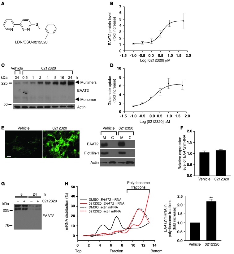
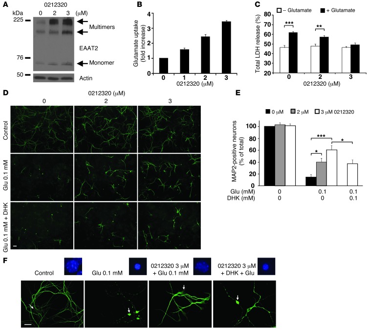
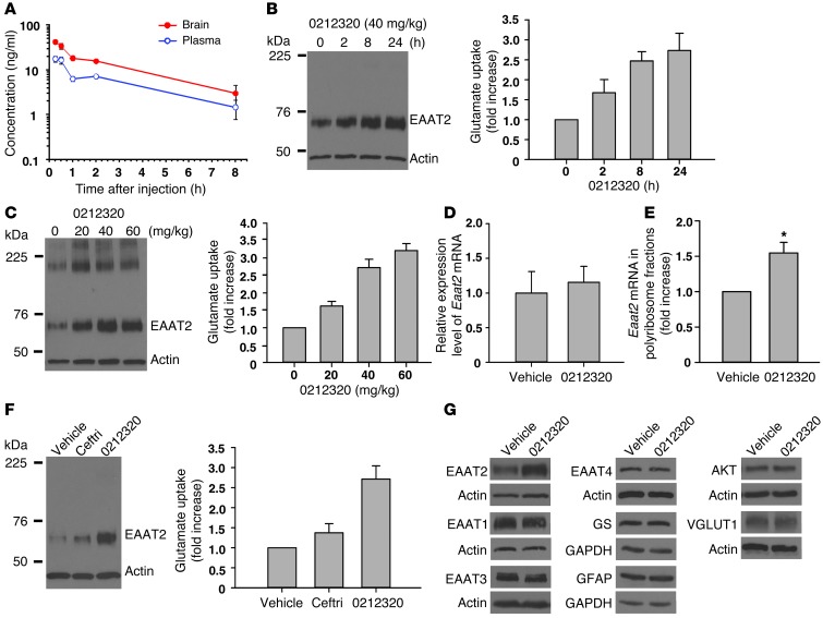
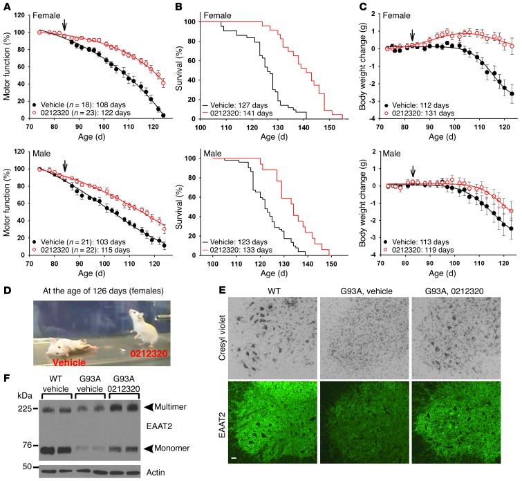
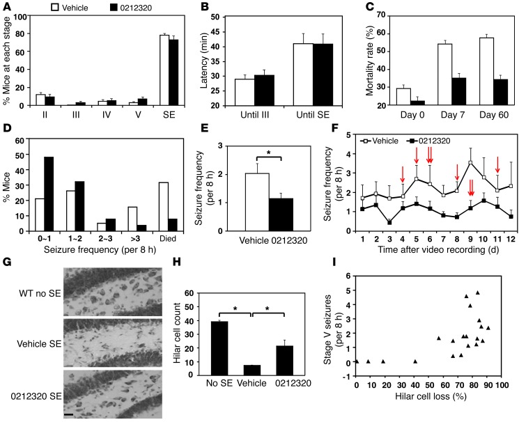
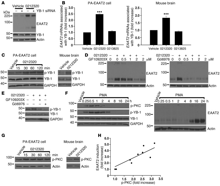
Glial glutamate transporter EAAT2 plays a major role in glutamate clearance in synaptic clefts. Several lines of evidence indicate that strategies designed to increase EAAT2 expression have potential for preventing excitotoxicity, which contributes to neuronal injury and death in neurodegenerative diseases. We previously discovered several classes of compounds that can increase EAAT2 expression through translational activation. Here, we present efficacy studies of the compound LDN/OSU-0212320, which is a pyridazine derivative from one of our lead series. In a murine model, LDN/OSU-0212320 had good potency, adequate pharmacokinetic properties, no observed toxicity at the doses examined, and low side effect/toxicity potential. Additionally, LDN/OSU-0212320 protected cultured neurons from glutamate-mediated excitotoxic injury and death via EAAT2 activation. Importantly, LDN/OSU-0212320 markedly delayed motor function decline and extended lifespan in an animal model of amyotrophic lateral sclerosis (ALS). We also found that LDN/OSU-0212320 substantially reduced mortality, neuronal death, and spontaneous recurrent seizures in a pilocarpine-induced temporal lobe epilepsy model. Moreover, our study demonstrated that LDN/OSU-0212320 treatment results in activation of PKC and subsequent Y-box-binding protein 1 (YB-1) activation, which regulates activation of EAAT2 translation. Our data indicate that the use of small molecules to enhance EAAT2 translation may be a therapeutic strategy for the treatment of neurodegenerative diseases.


Figures

Figure 1
Figure 1. Characterization of LDN/OSU-0212320 in PA-EAAT2 cells.
(A) Structure of LDN/OSU-0212320. (B) Dose response experiments. Cells were treated with compound for 24 hours. EC50 = 1.83 ± 0.27 μM. n = 5. (C) Time course experiments. Cells were treated with compound at 10 μM. Compound increased EAAT2 protein levels in a short amount of time. (D) [3H] glutamate uptake experiments. Glutamate uptake activity correlated with increased EAAT2 protein levels. Cells were treated with compound for 24 hours. n = 6. (E) Immunofluorescence staining and subcellular fractionation analysis. Induced EAAT2 was properly localized in the plasma membrane. Cells were treated with compound at a concentration of 3.3 μM for 24 hours. M, plasma membrane fraction. C, cytosolic fraction. Scale bar: 25 μm. (F) Real-time RT-PCR analysis. Compound treatment did not increase EAAT2 mRNA levels. Cells were treated with compound at a concentration of 10 μM for 6, 18, 24, and 48 hours. Results after 24 hours are shown. n = 5. (G) Pulse-chase analysis. Compound treatment did not affect the rate of EAAT2 protein degradation. Cells were preincubated with sulfo-NHS-SS-biotin to label surface EAAT2 proteins and were then treated with compound (10 μM). Cells were harvested at 8 and 24 hours to measure biotin-labeled EAAT2 protein levels. Equal protein loading was confirmed by Ponceau S staining. (H) Polyribosome analysis. Compound treatment increased EAAT2 mRNA translation activity. Cells were treated with compound at a concentration of 10 μM for 1 hour. Cell lysates were prepared and fractionated by a 15% to 60% sucrose gradient. RNAs were extracted from each fraction and analyzed by real-time RT-PCR. n = 4. **P < 0.01.
Figure 2
Figure 2. Characterization of LDN/OSU-0212320 in primary dissociated neuron and astrocyte mixed cultures.
Seven-day-old primary cultures were treated with the indicated doses of compound for 24 hours and then harvested for analyses. (A) Western blot analysis shows that compound increased EAAT2 expression in a dose-dependent manner. (B) [3H] glutamate uptake assay shows a dose-dependently increased glutamate uptake. n = 6. (C) Cultures were insulted with 0.5 mM glutamate for 2 hours to induce excitotoxicity. LDH assay shows a dose-dependent reduction of LDH release in compound-treated cultures. n = 6. (D) MAP2 immunostaining shows that compound treatment significantly prevented glutamate-mediated neuronal loss and degeneration. The protections were partially abolished by pretreatment of DHK (100 μM). Cultures were exposed to 0.1 mM glutamate for 2 hours. Scale bar: 50 μm. (E) Quantitative analysis of MAP2-positive neurons. MAP2-positive neurons were counted and quantified as described in the Methods. n = 15. (F) MAP2-immunolabeled neurons and their nuclear morphology (Hoechst 33342 staining) at a higher magnification. Arrows point to the corresponding neurons of the highlighted nuclei. Scale bar: 25 μm. *P < 0.05; **P < 0.01; ***P < 0.001.
Figure 3
Figure 3. Evaluation of LDN/OSU-0212320 in WT mice.
(A) Pharmacokinetic studies. After administration of a single i.p. dose of compound (3 mg/kg) to mice, plasma and brain concentrations were determined (n = 3/time point). Cmax = 42.1 ± 3.6 ng/l at 15 minutes, plasma t1/2 = 2.63 hours, brain t1/2 = 2.64 hours. (B) Time course studies. After a single dose of compound (40 mg/kg), the brains were harvested at the indicated times (n = 3/time point). Plasma membrane vesicles were prepared from forebrains to determine EAAT2 protein levels and measure [3H] glutamate uptake activity. Increased EAAT2 could be detected as early as 2 hours after injection. (C) Dose response studies. Mice received a single dose of compound (n = 3/dose), and brains were harvested at 24 hours after injection. Compound dose-dependently increased EAAT2 levels and glutamate uptake. (D) Quantitative real-time RT-PCR analysis. Eaat2 mRNA levels were not changed by compound treatment. Mice received compound (40 mg/kg), and brains were harvested at 8 and 24 hours. Results at 8 hours are shown. n = 5. (E) Polyribosome analysis. Compound treatment increased Eaat2 mRNA translation activity. Mice received compound (40 mg/kg), and brains were harvested at 1 hour. n = 4. (F) Comparison of ceftriaxone (Ceftri) and LDN/OSU-0212320. Mice received a single dose of ceftriaxone (200 mg/kg) or LDN/OSU-0212320 (40 mg/kg) (n = 3 each), and brains were harvested at 24 hours. LDN/OSU-0212320 is more potent than ceftriaxone. (G) Western blot analysis of other protein expression levels in LDN/OSU-0212320–treated (40 mg/kg) brains. LDN/OSU-0212320 did not induce global protein synthesis. *P < 0.05.
Figure 4
Figure 4. LDN/OSU-0212320 delays motor function decline and extends lifespan in SOD1(G93A) mice.
SOD1(G93A) mice received compound daily (i.p., 40 mg/kg) starting at 84 days of age (indicated by arrows in the figure) until death. (A) Motor function decline as assessed by grip strength measurement. Data were analyzed using dynamic fitting nonlinear regression analysis. The average number of days until a 50% grip strength decline occurred is indicated. (B) Survival results. Data were analyzed using Kaplan-Meier survival analysis. The average number of days of survival is indicated. (C) Body weight change. The average number of days until a 10% decline in initial body weight occurred is indicated. (D) Representative female mice at 126 days old. (E) Representative images showing cresyl violet staining and EAAT2 immunostaining of the ventral horn region of the spinal cord from 120-day-old female mice. Scale bar: 50 μm. (F) Western blot results show that LDN/OSU-0212320 restores EAAT2 protein levels. Spinal cords from 120-day-old mice were analyzed. Six animals in each group were analyzed for E and F.
Figure 5
Figure 5. LDN/OSU-0212320 reduces mortality rate, chronic seizure, and neuronal death following pilocarpine-induced SE.
Mice received compound (i.p., 40 mg/kg) and were treated with pilocarpine 3 hours later. Mice that reached SE received compound daily, and chronic seizures were recorded 4 weeks after SE for a 2-week period (15 independent experiments in a total of 153 vehicle- and 152 compound-treated mice). (A) Acute seizure severity (maximal seizure activity of each animal within 2 hours after pilocarpine injection). The percentage of mice that reached each seizure stage did not differ between the two groups. (B) Latency (time interval between pilocarpine injection and the indicated stage). Latency did not differ between the two groups. (C) Mortality rate (percentage of mice that died in each experiment, n = 15). A significant decrease was observed in the compound-treated group on days 7 and 60 after SE. (DF) Spontaneous recurrent seizures. (D) Percentage of mice (vehicle, n = 19; compound, n = 25) that developed <1, 1~2, 2~3, or >3 stage V seizures in an 8-hour period. (E) Average number of stage V seizures in an 8-hour period. (F) Frequency of stage V seizures during each day of recording. Chronic seizure frequency was significantly reduced in compound-treated mice. Arrows indicate that a mouse died on that day. (G and H) Hippocampal damage. Nine sets of brains 8 weeks after SE were analyzed with cresyl violet staining (15–20 sections/mouse). (G) Representative images of the dentate hilus. Scale bar: 20 μm. (H) Quantitative analysis of hilar cells. Compound treatment significantly attenuated cell damage. (I) A strong positive correlation was detected between hilar cell loss and chronic seizure frequency. *P < 0.05.
Figure 6
Figure 6. LDN/OSU-0212320 treatment results in activation of PKC, which subsequently activates YB-1, leading to enhancement of EAAT2 translation.
(A) Depletion of YB-1 by siRNA reduced compound-induced EAAT2 expression in PA-EAAT2 cells. Cells were transfected with YB-1 siRNA and were treated 24 hours later with compound (10 μM) for 24 hours. Lanes were run on the same gel but were noncontiguous. (B) Increased YB-1-EAAT2 mRNA interaction following compound treatment. PA-EAAT2 cells were treated with compound (10 μM) for 2 hours and then harvested for immunoprecipitation (anti–YB-1 antibodies). Coimmunoprecipitated EAAT2 mRNAs were quantified by real-time RT-PCR. Mice received a single dose of compound (40 mg/kg, i.p.), and brains were harvested at 2 hours for immunoprecipitation. n = 5. (C) Compound increased YB-1 phosphorylation (p-YB-1). PA-EAAT2 cells: 10 μM compound. Mouse brain: 40 mg/kg, harvested at 1 hour. (D) PKC inhibitors blocked compound-induced EAAT2 expression in PA-EAAT2 cells. Cells were pretreated with inhibitors for 1 hour and then treated with compound (10 μM) for 24 hours. (E) PKC inhibitors blocked compound-induced YB-1 phosphorylation in PA-EAAT2 cells. (F) PMA increased YB-1 phosphorylation concomitantly with PKC phosphorylation (p-PKC). Increased EAAT2 protein levels were not observed until 8 hours after treatment. (G) Compound increased PKC phosphorylation. PA-EAAT2 cells: 10 μM compound. Mouse brain: 40 mg/kg, harvested at 1 hour. (H) Compounds of different potencies were examined for their effects on PKC activation. p-PKC levels are highly correlated with EAAT2 induction levels. Each data point represents the mean of three experiments. ***P < 0.001.

Comment in

References

    1. Choi DW. Ionic dependence of glutamate neurotoxicity. J Neurosci. 1987;7(2):369–379. - PMC - PubMed
    1. Chaudhry FA, Lehre KP, van Lookeren Campagne M, Ottersen OP, Danbolt NC, Storm-Mathisen J. Glutamate transporters in glial plasma membranes: highly differentiated localizations revealed by quantitative ultrastructural immunocytochemistry. Neuron. 1995;15(3):711–720. doi: 10.1016/0896-6273(95)90158-2. - DOI - PubMed
    1. Rothstein JD, et al. Knockout of glutamate transporters reveals a major role for astroglial transport in excitotoxicity and clearance of glutamate. Neuron. 1996;16(3):675–686. doi: 10.1016/S0896-6273(00)80086-0. - DOI - PubMed
    1. Lin CG, Kong Q, Cuny GD, Glicksman MA. Glutamate transporter EAAT2: a new target for the treatment of neurodegenerative diseases. Future Med Chem. 2012;4(13):1689–1700. doi: 10.4155/fmc.12.122. - DOI - PMC - PubMed
    1. Rothstein JD, Van Kammen M, Levey AI, Martin LJ, Kuncl RW. Selective loss of glial glutamate transporter GLT-1 in amyotrophic lateral sclerosis. Ann Neurol. 1995;38(1):73–84. doi: 10.1002/ana.410380114. - DOI - PubMed

Publication types

MeSH terms