. 2014 Feb 25:5:3360.
doi: 10.1038/ncomms4360.

The origin of the bifurcated axial skeletal system in the twin-tail goldfish

Affiliations
Free PMC article

The origin of the bifurcated axial skeletal system in the twin-tail goldfish

Gembu Abe et al. Nat Commun. .
Free PMC article

Abstract

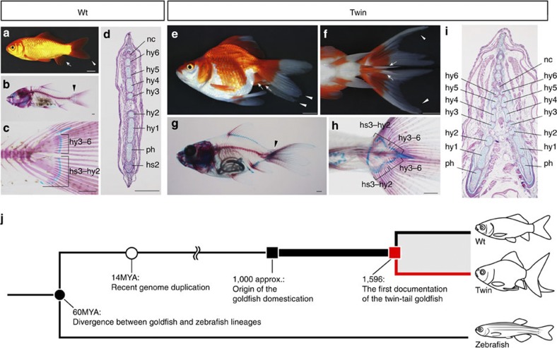
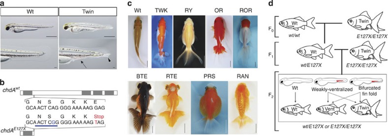
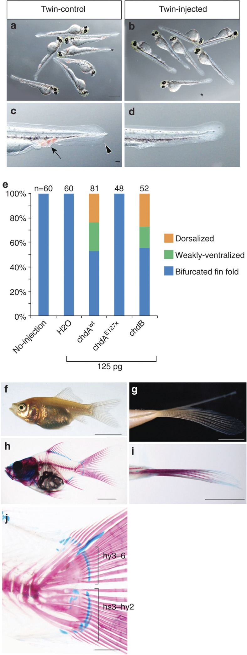
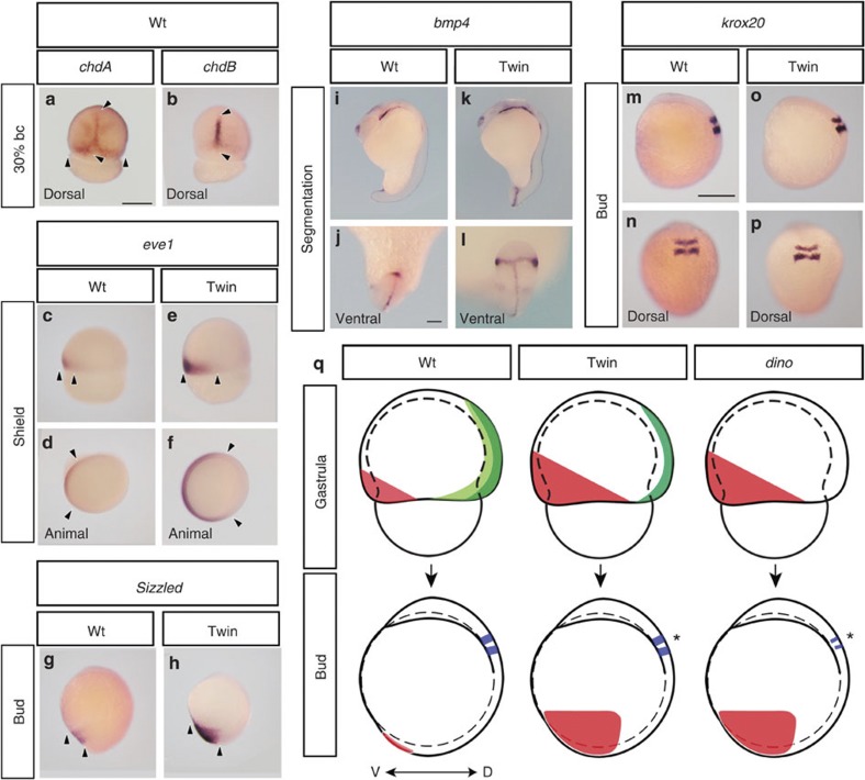
Twin-tail goldfish possess a bifurcated caudal axial skeleton. The scarcity of this trait in nature suggests that a rare mutation, which drastically altered the mechanisms underlying axial skeleton formation, may have occurred during goldfish domestication. However, little is known about the molecular development of twin-tail goldfish. Here we show that the bifurcated caudal skeleton arises from a mutation in the chordin gene, which affects embryonic dorsal-ventral (DV) patterning. We demonstrate that formation of the bifurcated caudal axial skeleton requires a stop-codon mutation in one of two recently duplicated chordin genes; this mutation may have occurred within approximately 600 years of domestication. We also report that the ventral tissues of the twin-tail strain are enlarged, and form the embryonic bifurcated fin fold. However, unlike previously described chordin-deficient embryos, this is not accompanied by a reduction in anterior-dorsal neural tissues. These results provide insight into large-scale evolution arising from artificial selection.


Figures

Figure 1
Figure 1. Evolution of goldfish morphology.
(a) Lateral view of the wild-type goldfish (Wt). (b) Skeletal structure of Wt goldfish. (c) Magnified view of the caudal region of the specimen in b. (d) Transverse section of the Wt at the caudal level. (e) Lateral view of the twin-tail goldfish (Twin). (f) Ventral view of the caudal region of the specimen in e. (g) Skeletal structure of Twin goldfish. (h) Magnified view of the ventral side of the specimen in g. (i) Transverse section of the Twin goldfish specimen. The approximate levels of sectioning are indicated by black arrowheads (b,g). Anal and caudal fins are indicated by white arrows and arrowheads, respectively (a,e,f). (j) Evolutionary events in the goldfish lineage. The black circle indicates the time of divergence between the goldfish and zebrafish lineages, and the white circle indicates the estimated time of the genome duplication in goldfish. The red line represents the twin-tail goldfish lineage. hs, haemal spines; hy, hypural; MYA, million years ago; nc, notochord; ph, parhypural. Scale bars, 1 cm (a,e,f), 1 mm (b,c,g,h), 100 μm (d,i).
Figure 2
Figure 2. The involvement of the chdA gene in the twin-tail phenotype.
(a) Lateral view of wild-type (Wt) and twin-tail (Twin) goldfish larvae. The lower panels are magnified views of the larval caudal regions. Blood cell accumulation and duplicated fin folds are indicated by the black arrow and arrowhead, respectively. (b) The goldfish chdAwt and chdAE127X amino-acid sequences. The positions of CR domains are indicated by grey boxes. The twin-tail-specific mutation changes a glutamic acid codon (GAG) to a stop codon (TAG) at amino-acid position 127 (the mutated nucleotide is indicated by the red ‘T’). A twin-tail-specific AvaI site is located near the stop codon (indicated by the blue line and ‘G’). (c) Dorsal views of the Wt and eight twin-tail goldfish strains. (d) Schematic detailing the phenotyping of backcross segregants. Scale bars, 1 mm (upper row in a), 0.1 mm (bottom row in a), approximately 1 cm (c).
Figure 3
Figure 3. Rescue of the twin-tail phenotype by mRNA microinjection.
(a,b) Larval phenotypes of twin-tail goldfish. Non-injected controls (a) or embryos injected with 125 pg chdAwt mRNA (b). (c,d) Magnified view of the caudal regions indicated by asterisks in a and b, respectively. Arrow and arrowhead indicate accumulated blood cells and bifurcated fin folds, respectively. (e) Proportion of rescued specimens following injection of embryos with the indicated mRNA. The number of larvae analysed is indicated above each bar. (fj) Twin-tail goldfish juvenile injected with chdAwt mRNA. (f) Lateral view. (g) Ventral view of the caudal level. (h) Lateral view of the skeletal structure. (j) Magnified view of the caudal region of h. The dorsal phenotype criteria were based on previous descriptions. Panels a and b, and c and d are at the same magnification, respectively. Scale bars, 1 mm (a,h,j), 100 μm (c), 5 mm (fi).
Figure 4
Figure 4. Comparison of gene expression patterns between wild-type and twin-tail goldfish.
(ap) Expression patterns of chdA (a), chdB (b), eve1 (cf), sizzled (g,h), bmp4 (il) and krox20 (mp). Unless otherwise noted, panels show lateral views of embryos. Black arrowheads indicate areas of gene expression. (j,l) Magnified views of i and k, respectively. (q) Schematic representation of the gene expression patterns in wild-type and twin-tail goldfish, and dino zebrafish mutants. Light green, dark green, red and blue regions represent areas positive for chdAwt, chdB, ventral markers (eve1, sizzled and bmp4) and krox20, respectively. Asterisks indicate krox20 expression areas in twin-tail goldfish and dino zebrafish. Panels ak, j and l, and mp are at the same magnification. Scale bars, 500 μm (a,m); 100 μm (j).

References

    1. Janvier P. Early Vertebrates Clarendon Press: Oxford, (1996).
    1. Kardong K. V. Vertebrates: Comparative Anatomy, Function, Evolution 6th edn McGraw-Hill: New York, (2011).
    1. Lauder G. V. & Liem K. F. The evolution and interrelationships of the Actinopterygian Fishes. Vol. 150 (1983).
    1. Schultze H.-P. & Arratia G. The composition of the caudal skeleton of teleosts (Actinopterygil: Osteichthyes). Zool. J. Linn. Soc-Lond. 97, 189–231 (1989).
    1. Chen S. C. A history of the domestication and the factors of the varietal formation of the common goldfish, Carassius auratus. Sci. Sinica 6, 89–116 (1956).

Publication types

MeSH terms

LinkOut - more resources