. 2014 Apr;164(4):1842-56.
doi: 10.1104/pp.114.236596. Epub 2014 Feb 25.

CELLULOSE SYNTHASE-LIKE A2, a glucomannan synthase, is involved in maintaining adherent mucilage structure in Arabidopsis seed

Affiliations

CELLULOSE SYNTHASE-LIKE A2, a glucomannan synthase, is involved in maintaining adherent mucilage structure in Arabidopsis seed

Li Yu et al. Plant Physiol. 2014 Apr.

Abstract

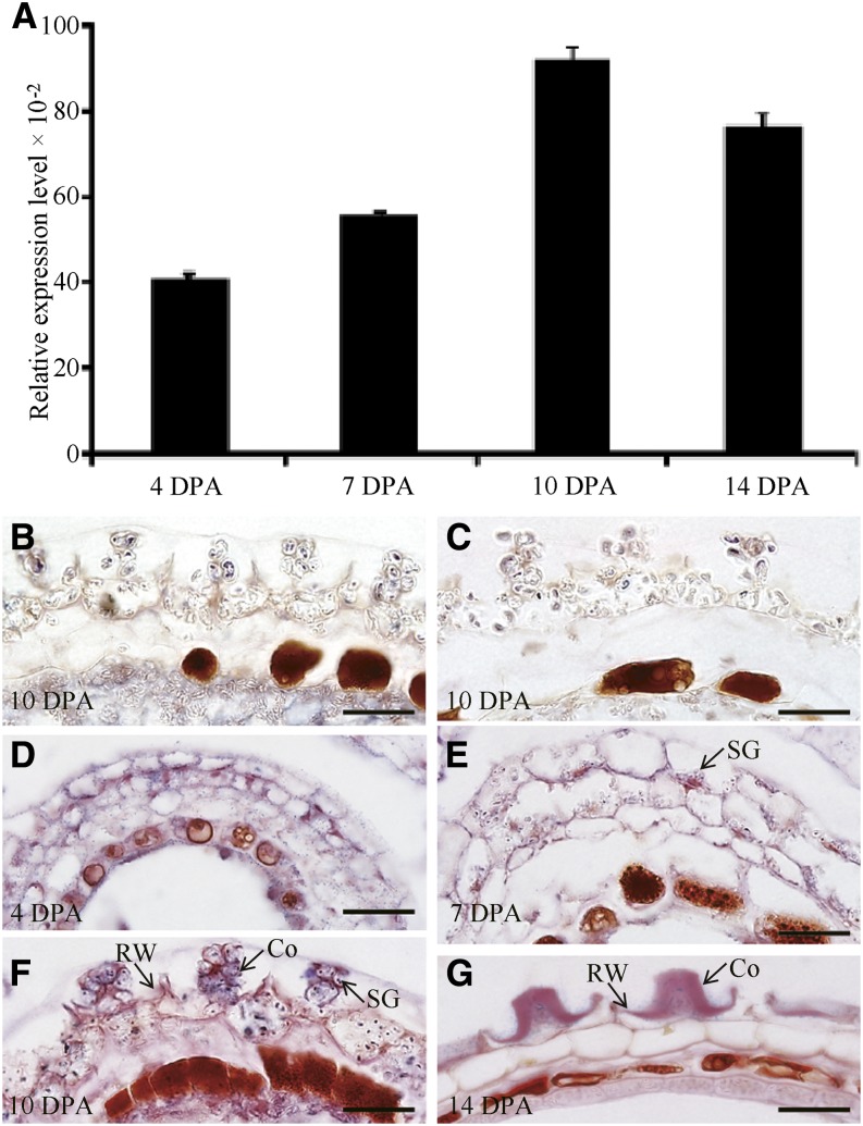
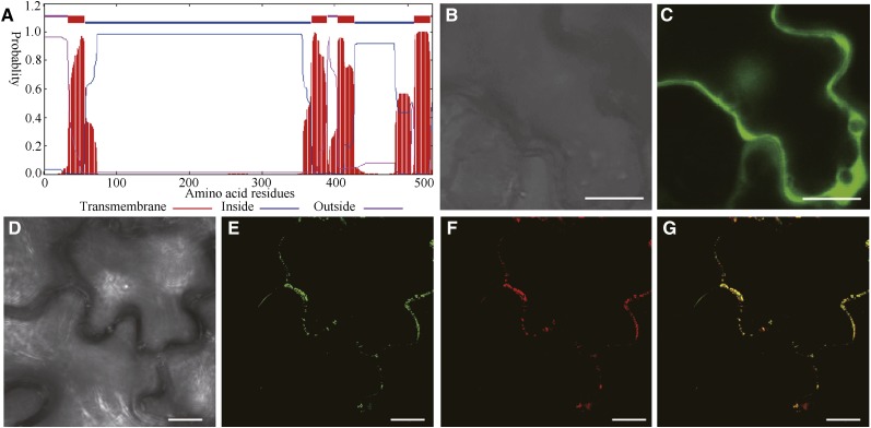
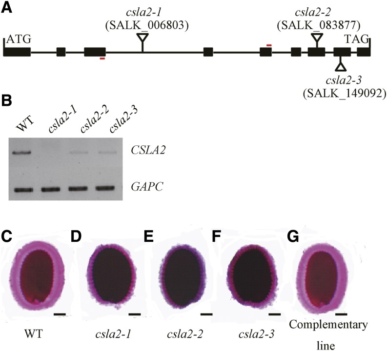
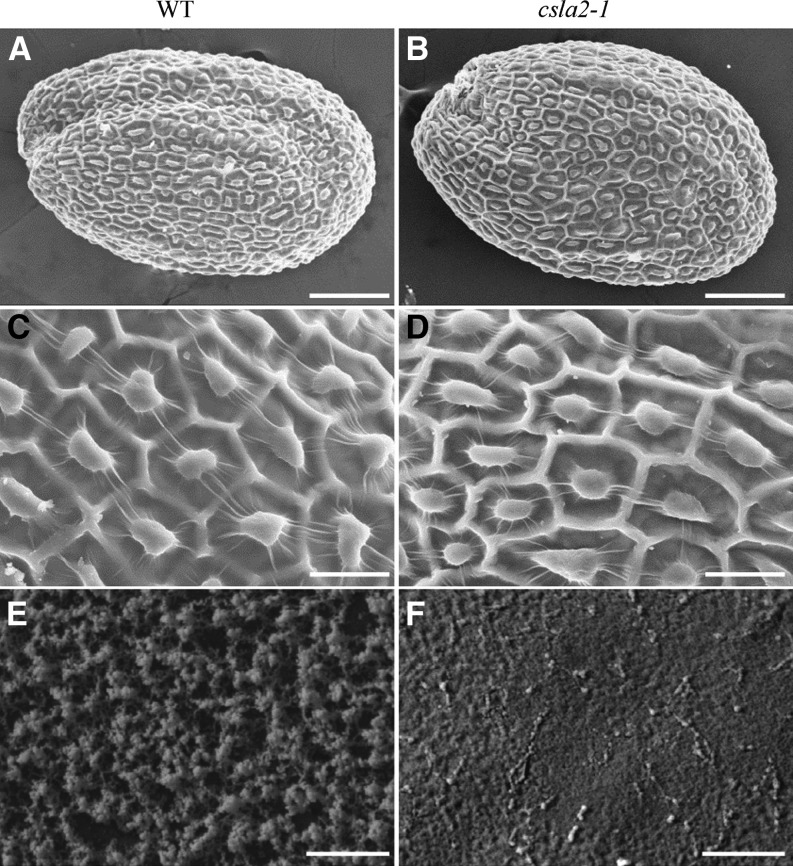
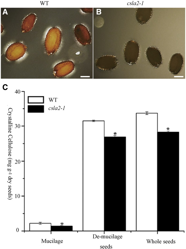
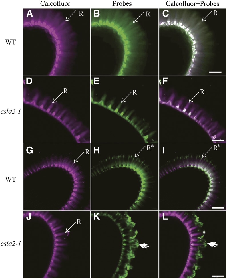
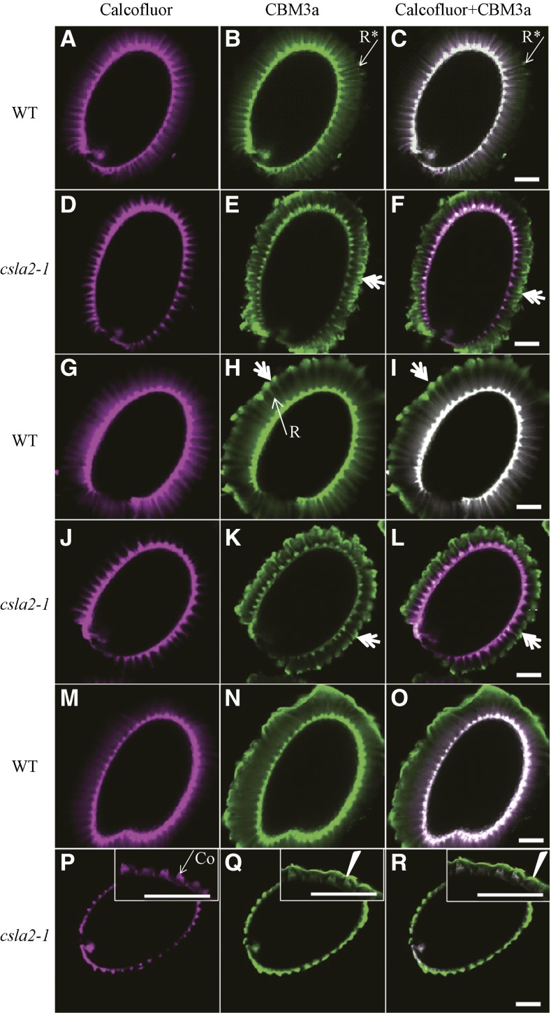
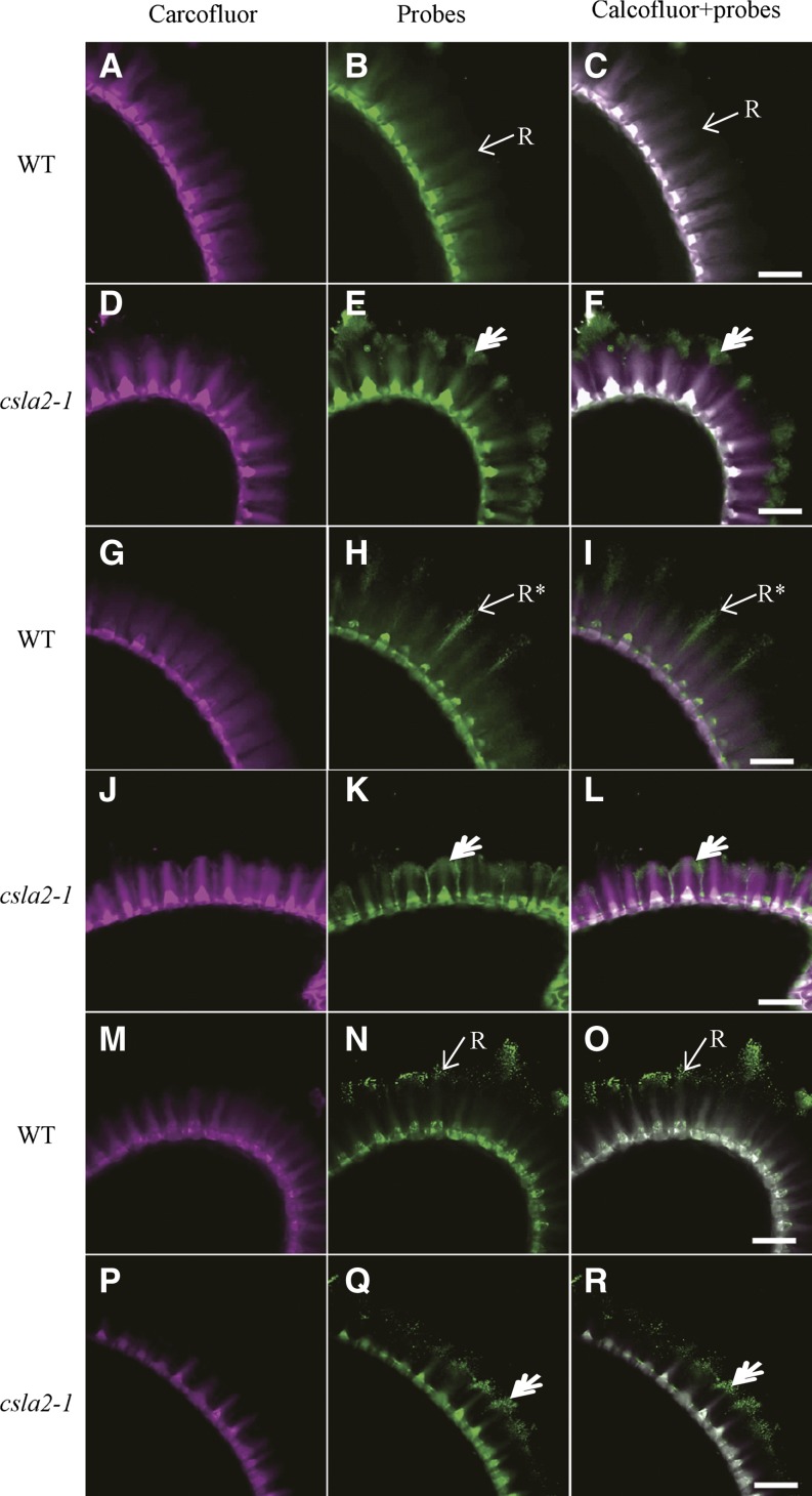
Mannans are hemicellulosic polysaccharides that are considered to have both structural and storage functions in the plant cell wall. However, it is not yet known how mannans function in Arabidopsis (Arabidopsis thaliana) seed mucilage. In this study, CELLULOSE SYNTHASE-LIKE A2 (CSLA2; At5g22740) expression was observed in several seed tissues, including the epidermal cells of developing seed coats. Disruption of CSLA2 resulted in thinner adherent mucilage halos, although the total amount of the adherent mucilage did not change compared with the wild type. This suggested that the adherent mucilage in the mutant was more compact compared with that of the wild type. In accordance with the role of CSLA2 in glucomannan synthesis, csla2-1 mucilage contained 30% less mannosyl and glucosyl content than did the wild type. No appreciable changes in the composition, structure, or macromolecular properties were observed for nonmannan polysaccharides in mutant mucilage. Biochemical analysis revealed that cellulose crystallinity was substantially reduced in csla2-1 mucilage; this was supported by the removal of most mucilage cellulose through treatment of csla2-1 seeds with endo-β-glucanase. Mutation in CSLA2 also resulted in altered spatial distribution of cellulose and an absence of birefringent cellulose microfibrils within the adherent mucilage. As with the observed changes in crystalline cellulose, the spatial distribution of pectin was also modified in csla2-1 mucilage. Taken together, our results demonstrate that glucomannans synthesized by CSLA2 are involved in modulating the structure of adherent mucilage, potentially through altering cellulose organization and crystallization.


Figures

Figure 1.
Figure 1.
Expression of CSLA2 in developing Arabidopsis seeds. A, qRT-PCR analysis of CSLA2 expression during seed development at the indicated time points. Error bars represent sd values (n = 3). B, In situ hybridization using a CSLA2-specific antisense probe to the csla2-1 seed coat. C, In situ hybridization using a CSLA2-specific sense probe to the wild-type seed coat. D to G, In situ hybridization using a CSLA2-specific antisense probe to the wild-type seed coat. Co, Columella; RW, Radial cell wall; SG, Starch granule. Bar = 20 μm.
Figure 2.
Figure 2.
Subcellular localization of fluorescence protein-tagged CSLA2. A, Sequence analysis using the TMHMM2.0 program for prediction of transmembrane helices of CSLA2 protein. B and C, Differential interference contrast (DIC) image (B) and the corresponding fluorescent signals (C) of tobacco leaf epidermal cells expressing GFP alone. D to G, DIC image (D) and the corresponding GFP-CSLA2 signals (E), mCherry-tagged Man 49 (F), and a merged image (G) of tobacco leaf epidermal cell expressing GFP-CSLA2 and the Golgi-localized mCherry-tagged Man 49. Note that the GFP-CSLA2 signals show a punctate pattern and co-localize with mCherry-tagged Man 49 signals. Bar = 20 μm in B and C; 10 μm in D to G.
Figure 3.
Figure 3.
CSLA2 is involved in maintaining the correct adherent mucilage structure. A, Schematic representation of the structure of CSLA2. The sites and orientation of insertion lines in csla2-1 (SALK_006803), csla2-2 (SALK_083877), and csla2-3 (SALK_149092) are indicated. Boxes and connecting lines represent exons and introns, respectively. Red bars indicate primers for RT-PCR amplification used in B. B, Expression of the CSLA2 gene as revealed by RT-PCR on siliques of wild-type (WT) and csla2 mutant plants. GAPC was amplified as the loading control. C to G, Arabidopsis seeds stained with ruthenium red after being shaken in water. C, Wild type. D to F, Three csla2 mutant alleles. G, Complemented csla2-1. Bar = 100 μm. [See online article for color version of this figure.]
Figure 4.
Figure 4.
Analysis of dry and hydrated seeds by SEM. A to D, The surface morphology of dry mature Arabidopsis wild-type (WT; A and C) and csla2-1 mutant (B and D) seeds viewed with SEM. E and F, Cryo-SEM of mucilage extruded from dry mature Arabidopsis wild-type (E) and csla2-1 mutant (F) seeds hydrated in water. Bar = 100 μm in A and B; 25 μm in C and D; 5 μm in E and F.
Figure 5.
Figure 5.
Crystalline cellulose content is reduced in the csla2-1 mutant. A and B, Observations of birefringence of polarized light by highly ordered cellulose microfibrils in adherent mucilage released from mature, imbibed Arabidopsis wild-type (WT; A) and csla2-1 (B) seeds. C, Crystalline cellulose content determined by the Updegraff (1969) method. Error bars represent sd (n = 3). Asterisks indicate a significant difference from the wild type (P < 0.005). Similar results were obtained in two biological repeats. Bar = 200 μm. [See online article for color version of this figure.]
Figure 6.
Figure 6.
In situ localization of mannan and crystalline cellulose in adherent mucilage released from wild-type (WT) and csla2-1 seeds. A, D, G, and J, Staining of β-glycans with Calcofluor White. B and E, Indirect immunofluorescence detection of LM21 binding to heteromannan. C and F, Composite images of double labeling with calcofluor and LM21. H and K, Indirect immunofluorescence detection of His-tagged CBM3a with high affinity to crystalline cellulose. I and L, Composite images of double labeling with calcofluor and CBM3a. Asterisks indicate CBM3a labeling of mucilage not overlapped with calcofluor at the ends of the rays in the wild type. Double arrows indicate CBM3a binding of the external domain of the adherent mucilage-like mushroom-cap in csla2-1. R, Ray. Bar = 50 μm.
Figure 7.
Figure 7.
Detection of crystalline cellulose with CBM3a in enzyme-treated Arabidopsis seed mucilage. A to F, Seeds without enzyme treatment were labeled as controls. G to L, Seeds were labeled after mannanase treatment. M to R, Seeds were labeled after EβG treatment. A, D, G, J, M, and P, Calcofluor labeling. B, E, H, K, N, and Q, CBM3a labeling. C, F, I, L, O, and R, Composite images of double labeling with Calcofluor and CBM3a. Asterisks indicate CBM3a labeling of mucilage not overlapped with Calcofluor at the ends of rays in the wild type. Double arrows indicate CBM3a binding of the external domain of the adherent mucilage-like mushroom-cap. Arrowheads indicate mucilage collapse in csla2-1. Co, Columella; R, Ray. Bar = 100 μm.
Figure 8.
Figure 8.
Labeling of pectin in adherent mucilage released from wild type and csla2-1 seeds. A, D, G, J, M, and P, Calcofluor labeling. B and E, Indirect immunofluorescence detection of CCRC-M14 binding to unbranched RG-I. H and K, Indirect immunofluorescence detection of JIM5 binding to low methylesterified HG. N and Q, Indirect immunofluorescence detection of JIM7 binding to high methylesterified HG. C, F, I, L, O, and R, Composite images of double labeling with calcofluor and pectin probes. Asterisks indicate JIM5 labeling of mucilage not overlapped with calcofluor at the ends of rays in the wild type; double arrows indicate pectin probe binding to the external domain of the adherent mucilage-like mushroom-cap in csla2-1. R, Ray. Bar = 50 μm.

References

    1. Arsovski AA, Popma TM, Haughn GW, Carpita NC, McCann MC, Western TL. (2009) AtBXL1 encodes a bifunctional β-D-xylosidase/α-L-arabinofuranosidase required for pectic arabinan modification in Arabidopsis mucilage secretory cells. Plant Physiol 150: 1219–1234 - PMC - PubMed
    1. Beeckman T, De Rycke R, Viane R, Inzé D. (2000) Histological study of seed coat development in Arabidopsis thaliana. J Plant Res 113: 139–148
    1. Blake AW, McCartney L, Flint JE, Bolam DN, Boraston AB, Gilbert HJ, Knox JP. (2006) Understanding the biological rationale for the diversity of cellulose-directed carbohydrate-binding modules in prokaryotic enzymes. J Biol Chem 281: 29321–29329 - PubMed
    1. Blumenkrantz N, Asboe-Hansen G. (1973) New method for quantitative determination of uronic acids. Anal Biochem 54: 484–489 - PubMed
    1. Bosch M, Hepler PK. (2005) Pectin methylesterases and pectin dynamics in pollen tubes. Plant Cell 17: 3219–3226 - PMC - PubMed

Publication types

MeSH terms