. 2014 May 1;127(Pt 9):2106-19.
doi: 10.1242/jcs.141440. Epub 2014 Feb 25.

Tuning the electrical properties of the heart by differential trafficking of KATP ion channel complexes

Affiliations

Tuning the electrical properties of the heart by differential trafficking of KATP ion channel complexes

Eric C Arakel et al. J Cell Sci. .

Abstract

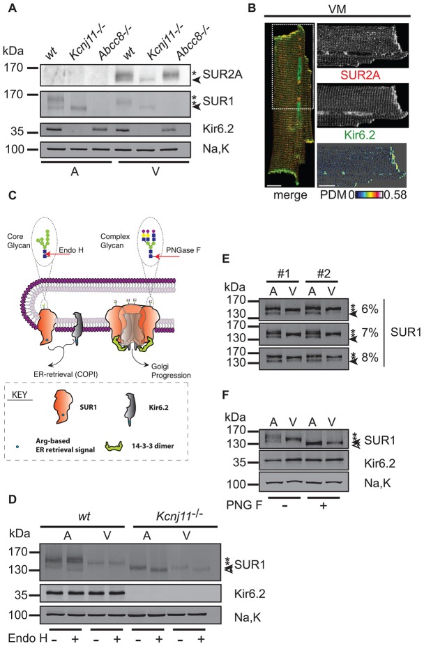
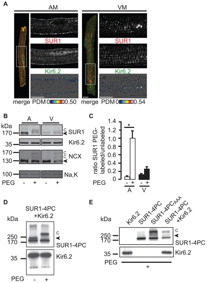
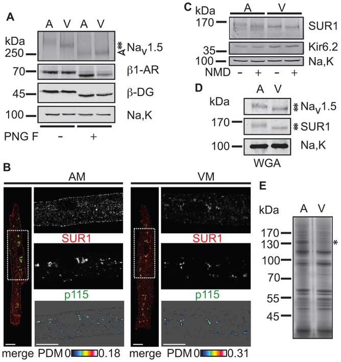
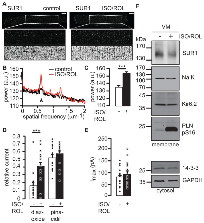
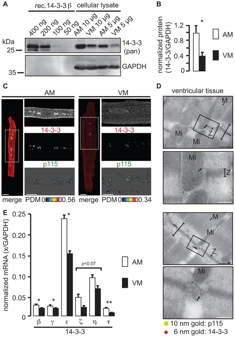
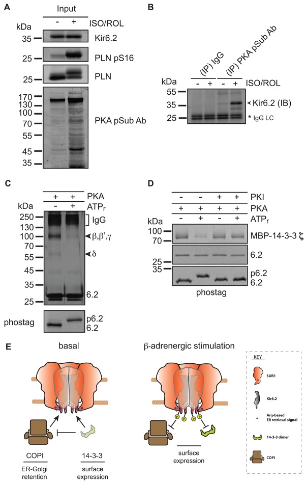
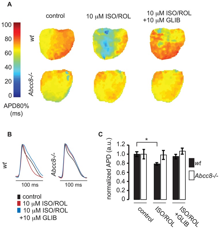
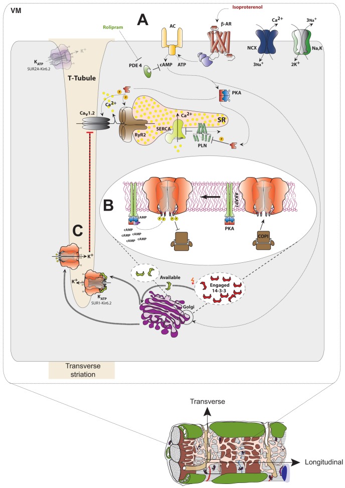
The copy number of membrane proteins at the cell surface is tightly regulated. Many ion channels and receptors present retrieval motifs to COPI vesicle coats and are retained in the early secretory pathway. In some cases, the interaction with COPI is prevented by binding to 14-3-3 proteins. However, the functional significance of this antagonism between COPI and 14-3-3 in terminally differentiated cells is unknown. Here, we show that ATP-sensitive K(+) (KATP) channels, which are composed of Kir6.2 and SUR1 subunits, are stalled in the Golgi complex of ventricular, but not atrial, cardiomyocytes. Upon sustained β-adrenergic stimulation, which leads to activation of protein kinase A (PKA), SUR1-containing channels reach the plasma membrane of ventricular cells. We show that PKA-dependent phosphorylation of the C-terminus of Kir6.2 decreases binding to COPI and, thereby, silences the arginine-based retrieval signal. Thus, activation of the sympathetic nervous system releases this population of KATP channels from storage in the Golgi and, hence, might facilitate the adaptive response to metabolic challenges.

Keywords: 14-3-3; ATP-sensitive K+ channels; Arg-based retrieval signal; COPI; Cardiomyocyte; Coatomer; KATP; PKA; Protein kinase A; Trafficking.


Figures

Fig. 1.
Fig. 1.
Biochemical analysis of KATP channel subunits in atria and ventricles. (A) Western blotting (see supplementary material Table S1 for antibodies) for SUR2A, SUR1, Kir6.2 and the α1 subunit of the Na+/K+-ATPase (Na,K) in membranes from mouse atrial (A) and ventricular tissue (V). Filled arrowheads and asterisks indicate core- and complex-glycosylated SUR proteins, respectively. The western blot is representative of three independent experiments. (B) Confocal analysis of immunostained mouse ventricular myocytes (VM). SUR2A (red) and Kir6.2 (green) signals are shown by the region of interest (ROI) that is indicated in the merged whole cell image (dashed white box). Kir6.2 nuclear staining is unspecific (see supplementary material Fig. S2 for knockout control). PDM denotes the product of differences from the mean, indicating colocalization by intensity correlation analysis (Li et al., 2004). Values of intensity correlation quotient between 0 and 0.5 indicate co-dependent staining and were 0.16±0.005 for VM (mean±s.e.m., n = 11). Scale bars: 10 µm. (C) Representation of SUR1 and Kir6.2 as cargo proteins of the early secretory pathway and the rationale of glycan analysis using Endo H and PNGase F. Shapes and symbols are identified by the boxed key. (D) Western blotting for SUR1, Kir6.2 and Na,K in membranes from atrial (A) and ventricular (V) tissues from wild-type or Kcnj11-/- mice. Treatment with Endo H was as indicated; open arrowhead, filled arrowhead and asterisks mark deglycosylated, core-glycosylated, and complex-glycosylated forms of SUR1, respectively. Na,K serves as loading control. The western blot is representative of six independent experiments. (E) Western blotting for SUR1 in membranes from rat atrial (A) and ventricular (V) tissue demonstrating differing migration behaviors. The filled arrowheads indicate the core-glycosylated form of SUR1, and the asterisks denote the two complex-glycosylated forms of SUR1. The panel shows three technical replicates (the proteins were resolved on gels of increasing percentages – 6%, 7% and 8%) of two biological replicates (from two rats, #1 and #2). The western blot is representative of eight independent experiments. (F) Treatment with PNGase F glycosidase (PNG F) of solubilized membranes from rat atrial (A) or ventricular (V) tissue to probe whether differences in SUR1 migratory behavior (asterisks) were caused by differential complex glycosylation. The difference in migration was lost upon deglycosylation (indicated by the open arrowhead). The filled arrowhead indicates the core-glycosylated form of SUR1, and the asterisks indicate the complex-glycosylated forms of SUR1. The western blot is representative of eleven independent experiments.
Fig. 2.
Fig. 2.
SUR1–Kir6.2 KATP channels are localized differently in atrial and ventricular myocytes. (A) Confocal analysis of immunostained mouse atrial (AM) or ventricular (VM) myocytes. SUR1 (red) and Kir6.2 (green) immunofluorescence signals are shown by the ROI as indicated in the whole cell image (merge, dashed white box). The values of intensity correlation quotient for AM were 0.20±0.007 (mean±s.e.m., n = 7) and VM 0.14±0.008 (mean±s.e.m., n = 4) confirming colocalization of Kir6.2 and SUR1 subunits. Scale bars: 10 µm. Refer to Fig. 8 for an illustration of cardiac myocyte morphology. (B) Cell surface PEGylation analysis in intact mouse hearts indicating cell surface expression of SUR1 in atrial, but not ventricular, myocytes. Modified bands are marked by ‘C’, the Na+/Ca2+ exchanger (NCX) is shown as a positive control for cell surface PEGylation. Kir6.2 and Na,K are not PEGylated and serve as negative controls. The western blot is representative of three independent experiments quantified in C. Note that the PEG modification is sensitive to reducing agents; hence, non-reducing conditions were employed in contrast with all other figures. Therefore, the core- and complex-glycosylated forms of SUR1 were not resolved. (C) The ratio of intensity of the labeled PEGylated SUR1 species (‘C’ in Fig. 2B) and unlabeled SUR1 (black arrowhead in Fig. 2B) for atria (open bars) and ventricles (filled bars). The increased PEGylation in atria was statistically significant (*P<0.05), whereas the ratio of PEGylated species was not significantly increased in ventricular membranes. (D) PEGylation of epitope-tagged SUR1-4PC (4× Protein C tags) and Kir6.2 in HEK293 cells. Western blotting for the PC-epitope is shown for PEG-maleimide treated sample and untreated control. ‘C’ indicates the position of PEGylated SUR1 species. The arrowhead indicates complex glycosylated SUR1. (E) SUR1 is only PEGylated upon coexpression with Kir6.2 or when the Arg-based retrieval signal is inactivated by site-directed mutagenesis (SUR1-4PCAAA), which is consistent with Arg-based retrieval signals preventing unassembled subunits from reaching the cell surface. The arrowhead indicates unmodified complex glycosylated SUR1.
Fig. 3.
Fig. 3.
SUR1–Kir6.2 KATP channels are retained in the Golgi of ventricular myocytes. (A) Treatment with PNGase F glycosidase ‘PNG F’ of solubilized membranes from rat atrial (A) or ventricular (V) tissue. Like SUR1, the Na+ channel α-subunit NaV1.5 displayed differing migratory behavior (asterisks) between atria and ventricles, whereas the β-adrenergic receptor (β1-AR) and β-dystroglycan (β-DG) migration was indistinguishable in the two tissues. Na,K is not glycosylated and serves as a loading control. All three glycoproteins were fully deglycosylated by PNGase F (open arrowhead) leading to loss of the different migration patterns. The western blot is representative of three independent experiments. (B) Confocal analysis of immunostained mouse atrial (AM) or ventricular (VM) myocytes. SUR1 (red) and p115 (green) immunofluorescence signals are shown by the ROI in the whole cell image (merge, dashed white box). For an explanation of PDM analyses, see Fig. 1. Scale bars: 10 µm. Refer to Fig. 8 for an illustration of cardiac myocyte morphology. (C) Treatment of rat atrial and ventricular membranes with neuraminidase (NMD), as indicated, to probe for the extent of sialylation of SUR1. The western blot is representative of five independent experiments. (D) SDS eluates from a wheat germ agglutinin column (WGA) were probed with the indicated antibodies. Asterisks indicate the different migration of NaV1.5 and SUR1. The western blot is representative of three independent experiments. (E) Silver-stained eluates from WGA. The asterisk indicates a protein that was identified as peptidylglycine α-amidating monooxygenase, the staining of which was prominently different between atrial and ventricular tissue. The experiments shown in C,D,E were performed on rat cardiac tissue.
Fig. 4.
Fig. 4.
β-adrenergic stimulation deploys Golgi-stalled SUR1–Kir6.2 channels to T-tubule membrane invaginations at striations of ventricular myocytes. (A) Confocal analysis of mouse ventricular myocytes that were immunostained for SUR1 in the absence or presence of 10 µM isoproterenol and 10 µM rolipram (ISO/ROL). Dashed boxes indicate the magnified (2×) intracellular ROI (middle) and the binary inverse contrasted signal (bottom). Scale bars: 10 µm. Refer to Fig. 8 for a summary of cardiac myocyte morphology. (B) Power spectrum (Fourier analysis) of 20 untreated and 18 treated myocytes, the first peak indicates the degree of periodicity of the striated signal (arrowhead). (C) The average change in power at the first peak marked in B, error bars show the s.e.m., ***P<0.0005, a.u., arbitrary units. (D) Inside–out patch clamp recordings of mouse ventricular myocytes that were untreated or had been treated for 1 h and during recordings. ‘Relative current’ refers to the fraction of the current under conditions of no ATP that is activated by diazoxide or pinacidil. Individual data points are shown as circles (untreated) or squares (treated), ***P<0.0005, error bars reflect the s.e.m., n = 16 or 17 cells. (E) Inside–out patch clamp recordings of mouse ventricular myocytes that were untreated or had been treated with 10 µM isoproterenol and 10 µM rolipram each for 1 h and during recordings. Imax, the maximum current under conditions lacking ATP. (F) Western blotting for SUR1, Na,K, Kir6.2 and the phosphorylated form of phospholamban (phosphorylated at serine residue 16, PLN pS16) in membranes (top panel), and 14-3-3 proteins and GAPDH in the cytosol (bottom panel) from mouse ventricular myocytes (VM) that were untreated or treated as in A–E (±ISO/ROL). The western blot is representative of three independent experiments.
Fig. 5.
Fig. 5.
Ventricular myocytes contain significantly lower amounts of 14-3-3. Western blotting (A) and quantification (B) of three independent experiments using a pan-reactive antibody cocktail against 14-3-3 (supplementary material Table S1, antibodies 1b and 1c). The indicated amounts of soluble cellular lysate from rat atrial (AM) or ventricular (VM) myocytes were loaded. Varying concentrations of recombinant 14-3-3β (rec. 14-3-3 β) were used to approximate the detection threshold of the pan-reactive antibody. Glyceraldehyde-3-phosphate dehydrogenase (GAPDH) was detected as a loading control. *P<0.05. (C) Confocal analysis of immunostained mouse atrial (AM) or ventricular (VM) myocytes. 14-3-3 (red) and p115 (green) immunofluorescence signals are shown in the ROI indicated in the whole cell image (merge, dashed white box). Scale bars: 10 µm. Refer to Fig. 8 for an illustration of cardiac myocyte morphology. (D) Two different examples of immuno-electron microscopy that were performed on fixed cryosections from the left ventricle of mouse hearts. The upper picture indicates the inset that is shown in the lower panel. I, M and Z indicate the respective bands of the cardiac muscle. Mi, mitochondria and arrows point to the gold label (14-3-3, 6 nm and p115, 10 nm). Scale bars: 500 nm (black), 200 nm (white, the boxed inset magnification). (E) RT-PCR analysis of six 14-3-3 isoforms using mRNA from isolated rat atrial (AM) and ventricular (VM) myocytes. mRNA abundance is normalized to the message encoding GAPDH. The means were derived from three biological replicates. *P<0.05, **P<0.01, a non-significant value of P<0.07 is indicated for two isoforms.
Fig. 6.
Fig. 6.
Phosphorylation of the Kir6.2 C-terminus by PKA reduces COPI and 14-3-3 binding. (A) Western blotting for Kir6.2, Phospholamban (PLN), a phosphorylated form of phospholamban (at serine residue 16, PLN pS16) and substrates phosphorylated by PKA (PKA pSub Ab) in hearts perfused in the presence (+) or absence (−) of 10 µM isoproterenol and 10 µM rolipram (ISO/ROL). (B) Phosphorylated Kir6.2 was immunoprecipitated (IP) using an antibody that binds PKA phosphorylated substrates (PKA pSub Ab). Solubilized membranes that had been prepared from treated (+) or untreated (−) hearts, as in A, were used. The blots were probed for Kir6.2 (IB, immunoblot). Purified Rabbit IgG (IgG) was used as a negative control. IgG LC refers to the antibody light chain and serves as a loading control. (C) Silver-stained eluates from a COPI binding assay reveals the reduction of COPI binding after phosphorylation of the C-terminus of Kir6.2. Compare supplementary material Fig. S4 for the quantification of three independent experiments. β, β', γ and δ refers to four subunits of coatomer. IgG refers to the co-eluted antibody from the IgG sepharose affinity matrix. ATPr, ATP regeneration system. The lower panel compares the electrophoretic mobility shift of the unphosphorylated (6.2) and phosphorylated (p6.2) Kir6.2 C-terminal peptide on a Phostag-polyacrylamide gel. (D) Coomassie-stained eluates from a 14-3-3 binding assay revealed a reduction of 14-3-3 binding after phosphorylation of the C-terminus of Kir6.2. The gel is representative of three independent experiments. PKI, protein kinase A inhibitor. (E) The release of SUR1-containing KATP channels from the antagonistic actions of COPI and 14-3-3 after ;phosphorylation.
Fig. 7.
Fig. 7.
Action potential shortening during sustained β-adrenergic stimulation requires SUR1. (A) Representative APD maps from a wild-type (wt, top row) and Abcc8−/− (bottom row) ventricle were constructed from optical mapping recordings under control conditions (left column), 20 min after administration of 10 µM ISO and 10 µM ROL (center column), and 10 min after the addition of 10 µM glibenclamide (GLIB, right column). Each pixel in the map represents the APD80% from that region of the ventricular myocardium according to the color bar on the left. (B) The signal-averaged action potential traces from representative WT and Abcc8−/− ventricles are shown. (C) The normalized APD from WT hearts (n = 5) shows significant shortening (at 80% repolarization) upon treatment with ISO and ROL, and clear reversal with glibenclamide. However, the normalized APD from Abcc8−/− hearts (n = 5) shows no significant changes throughout the experiment. *P<0.05, means±s.e.m. a.u., arbitrary units.
Fig. 8.
Fig. 8.
Model of the regulated deployment of SUR1–Kir6.2 KATP channels in ventricular myocytes. (A) Agonist (isoproterenol) binding to β -adrenergic receptors (β-AR) triggers the activation of the adenylyl cyclase (AC) through a β-AR-coupled G-protein, resulting in elevation of cAMP and the activation of PKA. Attenuation of signal transduction through degradation of cAMP by phosphodiesterase (PDE) was inhibited using the PDE4-specific inhibitor rolipram. Known PKA targets include the voltage-gated Ca2+ channel (CaV1.2) and the Ryanodine receptor (RyR2), culminating in elevation of cytosolic Ca2+ [by release from the sarcoplasmic reticulum (SR) Ca2+ stores and by influx of extracellular Ca2+]. The phosphorylation of phospholamban (PLN) by PKA relieves its inhibitory effect on the SR Ca2+ pump (SERCA). For simplicity, the relevant PKA holoenzyme has been depicted as being cytosolic and not membrane associated. (B) PKA dependent phosphorylation of S372 (adjacent to the Arg-based ER retrieval signal) in Kir6.2 releases Golgi-stored SUR1–Kir6.2 KATP channels from COPI binding, thus, facilitating Golgi exit. An unknown kinase anchoring protein (AKAP), conceivably, localizes PKA to the vicinity of KATP channels. (C) Deployment of SUR1-containing KATP channels from the Golgi to the T-tubular plasma membrane. Hypothetically, signal transduction might affect the available pool of 14-3-3 proteins, in addition to direct phosphorylation of cargo proteins, by shifting the equilibrium between an engaged (substrate bound) and available (substrate free) pool, thus, overcoming the limitations of cell surface expression of 14-3-3 substrates by the limited availability of 14-3-3 proteins.

Similar articles

Cited by

References

    1. Abe K., Puertollano R. (2011). Role of TRP channels in the regulation of the endosomal pathway. Physiology (Bethesda) 26, 14–22 10.1152/physiol.00048.2010 - DOI - PMC - PubMed
    1. Babenko A. P., Gonzalez G., Aguilar-Bryan L., Bryan J. (1998). Reconstituted human cardiac KATP channels: functional identity with the native channels from the sarcolemma of human ventricular cells. Circ. Res. 83, 1132–1143 10.1161/01.RES.83.11.1132 - DOI - PubMed
    1. Béguin P., Nagashima K., Nishimura M., Gonoi T., Seino S. (1999). PKA-mediated phosphorylation of the human K(ATP) channel: separate roles of Kir6.2 and SUR1 subunit phosphorylation. EMBO J. 18, 4722–4732 10.1093/emboj/18.17.4722 - DOI - PMC - PubMed
    1. Bezzerides V. J., Ramsey I. S., Kotecha S., Greka A., Clapham D. E. (2004). Rapid vesicular translocation and insertion of TRP channels. Nat. Cell Biol. 6, 709–720 10.1038/ncb1150 - DOI - PubMed
    1. Bogan J. S. (2012). Regulation of glucose transporter translocation in health and diabetes. Annu. Rev. Biochem. 81, 507–532 10.1146/annurev--biochem--060109--094246 - DOI - PubMed

Publication types

LinkOut - more resources