. 2014 Apr 11;289(15):10466-10475.
doi: 10.1074/jbc.M113.482299. Epub 2014 Feb 25.

Neuroendocrine signaling via the serotonin transporter regulates clearance of apoptotic cells

Affiliations

Neuroendocrine signaling via the serotonin transporter regulates clearance of apoptotic cells

Takeshi Tanaka et al. J Biol Chem. .

Abstract

Serotonin (5-hydroxytryptamine; 5-HT) is a CNS neurotransmitter increasingly recognized to exert immunomodulatory effects outside the CNS that contribute to the pathogenesis of autoimmune and chronic inflammatory diseases. 5-HT signals to activate the RhoA/Rho kinase (ROCK) pathway, a pathway known for its ability to regulate phagocytosis. The clearance of apoptotic cells (i.e. efferocytosis) is a key modulator of the immune response that is inhibited by the RhoA/ROCK pathway. Because efferocytosis is defective in many of the same illnesses where 5-HT has been implicated in disease pathogenesis, we hypothesized that 5-HT would suppress efferocytosis via activation of RhoA/ROCK. The effect of 5-HT on efferocytosis was examined in murine peritoneal and human alveolar macrophages, and its mechanisms were investigated using pharmacologic blockade and genetic deletion. 5-HT impaired efferocytosis by murine peritoneal macrophages and human alveolar macrophages. 5-HT increased phosphorylation of myosin phosphatase subunit 1 (Mypt-1), a known ROCK target, and inhibitors of RhoA and ROCK reversed the suppressive effect of 5-HT on efferocytosis. Peritoneal macrophages expressed the 5-HT transporter and 5-HT receptors (R) 2a, 2b, but not 2c. Inhibition of 5-HTR2a and 5-HTR2b had no effect on efferocytosis, but blockade of the 5-HT transporter prevented 5-HT-impaired efferocytosis. Genetic deletion of the 5-HT transporter inhibited 5-HT uptake into peritoneal macrophages, prevented 5-HT-induced phosphorylation of Mypt-1, reversed the inhibitory effect of 5-HT on efferocytosis, and decreased cellular peritoneal inflammation. These results suggest a novel mechanism by which 5-HT might disrupt efferocytosis and contribute to the pathogenesis of autoimmune and chronic inflammatory diseases.

Keywords: Autoimmune Diseases; Efferocytosis; Inflammation; Lung; Phagocytosis; Rho Kinase; RhoA; Serotonin Receptors.


Figures
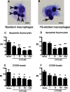
FIGURE 1.
FIGURE 1.
5-HT impairs ingestion of apoptotic cells and carboxylated beads by murine peritoneal macrophages. A and B, photomicrographs (40x original magnification, bar = 5 μm) show binding (arrows) and ingestion (arrowheads) of apoptotic thymocytes by murine peritoneal macrophages. C and D, murine resident (C) and TG-elicited (D) peritoneal macrophages were treated with 5-HT at 1, 10, 100, and 1,000 nm for 24 h then apoptotic thymocytes were co-cultured with macrophages at a 10:1 ratio for 1 h in 10% CO2 at 37 °C. Uningested apoptotic thymocytes were then washed off and a PI was determined by blinded visual inspection. Data represent the mean ± S.E. for n = 3 per group. *, one-way ANOVA, p < 0.05 versus media control (Dunnett's Test); **, p < 0.01 versus media control (Dunnett's test). E and F, murine resident (E) and TG-elicited (F) peritoneal macrophages were treated with 5-HT at 1, 10, 100, and 1,000 nm for 24 h, then carboxylated beads were co-cultured with macrophages at a 2:1 ratio for 20 min in 10% CO2 at 37 °C. Uningested beads were washed off and a PI was determined by blinded visual inspection. Data represent the mean ± S.E. for n = 3 per group. *, one-way ANOVA, p < 0.05, p < 0.05 versus media control (Dunnett's test).
FIGURE 2.
FIGURE 2.
5-HT impairs phagocytosis of latex beads by murine peritoneal macrophages, but has no effect on uptake of IgG-opsonized erythrocytes through the Fcγ receptor. A and B, mouse resident (A) and TG-elicited (B) peritoneal macrophages were treated with 5-HT at 1, 10, 100, and 1,000 nm for 24 h, then latex beads were co-cultured at a 1:1 ratio for 10 min in 10% CO2 at 37 °C. Uningested beads were washed off and a PI was determined by blinded visual inspection. Data represent the mean ± S.E. for n = 3 per group, *, one-way ANOVA, p < 0.05 versus media control (Dunnett's Test). **, p < 0.01 versus media control (Dunnett's test). C and D, mouse resident (C) and TG-elicited (D) peritoneal macrophages were treated with 5-HT at 1, 10, 100, and 1,000 nm for 24 h then co-cultured with IgG-opsonized erythrocytes (RBCs) at a 10:1 ratio in 10% CO2 at 37 °C. Uningested RBCs were then washed off and a PI was determined by blinded visual inspection. Data represent the mean ± S.E. for n = 3 per group.
FIGURE 3.
FIGURE 3.
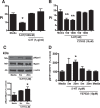
5-HT impairs efferocytosis through activation of the RhoA/Rho kinase pathway. A, murine TG-elicited macrophages were treated with 5-HT at 1,000 nm for 24 h. The RhoA inhibitor, C3 transferase (1 μg/ml), was added either alone or in the presence of 5-HT for 3 h prior to the assay. Apoptotic thymocytes were co-cultured with macrophages at a 10:1 ratio for 1 h in 10% CO2 at 37 °C. Uningested apoptotic thymocytes were washed off and a PI was determined by blinded visual inspection. Data represent the mean ± S.E. for n = 3 per group. *, one-way ANOVA, p < 0.05 versus media control (Dunnett's test). B, murine TG-elicited macrophages were treated with 5-HT at 100 or 1,000 nm for 24 h. The ROCK inhibitor, Y-27632 (10 μm), was added either alone or in the presence of 5-HT for 3 h prior to the assay. Apoptotic thymocytes were co-cultured with macrophages at a 10:1 ratio for 1 h in 10% CO2 at 37 °C. Uningested apoptotic thymocytes were washed off and a PI was determined by blinded visual inspection. Data represent the mean ± S.E. for n = 3 per group. **, one-way ANOVA, p < 0.05, p < 0.01 versus media control (Dunnett's test). C, murine TG-elicited peritoneal macrophages were stimulated with 5-HT (1 μm) for the indicated times. Phosphorylation of myosin phosphatase 1 (Mypt1) at Thr-696 increased upon 5-HT stimulation. Representative immunoblots are shown in the upper panel. In the lower panel, densitometry was used to determine the ratio of phospho-Mypt1 to total Mypt1. Data represent the mean ± S.E. as a percent of media control for n = 3 per group. Media control = 0.2545 ± 0.1182. *, F-test, p < 0.05 for significance of percent difference from media control (Bonferroni t statistic). D, murine TG-elicited peritoneal macrophage were stimulated with 5-HT (1 μm) for the indicated times in the presence and absence of the Rho kinase inhibitor, Y-27632 (10 μm), and examined for the ratio of phospho-Mypt-1 to total Mypt1 by Western blot. Data represent the mean ± S.E. as percent of media control for n = 5 per group. Media control = 0.1807 ± 0.0286. *, F-test, p < 0.05 for significance of percent control from media control (Bonferroni t statistic).
FIGURE 4.
FIGURE 4.
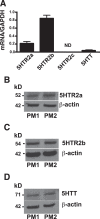
5-HT receptor 2a, 2b, and the 5-HT transporter are expressed in mouse peritoneal macrophages. A, real-time PCR was performed to measure the expression by mRNA. 5-HT receptor 2a (5HTR2a), 5-HT receptor 2b (5HTR2b), and the 5-HTT were expressed in TG-elicited peritoneal macrophages, but 5-HT receptor 2c (5HTR2c) was not detected. Data represent the mean ± S.E. for n = 3 per group. B–D, Western blotting is shown for two representative peritoneal macrophage lysates (PM1 and PM2) evaluating expression of 5-HT receptors and transporter. B, HTR2a (n = 4); C, HTR2b (n = 10); and D, 5-HTT (n = 8) were all detected in mouse peritoneal macrophages.
FIGURE 5.
FIGURE 5.
5-HT utilizes the 5-HT transporter, but not 5-HT receptors 2a or 2b, to regulate efferocytosis by murine peritoneal macrophages. A and B, TG-elicited macrophages were treated with 5-HT at 1,000 nm in the presence and absence of: A, the 5-HT receptor 2b inhibitor, R-96544; B, the 5-HT receptor 2b inhibitor, RS-127445; or C, the 5-HT transporter inhibitor, fluoxetine, at the indicated concentrations for 24 h. Apoptotic thymocytes were co-cultured with macrophages at a 10:1 ratio for 1 h in 10% CO2 at 37 °C. Uningested apoptotic thymocytes were washed off and a PI was determined by blinded visual inspection. 5-HT does not inhibit efferocytosis via 5HTR2a- or 5HTR2b-dependent pathways. Data represent the mean ± S.E. for n = 4–5 replicates per group, *, one-way ANOVA, p < 0.05 versus DMSO control (Dunnett's test); **, p < 0.01 versus DMSO control (Dunnett's test). C, TG-elicited macrophages were treated with 5-HT at 1.0 μm for 24 h in the presence and absence of the 5-HT transporter inhibitor, fluoxetine, at 0.1 and 1.0 μm. Apoptotic thymocytes were then co-cultured with macrophages at a 10:1 ratio for 1 h in 10% CO2 at 37 °C. Uningested apoptotic thymocytes were washed off and a PI was determined by blinded visual inspection. Data represent the mean ± S.E. for n = 5 per group, *, one-way ANOVA, p < 0.05 versus media control (Dunnett's test).
FIGURE 6.
FIGURE 6.
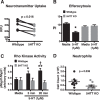
Effect of 5-HTT deficiency on 5-HT uptake, efferocytosis, Rho kinase activity, and peritoneal inflammation. A, function of the 5-HTT in TG-elicited peritoneal macrophages was investigated using a neuroendocrine uptake assay, where a tracer becomes fluorescent upon internalization. The tracer can be internalized by the 5-HT, dopamine, or the norepinephrine transporters, so specificity depends on genetic deletion of a given transporter. TG-elicited peritoneal macrophages were isolated from 5-HTT KO (gray circles) and wild-type control mice (black circles), and uptake of the fluorescent tracer was assessed and expressed as relative fluorescent units (RFU). Lines indicate paired experiments. p = 0.016, wild-type versus 5-HTT KO mice (paired t test). B, TG-elicited peritoneal macrophages from wild-type mice (black columns) and 5-HTT KO mice (gray columns) were treated with media alone or media containing 5-HT (1 μm) for 24 h. Apoptotic thymocytes were co-cultured with macrophages at a 10:1 ratio for 1 h in 10% CO2 at 37 °C. Uningested apoptotic thymocytes were washed off and a PI was determined by blinded visual inspection. 5-HT decreased efferocytosis in wild-type macrophages. Data represent the mean ± S.E. for n = 8 per group. Two-way ANOVA, p < 0.001 for interaction, and p < 0.01 for treatment effect (media versus 5-HT treatment). *, Tukey-Kramer post hoc analysis, p < 0.001 for wild-type 5-HT treatment versus wild-type media control; p < 0.01 for wild-type 5-HT treatment versus 5-HTT KO 5-HT treatment; p < 0.05 for wild-type 5-HT treatment versus 5-HTT KO media control. C, murine TG-elicited peritoneal macrophages from wild-type (black columns) and 5-HTT KO mice (gray columns) were stimulated with 5-HT (1 μm) for the indicated times. Densitometry was used to determine the ratio of phosphomyosin phosphatase 1 (pMypt1) to total Mypt1 by arbitrary units. 5-HT increased phosphorylation of Mypt1 at Thr-696 in wild-type but not in 5-HTT KO macrophages. Data represent the mean ± S.E. for n ≤ 13 per group. Two-way ANOVA, p = 0.03 for interaction, and p < 0.01 for genotype effect (wild-type versus 5-HTT KO). *, Tukey-Kramer post hoc analysis p < 0.03 for wild-type media control versus wild-type 5-HT treatment for 5 min; **, p < 0.01 for wild-type media control versus wild-type 5-HT treatment for 20 min; †, p < 0.03 for wild-type 5-HT treatment for 20 min versus 5-HTT KO 5-HT treatment for 20 min. D, 5-HTT KO (gray circles) and wild-type (black circles) mice were injected with TG. Four days later peritoneal lavage was performed and total cell counts and differentials were taken. Neutrophils were decreased in 5-HTT KO mice compared with wild-type mice. Data represent the mean ± S.E. for n = 8–10 mice per group. p = 0.036, wild-type versus 5-HTT KO mice (Mann-Whitney test).
FIGURE 7.
FIGURE 7.
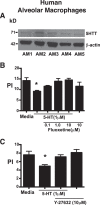
5-HT inhibits efferocytosis by human alveolar macrophages via a mechanism that involves the 5-HT transporter and Rho kinase. A, Western blots were performed on lysates from human alveolar macrophages from n = 5 normal subjects and probed for the presence of the 5-HT transporter. B, human alveolar macrophages were treated with 5-HT at 1.0 μm for 24 h in the presence and absence of the 5-HT transporter inhibitor, fluoxetine, at 0.1, 1.0, and 10 μm. Apoptotic human Jurkat T-cells were then co-cultured with macrophages at a 10:1 ratio for 3 h in 10% CO2 at 37 °C. Uningested apoptotic cells were washed off and PI was determined by visual inspection. Data represent the mean ± S.E. for n = 3 per group, *, one-way ANOVA, p < 0.05 versus media control (Dunnett's test). C, human alveolar macrophages were treated with 5-HT at 1.0 μm for 24 h in the presence and absence of the ROCK inhibitor, Y-27632, at 10 μm. Apoptotic human Jurkat T-cells were then co-cultured with macrophages at a 10:1 ratio for 3 h in 10% CO2 at 37 °C. Uningested apoptotic cells were washed off and a PI was determined by visual inspection. Data represent the mean ± S.E. for n = 4 per group, *, one-way ANOVA, p < 0.05 versus media control (Dunnett's test).
FIGURE 8.
FIGURE 8.
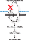
Proposed model for the ability of 5-HT to suppress efferocytosis. 5-HT impairs efferocytosis by activating RhoA and ROCK. 5-HT appears to exert its effect on efferocytosis through the 5-HT transporter and not through 5-HT receptor 2a or 5-HT receptor 2b (red X).

Similar articles

Cited by

References

    1. Mössner R., Lesch K. P. (1998) Role of serotonin in the immune system and in neuroimmune interactions. Brain Behav. Immun. 12, 249–271 - PubMed
    1. Walther D. J., Stahlberg S., Vowinckel J. (2011) Novel roles for biogenic monoamines: from monoamines in transglutaminase-mediated post-translational protein modification to monoaminylation deregulation diseases. FEBS J. 278, 4740–4755 - PubMed
    1. Paulmann N., Grohmann M., Voigt J. P., Bert B., Vowinckel J., Bader M., Skelin M., Jevsek M., Fink H., Rupnik M., Walther D. J. (2009) Intracellular serotonin modulates insulin secretion from pancreatic beta-cells by protein serotonylation. PLoS Biol. 7, e1000229. - PMC - PubMed
    1. Chabbi-Achengli Y., Coudert A. E., Callebert J., Geoffroy V., Côté F., Collet C., de Vernejoul M. C. (2012) Decreased osteoclastogenesis in serotonin-deficient mice. Proc. Natl. Acad. Sci. U.S.A. 109, 2567–2572 - PMC - PubMed
    1. Walther D. J., Peter J. U., Winter S., Höltje M., Paulmann N., Grohmann M., Vowinckel J., Alamo-Bethencourt V., Wilhelm C. S., Ahnert-Hilger G., Bader M. (2003) Serotonylation of small GTPases is a signal transduction pathway that triggers platelet α-granule release. Cell 115, 851–862 - PubMed

Publication types

MeSH terms

Substances

LinkOut - more resources