. 2014 Apr 11;289(15):10637-10649.
doi: 10.1074/jbc.M113.491506. Epub 2014 Feb 25.

DNA promoter methylation-dependent transcription of the double C2-like domain β (DOC2B) gene regulates tumor growth in human cervical cancer

Affiliations

DNA promoter methylation-dependent transcription of the double C2-like domain β (DOC2B) gene regulates tumor growth in human cervical cancer

Shama Prasada Kabekkodu et al. J Biol Chem. .

Abstract

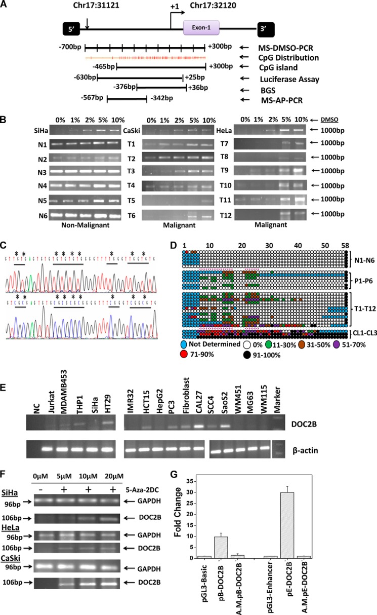
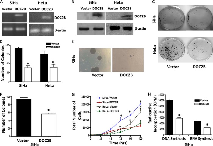
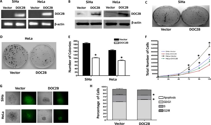
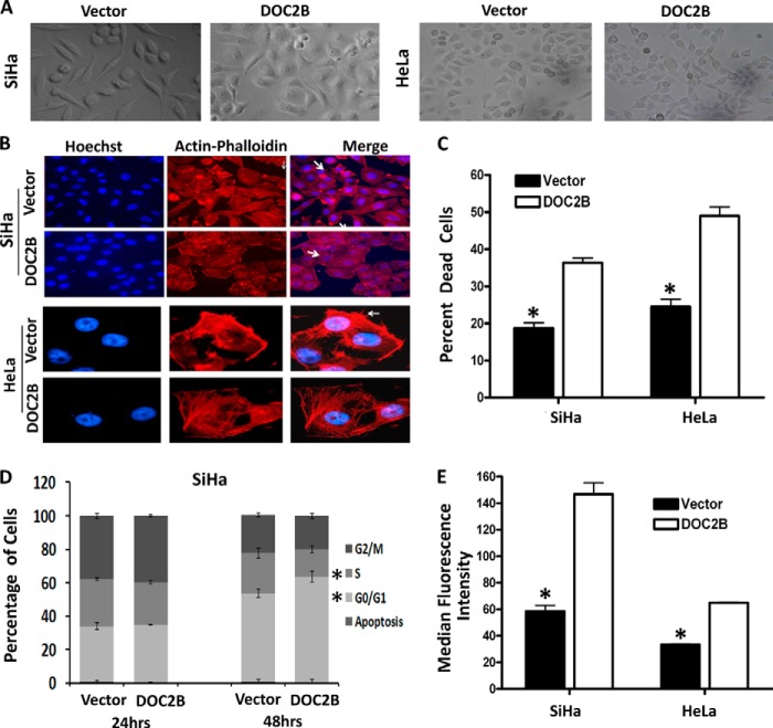
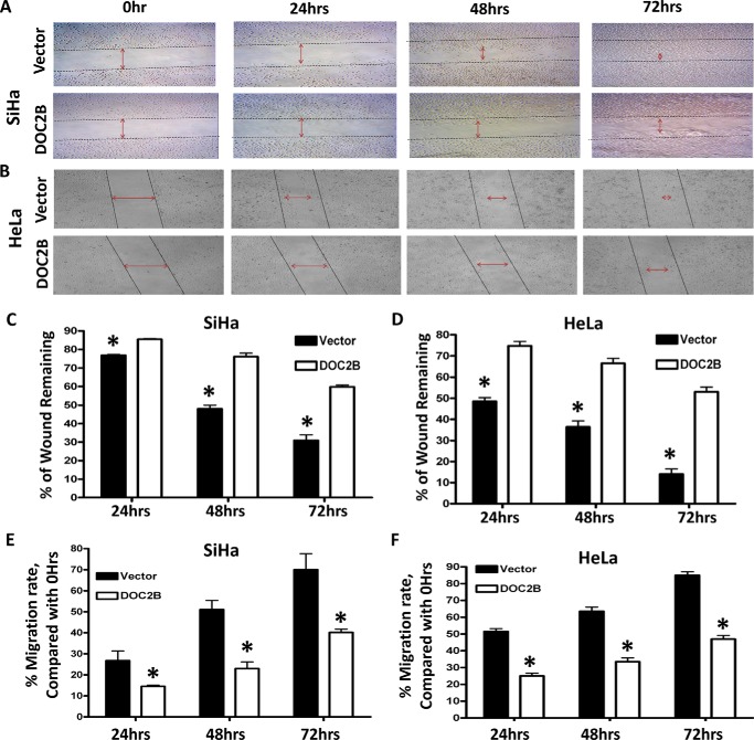
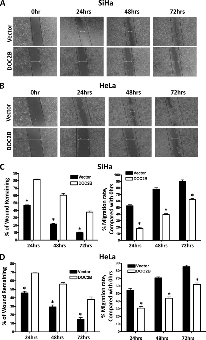
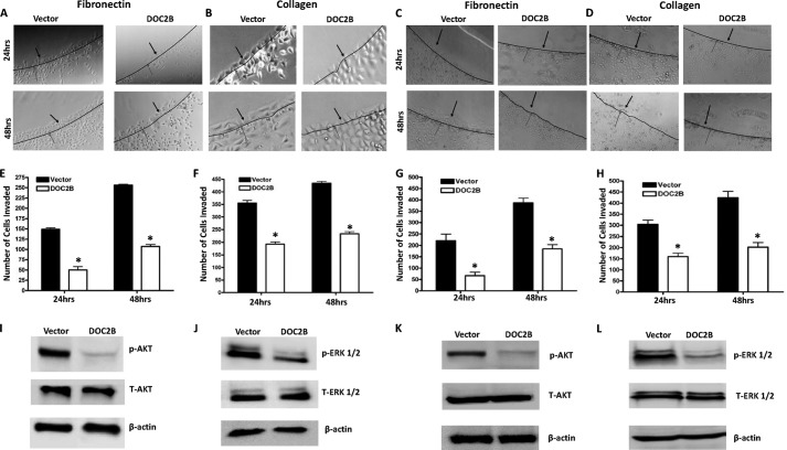
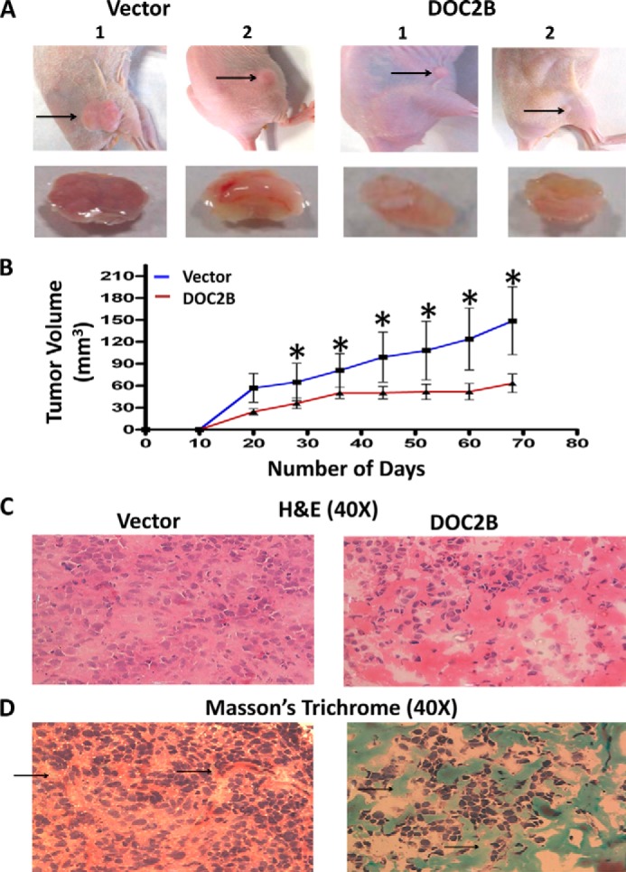
Double C2-like domain β (DOC2B) gene encodes for a calcium-binding protein, which is involved in neurotransmitter release, sorting, and exocytosis. We have identified the promoter region of the DOC2B gene as hypermethylated in pre-malignant, malignant cervical tissues, and cervical cancer cell lines by methylation-sensitive dimethyl sulfoxide-polymerase chain reaction and bisulfite genome sequencing; whereas, it was unmethylated in normal cervical tissues (p < 0.05). The promoter hypermethylation was inversely associated with mRNA expression in SiHa, CaSki, and HeLa cells and treatment with demethylating agent 5-aza-2-deoxycytidine restored DOC2B expression. The region -630 to +25 bp of the DOC2B gene showed robust promoter activity by a luciferase reporter assay and was inhibited by in vitro artificial methylation with Sss1 methylase prior to transient transfections. Overexpression of the DOC2B gene in SiHa cells when compared with controls showed significantly reduced colony formation, cell proliferation, induced cell cycle arrest, and repressed cell migration and invasion (p < 0.05). Ectopic expression of DOC2B resulted in anoikis-mediated cell death and repressed tumor growth in a nude mice xenograft model (p < 0.05). DOC2B expressing cells showed a significant increase in intracellular calcium level (p < 0.05), impaired AKT1 and ERK1/2 signaling, and induced actin cytoskeleton remodeling. Our results show that promoter hypermethylation and silencing of the DOC2B gene is an early and frequent event during cervical carcinogenesis and whose reduced expression due to DNA promoter methylation may lead to selective cervical tumor growth.

Keywords: Actin Remodeling; Akt; Cell Growth; Cell Invasion; Cell Motility; Cervical Cancer; DNA Promoter Methylation; DOC2B; ERK; Human Papillomavirus.


Figures

FIGURE 1.
FIGURE 1.
Methylation profiling of the Frag-13 fragment in cervical samples. A, schematic representation of the region selected for validation of Frag-13 fragment by MS-AP-PCR, MS-DMSO-PCR, BGS, and characterization of the promoter region of the DOC2B gene by a luciferase assay. B, MS-DMSO-PCR showed that DNA methylation changed the sensitivity of amplification to the DMSO concentration in the PCR mixture. In the case of methylated samples, amplification was detected at a DMSO concentration of 2% or more, whereas unmethylated or less methylated samples showed amplification even in the absence of DMSO. N and T represent non-malignant and tumor samples, respectively. C, representative electropherogram of a portion of the 412-bp region of non-malignant and tumor samples showing methylated and unmethylated CpG sites. The differentially methylated CpG sites were highlighted by asterisks (*). D, methylation map of the DOC2B promoter fragment in non-malignant, pre-malignant, malignant, and cervical cancer cell lines. The open and filled circles represent the unmethylated and methylated CpG sites, respectively. The partially methylated CpG sites were filled with different colors depending on the extent of methylation. Each horizontal line represents single samples, and circles representing single CpG sites. N1-N6, P1-P6, T1-T12, and CL1-CL3 represent 6 non-malignant, 6 pre-malignant, 12 malignant cervical samples, and 3 cervical cancer cell lines namely SiHa, CaSki, and HeLa, respectively. E, expression analysis of DOC2B in various cell lines by semi-quantitative RT-PCR with β-actin as control. F, demethylation by 5-aza-2DC and subsequent reactivation of mRNA of the DOC2B gene. Cervical cancer cell lines SiHa, CaSki, and HeLa were used for demethylation and reactivation experiments. In all three cell lines, reactivation was observed at 5 μm or above of 5-aza-2-DC treatment. GAPDH was used as an internal control for the integrity of the cDNA. RT-PCR analysis shows the loss of DOC2B expression and treatment with the demethylating agent 5-aza-2-DC restores its expression. G, characterization of promoter activities of the DOC2B gene by transient transfection experiments using DOC2B promoter constructs in pGL3-Basic and pGL3-Enhancer vectors. Histograms represent mean ± S.D. for at least two independent experiments. pB-DOC2B and pE-DOC2B represent the DOC2B promoter constructs cloned in pGL3-Basic and pGL3-Enhancer vectors, whereas A.M.pE-DOC2B represents corresponding artificially methylated constructs as discussed under ”Experimental Procedures.“
FIGURE 2.
FIGURE 2.
Effect of ectopic expression of DOC2B on cell growth and proliferation. A and B, representative figures showing expression of the DOC2B gene upon transfection by RT-PCR and Western blot, respectively. DOC2B was detected by anti-DDK tag antibody. β-Actin was used as an internal control. C, DOC2B inhibits tumor growth in vitro in SiHa and HeLa cells, respectively. D, quantitative analysis of colony forming assay represented as mean ± S.D., *, p < 0.05, shows a significant decrease in colony number after ectopic expression of DOC2B. E, representative image of soft agar colony forming assay. F, quantitative analysis of colony number represented as mean ± S.D.; *, p < 0.05. G, represents the cell proliferation rate in DOC2B expressing stable clones in comparison with vector control. Ectopic expression of DOC2B significantly inhibited cell proliferation resulting in delayed cell doubling time. The cell doubling analysis was performed using the cell doubling time calculator. H, cell proliferation rate was significantly inhibited at both DNA and RNA levels in DOC2B expressing cells when compared with control cells by [3H]thymidine and [3H]uridine incorporation assays, respectively (mean ± S.D. from 3 independent experiments in duplicates). *, represents p < 0.05.
FIGURE 3.
FIGURE 3.
Ectopic expression of DOC2B suppresses growth and proliferation in SiHa and HeLa cells. A and B, the DOC2B ORF was cloned into retroviral vector pMX-IRES-GFP to generate pMX-DOC2B-IRES-GFP and used to transduce SiHa and HeLa cells. The expression of DOC2B upon ectopic expression was confirmed by both RT-PCR (A) and Western blot by anti-DOC2B antibody (B). β-Actin was used as internal control. C and D, representative image of the colony formation assay in SiHa and HeLa, respectively. E, representive quantitative analysis of the colony numbers in SiHa and HeLa cells, respectively. The ectopic expression of DOC2B significantly reduced the colony numbers in both SiHa and HeLa cells, respectively. F, ectopic expression of DOC2B inhibits SiHa and HeLa cell proliferation, respectively. G, representative colonies from SiHa and HeLa, respectively. Both colony number and size decreased upon DOC2B expression. H, DOC2B expression induced arrest at the G0/G1 and S phases of the cell cycle. *, p < 0.05 by independent Student's t test was considered as statistically significant. The experiments were repeated 3 times in duplicate.
FIGURE 4.
FIGURE 4.
DOC2B expression changes cell morphology, induces remodeling of cytoskeletons, inhibits cell division, and increases intracellular calcium flux. A, representative cell morphology of DOC2B expressing and control cells in SiHa and HeLa cells, respectively (×40 magnification). B, representative images of actin staining showing rearrangement of actin fibers leading to increased cell to cell adhesion and decreased lamellipodia (indicated by arrows) in DOC2B expressing cells when compared with control cells (×400 magnifications). C, representative anoikis analysis for empty vector- and DOC2B-transfected SiHa and HeLa cells. Cells were cultured on poly-HEMA-coated tissue culture plates and cell death was analyzed by measuring the sub-G1 population of cells by propidium iodide staining and FACS analysis. Ectopic expression of DOC2B showed a significantly higher sub-G1 population of 18.66 versus 36.33% and 24.47 versus 48.96% between DOC2B and empty vector-transfected cells in both SiHa and HeLa cells, respectively. D, quantification of DNA content and cell cycle phase distribution in empty vector- and DOC2B-transfected SiHa cells at different time points as analyzed by a BrdU pulse-chase experiment. Ectopic expression of the DOC2B gene resulted in a significant cell cycle arrest at G0/G1 and S phases of the cell cycle. E, representative figures showing an increase in intracellular Ca2+ flux when compared with control cells. There was a significant increase in intracellular Ca2+ upon ectopic expression of DOC2B. Values were the median fluorescence intensities ± S.D. of at least three independent experiments performed in duplicates. The bar graph represents mean ± S.D. of triplicate experiments performed in duplicates. *, p < 0.05 by independent Student's t test. p value <0.05 was considered statistically significant.
FIGURE 5.
FIGURE 5.
DOC2B expression significantly suppresses the motility of cervical cancer cells. A and B, scratch assay was performed to assess the migration rate of SiHa and HeLa cells transfected with either vector or pCMV6-Entry-DOC2B. Photomicrographs were taken at the indicated time points under ×100 magnification. C and D, the percentage of wound closure in relationship to the time 0 h separation in SiHa and HeLa, respectively. E and F, the graph showing the rate of cell migration (migration index) in relationship to time 0 h separation in SiHa and HeLa, respectively. The migration index was calculated and quantified by measuring the area of the injured region in relationship to 0 h. The bar graph represents mean ± S.D. of triplicates experiments performed in duplicates. *, p < 0.05 by independent Student's t test. p value <0.05 was considered as statistically significant.
FIGURE 6.
FIGURE 6.
DOC2B expression inhibits migration of SiHa and HeLa cells in vitro. Wound healing assay was performed to identify the effect of DOC2B on cell migration. In brief cells were grown to confluence in 6-well plates, serum starved for 24 h, and a scratch was made using P-200 tip and would closure was monitored 72 h. A and B, representative image of wound healing assay. Our results show that the wound is completely closed in control cells by 72 h both in SiHa (left) and HeLa (right) cells, respectively. However, in DOC2B expressing SiHa and HeLa cells the wound was still intact even at the end of 72 h. C and D, representative graph showing the quantitative analysis of migration. Our experiments show that DOC2B expression inhibits migration of SiHa and HeLa cells. *, p < 0.05 by independent Student's t test was considered as statistically significant. The experiments were repeated 3 times in duplicates.
FIGURE 7.
FIGURE 7.
DOC2B inhibits invasion and phosphorylation of AKT-1 and ERK1/2. A–D, representative images showing the inhibition of invasion onto the agarose spot containing fibronectin and type I collagen upon ectopic expression of DOC2B in a single cell clone (A and B) and retrovirally transduced polyclonal cells (C and D). E–H, quantitative analysis of a number of tumor cells invading fibronectin and type 1 collagen in a single cell clone (E and F) and retrovirally transduced polyclonal cells (G and H), respectively. The bar graph represents mean ± S.D. of triplicate experiments performed in duplicates. *, p < 0.05 by independent Student's t test was considered as statistically significant. I–L, ectopic expression of DOC2B regulated both AKT and ERK signaling. A Western blot was performed using antibodies against total AKT, phospho-AKT, total ERK1/2, and phospho-ERK1/2. β-Actin was used as internal control. DOC2B expression inhibited AKT phosphorylation, whereas there was a decrease in ERK1/2 phosphorylation when compared with vector-transfected control SiHa cells in both single cell clone (I and J) and polyclonal cells (K and L), respectively. Relative phosphorylation levels of AKT and ERK1/2 were determined by normalization with total AKT and total ERK1/2, respectively.
FIGURE 8.
FIGURE 8.
DOC2B inhibits cervical cancer growth in vivo in nude mice. SiHa cells (1 × 10⁸ cells) transfected with pCMV-6-Entry vector or pCMV-6-DOC2B were injected subcutaneously into the flanks of female athymic nude (3 to 4 weeks old) mice (n = 6 each group). A, representative figure of tumor growth in nude mice inoculated subcutaneously with empty vector or DOC2B-transfected cells. B, tumor growth curves of DOC2B-expressing cells in nude mice were compared with control cells by a tumor xenograft experiment. The asterisk indicates statistical significance (*, p < 0.05). C, representative image of H&E staining of tumor xenografts with and without DOC2B expression. The H&E staining of DOC2B expressing cells showed a decreased nucleus to cytoplasmic ratio, less abnormal nucleus, and decreased tumor cell density. Furthermore, the cells were densely aggregated less in pleomorphic (atypical) cells and reduced in the number of abnormal mitosis. Stroma was also visible. In contrast to this, DOC2B negative cells were predominantly/increasingly pleomorphic, spindle morphology cells showing prominent nuclei with altered nucleus to cytoplasmic ratio and abnormal mitosis. D, representative image of Masson's trichrome staining of tumor xenografts with and without DOC2B expression. The Masson's trichrome staining showed significant degradation of collagen in the absence of DOC2B expression.

Similar articles

Cited by

References

    1. Shen H., Laird P. W. (2013) Interplay between the cancer genome and epigenome. Cell 153, 38–55 - PMC - PubMed
    1. Dueñas-González A., Lizano M., Candelaria M., Cetina L., Arce C., Cervera E. (2005) Epigenetics of cervical cancer. An overview and therapeutic perspectives. Mol. Cancer 4, 38. - PMC - PubMed
    1. Esteller M. (2008) Epigenetics in cancer. N. Engl. J. Med. 358, 1148–1159 - PubMed
    1. Duncan R. R., Shipston M. J., Chow R. H. (2000) Double C2 protein. A review. Biochimie 82, 421–426 - PubMed
    1. Orita S., Sasaki T., Naito A., Komuro R., Ohtsuka T., Maeda M., Suzuki H., Igarashi H., Takai Y. (1995) Doc2: a novel brain protein having two repeated C2-like domains. Biochem. Biophys. Res. Commun. 206, 439–448 - PubMed

Publication types

MeSH terms