Review
. 2014 Oct;21(5):543-52.
doi: 10.1007/s10140-014-1206-4. Epub 2014 Feb 26.

Anatomical variants and pathologies of the vermix

Affiliations
Review

Anatomical variants and pathologies of the vermix

Swati Deshmukh et al. Emerg Radiol. 2014 Oct.

Abstract

The appendix may demonstrate a perplexing range of normal and abnormal appearances on imaging exams. Familiarity with the anatomy and anatomical variants of the appendix is helpful in identifying the appendix on ultrasound, computed tomography, and magnetic resonance imaging. Knowledge of the variety of pathologies afflicting the appendix and of the spectrum of imaging findings may be particularly useful to the emergency radiologist for accurate diagnosis and appropriate guidance regarding clinical and surgical management. In this pictorial essay, we review appendiceal embryology, anatomical variants such as Amyand hernias, and pathologies from appendicitis to carcinoid, mucinous, and nonmucinous epithelial neoplasms.


Conflict of interest statement

Conflict of Interest The authors declare that they have no conflict of interest.

Figures

Fig. 1
Fig. 1
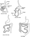
Embryology of the appendix. At 4 weeks, the midgut herniates into the umbilical cord. At 5 weeks, the prearterial segment of the midgut returns into the abdomen first as the gut rotates counterclockwise. By 12 weeks, the postarterial segment has already reduced and the cecum lies in the upper abdomen with a 270° gut rotation. The gut continues to elongate with fusion of parts of primitive mesentery to fix the duodenum, ascending and descending colon to the posterior abdominal wall. As the cecum forms at the distal segment of the umbilical loop, the appendix appears as a bud off the cecum (drawing by David Rini, MFA, Art as Applied to Medicine, Cellular and Molecular Medicine, Johns Hopkins School of Medicine 2012)
Fig. 2
Fig. 2
Origin of the appendix as visualized on CT near the ileocecal valve (arrow)
Fig. 3
Fig. 3
Unusual course of the appendix (arrows) in the upper abdomen into the hepatorenal fossa on a CT and b abdominal ultrasound
Fig. 4
Fig. 4
Amyand hernia, subtype 1. Axial (a) and coronal (b) IV and PO contrast-enhanced CT demonstrates a normal appearing appendix coursing into a right inguinal hernia (arrow)
Fig. 5
Fig. 5
Vascular supply to the appendix via the appendicular artery (arrows), demonstrated on 3D-reconstructed sagittal oblique contrast-enhanced CT
Fig. 6
Fig. 6
Normal appearance of the appendix on CT. The normal appendix (arrow) is filled with air or contrast and has thin walls and a <6-mm wall-to-wall diameter
Fig. 7
Fig. 7
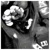
Normal ultrasound appearance of the appendix (arrows). A blind-ending tubular structure with five layered alternating bands of echogenicity that can be sonographically identified corresponding with the serosa, muscularis propria, submucosa, muscularis mucosa, and the epithelium (not shown). Normal diameter of the appendix measures less than 6 mm from serosa to serosa
Fig. 8
Fig. 8
Normal MRI appearance of the appendix, visualized as a T2 hypointense tubular structure, that measures less than 6 mm in diameter with wall thickness less than 2 mm, and surrounded by fat with normal homogeneous signal (arrow)
Fig. 9
Fig. 9
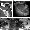
A 20-year-old woman with right lower quadrant pain. a Axial IV and PO contrast-enhanced CT demonstrates a dilated appendix (arrow) filled with fluid and coursing to the right adnexa, consistent with appendicitis. b Endovaginal transverse image of the right adnexa demonstrates a tubular, approximate 1-cm, blind-ending pouch with multilayered hypoechoic and hyperechoic bands and increased through-transmission due to fluid consistent with appendicitis (arrow). c Doppler ultrasound demonstrates hyperemia within the appendix wall (arrow)
Fig. 10
Fig. 10
Acute appendicitis on ultrasound. Sagittal (a) and transverse (b without compression and c with compression) transabdominal sonographic images demonstrating an approximate 1-cm noncompressible appendix (arrow). Note alternating bands of echogenicity. No periappendiceal abscess is present
Fig. 11
Fig. 11
Acute appendicitis in an Amyand hernia. a Axial IV and PO contrast CTscan demonstrates a dilated tubular mass in the right inguinal canal with adjacent fat stranding compatible with acute appendicitis (arrow). b Sagittal IV and PO contrast-enhanced CT demonstrates acute appendicitis (arrows) within an Amyand hernia with an associated large scrotal fluid collection (asterisk)
Fig. 12
Fig. 12
A 48-year-old man with right lower quadrant pain. a Coronal IV and PO contrast-enhanced CT demonstrates thickening of the appendix and periappendiceal inflammation (arrows). b Axial IV and PO contrast-enhanced CT demonstrates a discrete soft tissue nodule within the fluid-filled lumen (arrow), suspicious for neoplasm. Pathology reveals a carcinoid tumor
Fig. 13
Fig. 13
Primary appendiceal neoplasm of the carcinoid spectrum. a Axial IV and PO contrast-enhanced CT demonstrates a hypodense mass arising from the appendix and extending into the right adnexa (arrows). Sagittal (b) endovaginal ultrasound performed 8 months later for right adnexal pain demonstrates a 1.2-cm blind-ending heterogeneous tubular structure in the right adnexa (arrows). MRI (c) demonstrates a T1 hypointense, T2 hyperintense, heterogeneously enhancing mass extending to right adnexa (arrows). Pathology was compatible with mixed goblet cell carcinoid-carcinoma of the appendix
Fig. 14
Fig. 14
A 53-year-old female with right lower quadrant pain. a Right lower quadrant transabdominal ultrasound demonstrates an enlarged appendix (arrows). Axial (b) and coronal (c) IV contrast-enhanced CT scan demonstrates a tubular cystic mass in the right lower quadrant (arrow). Axial images from pelvic MRI demonstrate a T2 hyperintense (d), nonenhancing on T1 fat sat image (e) cystic mass in the right lower quadrant (arrow). Pathology was compatible with a mucinous cystadenoma
Fig. 15
Fig. 15
A 73-year-old female with right lower quadrant pain. Axial (a) and coronal (b) IV and PO contrast-enhanced CT demonstrated dilated fluid-filled appendix with extensive appendiceal inflammation (asterisk). There is an abrupt transition between the fluid in the lumen and soft tissue density at the base (arrow), suspicious for an obstructing mass. Pathology was compatible with colonic-type adenocarcinoma of the appendix
Fig. 16
Fig. 16
A 73-year-old male presenting for follow-up after periappendiceal abscess drainage. Coronal (a, b) and axial (c) IV and PO contrast-enhanced CT scan demonstrates circumferential thickening of the cecum (arrow), extending into the appendix (b). Diffuse periappendiceal inflammation is also present (asterisk). Findings are compatible with cecal adenocarcinoma invading the appendix with resultant appendiceal perforation

References

    1. Yeo . Shackelford’s surgery of the alimentary tract. 6. Saunders; Philadelphia: 2007.
    1. Schumpelick V, Dreuw B, Ophoff K, et al. Appendix and cecum: embryology, anatomy, and surgical applications. Surg Clinc North Am. 2000;80(1):295–318. - PubMed
    1. Long SS, Long C, Lai H, Macura KJ. Imaging strategies for right lower quadrant pain in pregnancy. AJR. 2011;196:4–12. - PubMed
    1. Spalluto LB, Woodfield CA, DeBenefectis CM, Lazarus E. MR imaging evaluation of abdominal pain during pregnancy: appendicitis and other nonobstetric causes. Radiographics. 2012;32(2):317–334. - PubMed
    1. Nikolaidis P, Hwang CM, Miller FH, Papanicolaou N. The nonvisualized appendix: incidence of acute appendicitis when secondary inflammatory changes are absent. Am J Roentgenol. 2004;183(4):889–892. - PubMed

LinkOut - more resources