. 2014 Jun;38(6):1173-81.
doi: 10.1007/s00264-014-2292-3. Epub 2014 Feb 26.

Accuracy of bony landmarks for restoring the natural joint line in revision knee surgery: an MRI study

Affiliations

Accuracy of bony landmarks for restoring the natural joint line in revision knee surgery: an MRI study

Günther Maderbacher et al. Int Orthop. 2014 Jun.

Abstract

Purpose: Restoring the joint line (JL) improves clinical and functional outcome in total knee arthroplasty (TKA). Therefore, anatomical landmarks to approximate the JL have been published. So far, the natural deviation of the JL 90° to the mechanical tibial axis has not been considered. Thus, we designed this study to: (1) determine the natural JL of knees in healthy persons in respect to the mechanical tibial axis, (2) validate and double-check intra-operative bony landmarks already been published in respect to the natural JL and (3) find possible correlations between distances from bony landmarks to the JL and femoral and tibial width.

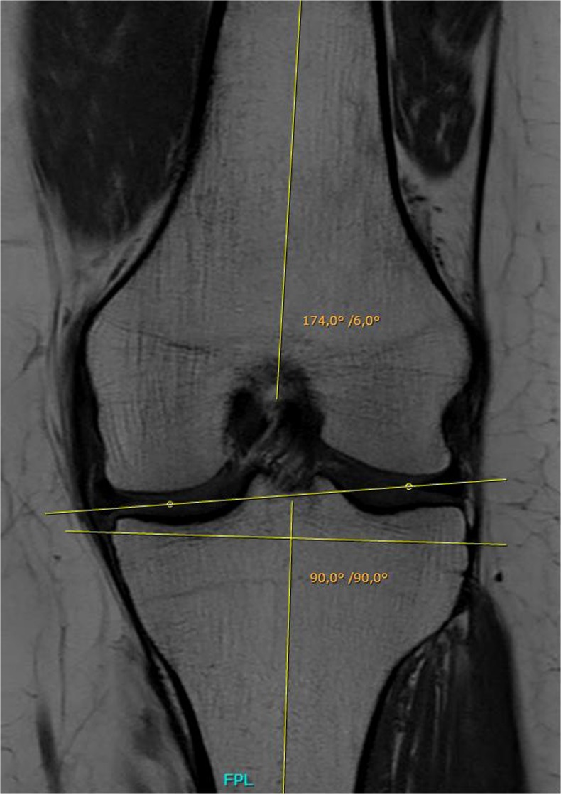
Methods: Eighty MRI scans of knees of healthy persons were examined by two independent observers. Distances from the tip of the fibular head (FH), the medial (ME) and lateral (LE) epicondyles and the adductor tubercle (AT) to the JL within the medial and lateral compartment were measured. Further, we determined the orientation of the JL in respect to the mechanical axis of the tibia. Interobserver correlations were calculated. Differences were analyzed using Student's t test. Linear regression models were calculated to analyze correlations.

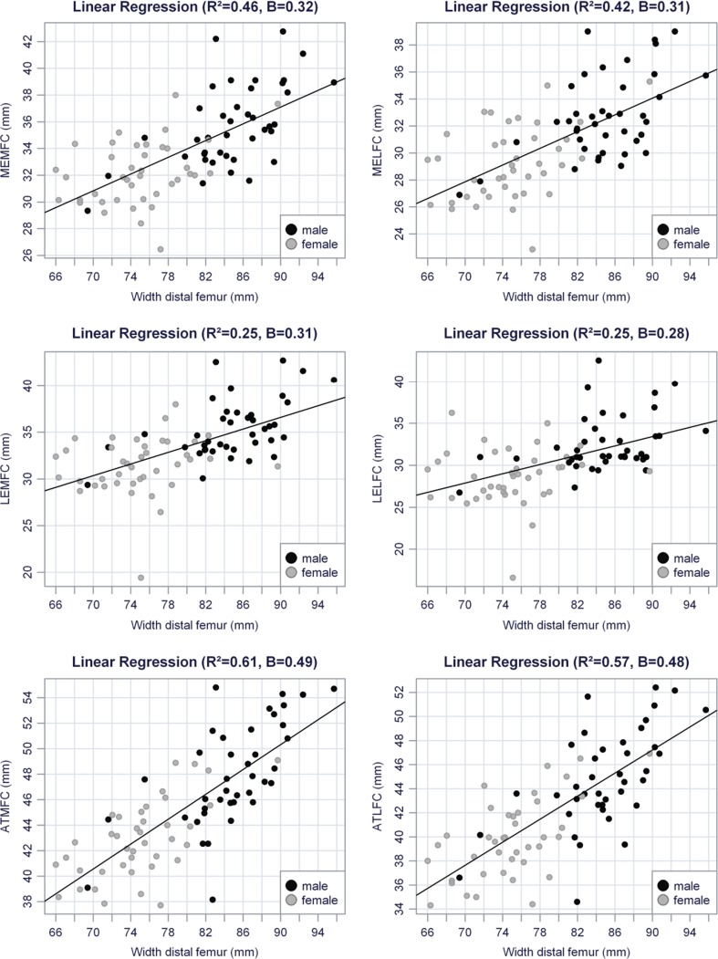
Results: Interobserver correlation was excellent. Mean JL deviation was 4.2° varus. Distance between the FH, ME, LE and AT to the JL within the medial compartment was 12.2, 33.9, 33.4 and 45.4 mm, respectively. Within in the lateral compartment, distances were 15.3, 31.0, 30.6 and 42.3 mm to the JL. Strong correlation was found between femoral width and distances from the AT, ME and LE to the JL.

Conclusion: In TKA, the JL is usually altered due to the classic resection technique, which does not respect the natural deviation of the JL. Estimating the natural JL by adding absolute values to bony landmarks, as proposed in the literature, is not recommended. According to our data, the JL can be best estimated by adding the calculated value: 6.40 + (width femur [mm] × 0.49) to the AT.


Figures

Fig. 1
Fig. 1
Femoral and tibial axes and Joint Line. The tibial mechanical axis is defined by connecting the midpoint of the tibial spines with a point determined as the centre of the tibial shaft. For the femoral mechanical axis a line from the midpoint of the distal femur to the midpoint of the centre of the shaft was drawn. The angle between the femoral and mechanical axes was measured. The articulation site between the femur and the tibia was determined by identifying the most distal point of the medial femoral condyle (MFC) and most distal point of the lateral femoral condyle (LFC) (see dots). The Joint Line was defined by connecting the MFC and LFC
Fig. 2
Fig. 2
Distance from the medial epicondyle (ME) to the Joint line. A line perpendicular to the mechanical axis of the tibia was parallelised and placed on the medial epicondyle. The distance between the parallelised line and the MFC (yellow dot) was measured and defined as the distance between the medial epicondyle to the JL within the medial compartment (MEMFC). In this case we measured 28.8 mm. The same procedure was repeated in order to obtain the distance from the medial epicondyle to the lateral compartment (MELFC). In this case we measured 24.3 mm. The angle between the mechanical femoral axis and the parallelised line was measured. In this case we found 6°
Fig. 3
Fig. 3
Regression of distances in millimetres to femoral bony landmarks (ME, LE, AT), to the joint line (JL) separately for the medial and lateral compartments (MFC, LFC) to the femoral width. Additionally, values were separated for male (black dots) and female (grey dots) patients. All regressions showed significance (p<0.001). ME medial epicondyle, LE lateral epicondyle, AT adductor tubercle
Fig. 4
Fig. 4
Distances (mm) between the fibular head (FH), medial epicondyle (ME), lateral epicondyle (LE) and adductor tubercle (AT), respectively, within the medial (FHMFC, MEMFC, LEMFC, ATMFC) and lateral (FHLFC, MELFC, LELFC, ATLFC) compartment. Red lines show the proposed values between the landmark and the joint line (JL) that could be found in the literature (FH to JL ∼10 mm [7], ME to JL=30.8 mm [9], LE to JL=25.3 [18] and AT to JL=48.7 [10]. FHMFC distance from the femoral head to the medial femoral compartment, MEMFC distance from the medial epicondyle to the medial femoral compartment, LEMFC distance from the lateral epicondyle to the medial femoral component, ATMFC distance from the adductor tubercle to the medial femoral component

References

    1. Singerman R, Davy DT, Goldberg VM. Effects of patella alta and patella infera on patellofemoral forces. J Biomech. 1994;27:1059. doi: 10.1016/0021-9290(94)90222-4. - DOI - PubMed
    1. Singerman R, Heiple KG, Davy DT, et al. Effect of tibial component position on patellar strain following total knee arthroplasty. J Arthroplasty. 1995;10:651. doi: 10.1016/S0883-5403(05)80210-4. - DOI - PubMed
    1. Martin JW, Whiteside LA. The influence of joint line position on knee stability after condylar knee arthroplasty. Clin Orthop. 1990;259:146. - PubMed
    1. Laskin RS. Joint line position restoration during revision total knee replacement. Clin Orthop Relat Res. 2002;404:169–171. doi: 10.1097/00003086-200211000-00029. - DOI - PubMed
    1. Figgie HE, III, Goldberg VM, Heiple KG, Moller HS, III, Gordon NH. The influence of tibial-patellofemoral location on function of the knee in patients with the posterior stabilized condylar knee prosthesis. J Bone Joint Surg Am. 1986;68:1035–1040. - PubMed