. 2014 Feb 25;5(2):e00856-14.
doi: 10.1128/mBio.00856-14.

Cell-type-specific effects of RNase L on viral induction of beta interferon

Cell-type-specific effects of RNase L on viral induction of beta interferon

Shuvojit Banerjee et al. mBio. .

Abstract

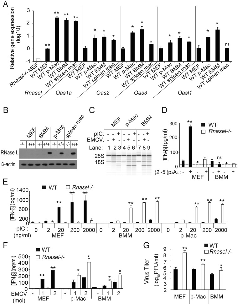
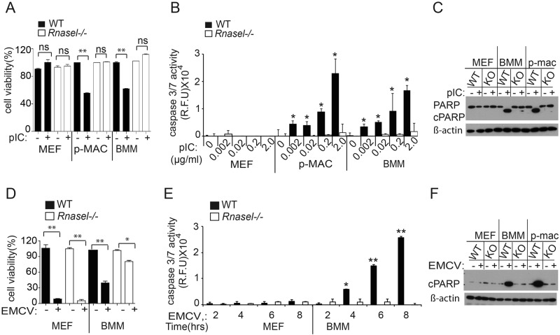
The interferon (IFN)-inducible antiviral state is mediated in part by the 2',5'-oligoadenylate (2-5A) synthetase (OAS)/RNase L system. 2-5A, produced from ATP by OAS proteins in response to viral double-stranded RNA, binds to and activates RNase L. RNase L restricts viral infections by degrading viral and cellular RNA, inducing autophagy and apoptosis, and producing RNA degradation products that amplify production of type I interferons (IFNs) through RIG-I-like receptors. However, the effects of the OAS/RNase L pathway on IFN induction in different cell types that vary in basal levels of these proteins have not been previously reported. Here we report higher basal expression of both RNase L and OAS in mouse macrophages in comparison to mouse embryonic fibroblasts (MEFs). In MEFs, RNase L gene knockout decreased induction of IFN-β by encephalomyocarditis virus infection or poly(rI):poly(rC) (pIC) transfection. In contrast, in macrophages, RNase L deletion increased (rather than decreased) induction of IFN-β by virus or pIC. RNA damage from RNase L in virus-infected macrophages is likely responsible for reducing IFN-β production. Similarly, direct activation of RNase L by transfection with 2-5A induced IFN-β in MEFs but not in macrophages. Also, viral infection or pIC transfection caused RNase L-dependent apoptosis of macrophages but not of MEFs. Our results suggest that cell-type-specific differences in basal levels of OAS and RNase L are determinants of IFN-β induction that could affect tissue protection and survival during viral infections. IMPORTANCE Type I interferons (IFNs) such as IFN-β are essential antiviral cytokines that are often required for animal survival following infections by highly pathogenic viruses. Therefore, host factors that regulate type I IFN production are critically important for animal and human health. Previously we reported that the OAS/RNase L pathway amplifies antiviral innate immunity by enhancing IFN-β production in mouse embryonic fibroblasts and in virus-infected mice. Here we report that high basal levels of OAS/RNase L in macrophages reduce, rather than increase, virus induction of IFN-β. RNA damage and apoptosis caused by RNase L were the likely reasons for the decreased IFN-β production in virus-infected macrophages. Our studies suggest that during viral infections, the OAS/RNase L pathway can either enhance or suppress IFN production, depending on the cell type. IFN regulation by RNase L is suggested to contribute to tissue protection and survival during viral infections.


Figures

FIG 1
FIG 1
Basal levels of OAS and RNase L regulate induction of IFN-β by virus or pIC. (A) Basal levels of Rnasel and Oas1a, Oas2, Oas3, and Oasl1 mRNAs were determined by qRT-PCR with total RNA isolated with mirVANA RNA kits (LifeTechnology) with Oas primers described previously (15) and with Rnasel forward primer: 5′ TAGGCGAACACATCAATGAGGA 3′ and reverse primer 5′ CTGCCTCTGGAACGCTGAG 3′. RNA expression relative to GAPDH (glyceraldehyde-3-phosphate dehydrogenase) mRNA was expressed as 2−ΔCT, where ΔCT represents the threshold cycle (CT) of the gene of interest − the CT of GAPDH. The open bar represents the baseline determined with RNA from Rnasel−/− MEFs. The data are shown as the means ± standard deviation (SD) calculated from a minimum of 3 (up to 6) biological replicates. MEFs immortalized with simian virus 40 (SV40) T antigen were cultured in RPMI 1640 with 10% fetal bovine serum (FBS) (5). Isolation and culture conditions for BMMs and thioglycolate-elicited p-Macs were as described previously (24, 25). Splenic macrophages were isolated by FACS with fluorescein isothiocyanate (FITC)-conjugated rat anti-mouse CD11b IgG2b (BD Pharmingen; catalog no. 557396, clone M1/70) and allophycocyanin-conjugated rat anti-mouse F4/80 IgG2b (Bio-Rad/Abd Serotec; product code MCA497APC) as described previously (25). Experiments involving mice were performed under an approved IACUC protocol from the Cleveland Clinic. (B) Western blots of the different cell-type extracts were performed with rabbit polyclonal antibody against murine RNase L (B. K. Jha, B. Dong, and R. H. Silverman, unpublished data) and antibody to β-actin (Sigma-Aldrich). (C) Cells were either infected with EMCV (a gift from Ian M. Kerr, London, United Kingdom) (MOI of 0.1) for 16 h or were Lipofectamine 2000 (Life Technologies) transfected with pIC (Sigma-Aldrich) at 2 µg/ml for 6 h. Total RNA was isolated with Trizol (Life Technologies), and rRNA degradation was monitored in RNA chips with an Agilent 2100 bioanalyzer. (D) Transfections of (2′-5′) p3A3 (10 µM) with Lipofectamine 2000 (Life Technologies) and mock transfections were done for 18 h. IFN-β levels in the cell supernatant were determined by ELISA (PBL Assay Science). The lower limit of detection of IFN-β in the ELISA was about 15 pg/ml (dashed line). (E and F) IFN-β concentrations were measured by ELISA from media of cells (E) transfected with pIC (at the indicated concentrations) for 8 h or mock transfected or (F) infected with EMCV (MOI of 1 or 2) for 16 h. Values are means from biological triplicate assays ± SD. (G) EMCV yields following infection (MOI of 0.1) at 8 h postinfection determined from the culture supernatant by plaque assay on Ifnar−/− MEF indicator cells as described previously (5). Results are shown as the means ± SD from three biological replicates. Two-tailed t tests were done. **, P < 0.001; *, P < 0.05.
FIG 2
FIG 2
Effects of pIC transfection or viral infection on cell viability and apoptosis. (A) Cells were transfected with pIC (2 µg/ml) for 12 h. Cell viability was quantified by MTS colorimetric assays (Promega). Results are means ± SD from three biological replicates. (B) Caspase 3/7 activation was determined with an Apo-ONE homogeneous caspase 3/7 kit (Promega) after pIC transfection (2 ng to 2 µg/ml for 8 h). Results are means ± SD from three biological replicates. (C) Western blots with antibody to uncleaved PARP and cleaved PARP (cPARP) (Cell Signaling; catalog no. 9542) or to β-actin (Sigma-Aldrich) from cells transfected with pIC (2 µg per ml) for 12 h. (D) Cells were infected with EMCV (MOI of 1.0) for 24 h, and cell viability was measured by MTS assays. (E) Activation kinetics for caspase 3/7 activity during EMCV infection (MOI of 1.0) at the indicated times postinfection. Results are means ± SD of three biological replicates. **, P < 0.0001; *, P < 0.05; ns, not significant. (F) PARP cleavage in response to EMCV infections at an MOI of 1.0 for 8 h as determined in Western blots.

References

    1. Silverman RH. 2007. Viral encounters with 2′,5′-oligoadenylate synthetase and RNase L during the interferon antiviral response. J. Virol. 81:12720–12729. 10.1128/JVI.01471-07 - DOI - PMC - PubMed
    1. Kristiansen H, Gad HH, Eskildsen-Larsen S, Despres P, Hartmann R. 2011. The oligoadenylate synthetase family: an ancient protein family with multiple antiviral activities. J. Interferon Cytokine Res. 31:41–47. 10.1089/jir.2010.0107 - DOI - PubMed
    1. Dong B, Silverman RH. 1995. 2-5A-dependent RNase molecules dimerize during activation by 2-5A. J. Biol. Chem. 270:4133–4137. 10.1074/jbc.270.8.4133 - DOI - PubMed
    1. Wreschner DH, McCauley JW, Skehel JJ, Kerr IM. 1981. Interferon action—sequence specificity of the ppp(A2′p)nA-dependent ribonuclease. Nature 289:414–417. 10.1038/289414a0 - DOI - PubMed
    1. Zhou A, Paranjape J, Brown TL, Nie H, Naik S, Dong B, Chang A, Trapp B, Fairchild R, Colmenares C, Silverman RH. 1997. Interferon action and apoptosis are defective in mice devoid of 2′,5′-oligoadenylate-dependent RNase L. EMBO J. 16:6355–6363. 10.1093/emboj/16.21.6355 - DOI - PMC - PubMed

Publication types

MeSH terms