

A Case of Esophageal Fibrovascular Polyp That Induced Asphyxia during Sleep
- PMID: 24570890
- PMCID: PMC3928480
- DOI: 10.5946/ce.2014.47.1.101
A Case of Esophageal Fibrovascular Polyp That Induced Asphyxia during Sleep
Abstract
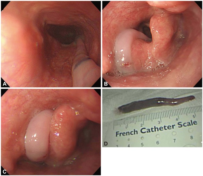
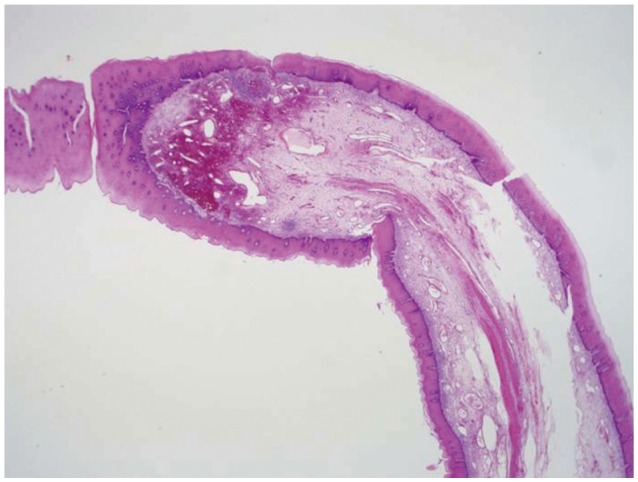
Esophageal fibrovascular polyps are rare, benign, submucosal tumors of the upper digestive tract that usually have an indolent course until the lesion attains a very large size. The most frequent complaints associated with these tumors include dysphagia and foreign body sensation. However, a long pedunculated polyp can regurgitate into the pharynx or oral cavity and cause asphyxia and sudden death if the larynx is occluded. We describe the case of a 51-year-old man who experienced snoring and occasional asphyxia during sleep. Upper endoscopy was performed, which indicated the presence of a pedunculated esophageal polyp that regurgitated into the vocal cords. The polyp was removed using a polypectomy snare and was confirmed to be a fibrovascular polyp based on pathologic examination findings. Three months after the excision of the polyp, the patient was found to be doing well without any further occurrence of asphyxia or sleep disturbances.
Keywords: Asphyxia; Esophagus; Polyps; Snoring.
Conflict of interest statement
The authors have no financial conflicts of interest.
Figures
References
-
- Sargent RL, Hood IC. Asphyxiation caused by giant fibrovascular polyp of the esophagus. Arch Pathol Lab Med. 2006;130:725–727. - PubMed
-
- LiVolsi VA, Perzin KH. Inflammatory pseudotumors (inflammatory fibrous polyps) of the esophagus. A clinicopathologic study. Am J Dig Dis. 1975;20:475–481. - PubMed
-
- Ginai AZ, Halfhide BC, Dees J, Zondervan PE, Klooswijk AI, Knegt PP. Giant esophageal polyp: a clinical and radiological entity with variable histology. Eur Radiol. 1998;8:264–269. - PubMed
-
- Fries MR, Galindo RL, Flint PW, Abraham SC. Giant fibrovascular polyp of the esophagus. A lesion causing upper airway obstruction and syncope. Arch Pathol Lab Med. 2003;127:485–487. - PubMed
-
- Avezzano EA, Fleischer DE, Merida MA, Anderson DL. Giant fibrovascular polyps of the esophagus. Am J Gastroenterol. 1990;85:299–302. - PubMed
LinkOut - more resources
Full Text Sources
Other Literature Sources
