

Diagnosis and treatment of hepatic angiomyolipoma
- PMID: 24570900
- PMCID: PMC3924623
- DOI: 10.3978/j.issn.2304-3881.2012.07.02
Diagnosis and treatment of hepatic angiomyolipoma
Abstract
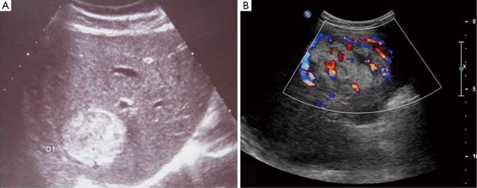
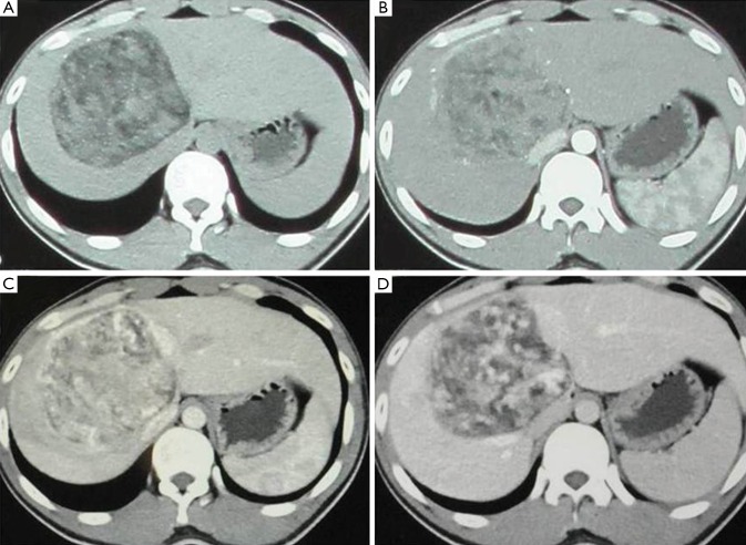
Background: Hepatic angiomyolipoma (HAML) is a rare liver tumor. This paper summarized the clinical, radiological and pathological features of HAML.
Methods: Seventeen cases of HAML were analyzed retrospectively. All patients were subjected to surgical resection of tumor, one of which was performed emergency surgery because of hemorrhage of tumor.
Results: There are 13 females and 4 males, most of whom were asymptotic except 4 had minimal abdominal discomfort. US, CT and/or MRI were taken and corresponding data was comprehensively analyzed with other clinical signs and symptoms. Correct preoperative diagnosis was able to be achieved in 9 patients. Pathological analysis and immunohistochemistry of HMB-45 was used as final diagnosis. All patients were followed up and survived without recurrence.
Conclusions: Preoperative diagnosis of HAML can be benefited from comprehensive analysis of clinical manifestations. The malignant potential and fast growth of tumor suggested surgical removal of tumor while it was diagnosed.
Keywords: Hepatic tumor; angiomyolipoma; diagnosis; surgery.
Figures

References
-
- Ishak KG. Mesenchymal tumors of the liver. In: Okuda K, Peters RL. Hepatocellular Carcinoma. New York: John Wiley &Sons,1976:247-307.
-
- Mizuguchi T, Katsuramaki T, Nobuoka T, et al. Growth of hepatic angiomyolipoma indicating malignant potential. J Gastroenterol Hepatol 2004;19:1328-30 - PubMed
-
- Flemming P, Lehmann U, Becker T, et al. Common and epithelioid variants of hepatic angiomyolipoma exhibit clonal growth and share a distinctive immunophenotype. Hepatology 2000;32:213-7 - PubMed
-
- Dalle I, Sciot R, de Vos R, et al. Malignant angiomyolipoma of the liver: a hitherto unreported variant. Histopathology 2000;36:443-50 - PubMed
LinkOut - more resources
Full Text Sources