. 2014 Jul;171(13):3182-95.
doi: 10.1111/bph.12659.

ABT-263 enhances sorafenib-induced apoptosis associated with Akt activity and the expression of Bax and p21((CIP1/WAF1)) in human cancer cells

Affiliations

ABT-263 enhances sorafenib-induced apoptosis associated with Akt activity and the expression of Bax and p21((CIP1/WAF1)) in human cancer cells

Jingru Li et al. Br J Pharmacol. 2014 Jul.

Abstract

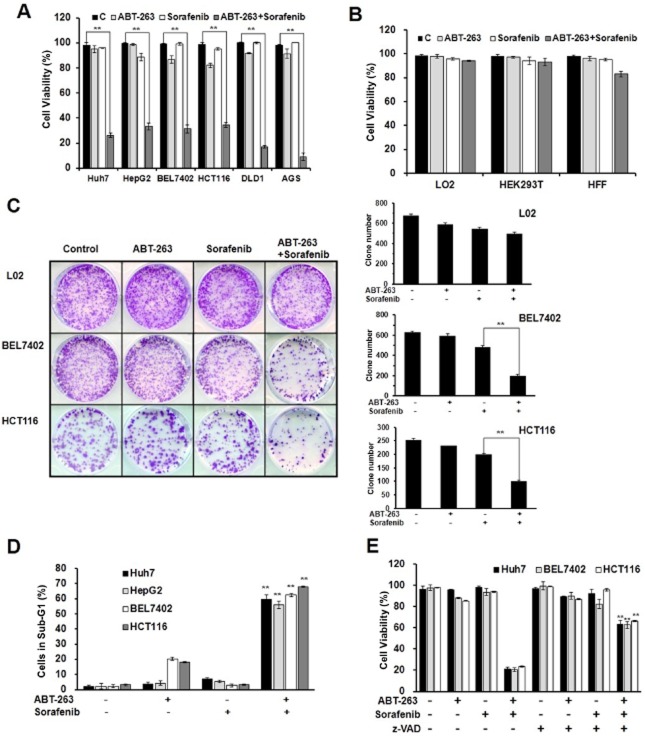
Background and purpose: Sorafenib, a potent inhibitor that targets several kinases associated with tumourigenesis and cell survival, has been approved for clinical treatment as a single agent. However, combining sorafenib with other agents improves its anti-tumour efficacy in various preclinical tumour models. ABT-263, a second-generation BH3 mimic, binds to the anti-apoptotic family members Bcl-2, Bcl-xL and Bcl-w, and has been demonstrated to enhance TNFSF10 (TRAIL)-induced apoptosis in human hepatocarcinoma cells. Hence, we investigated the effects of ABT-263 treatment combined with sorafenib.

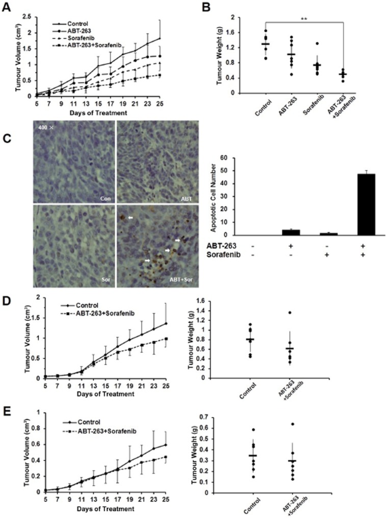
Experimental approach: The effects of ABT-263 combined with sorafenib were investigated in vitro, on cell viability, clone formation and apoptosis, and the mechanism examined using western blot and flow cytometry. This combination was also evaluated in vivo, in a mouse xenograft model; tumour growth, volume and weights were measured and a TUNEL assay performed.

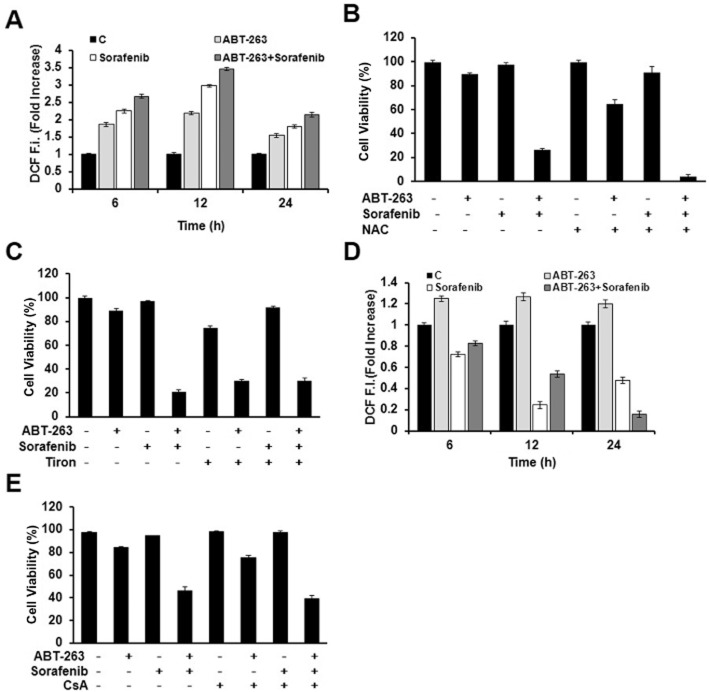
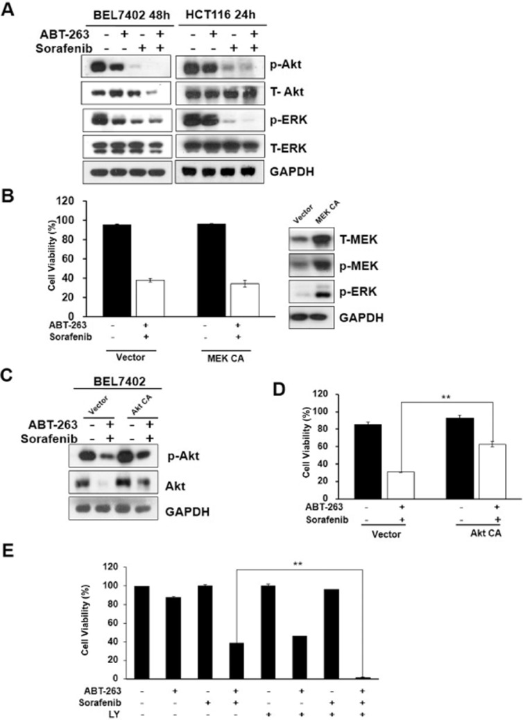
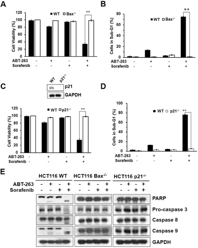
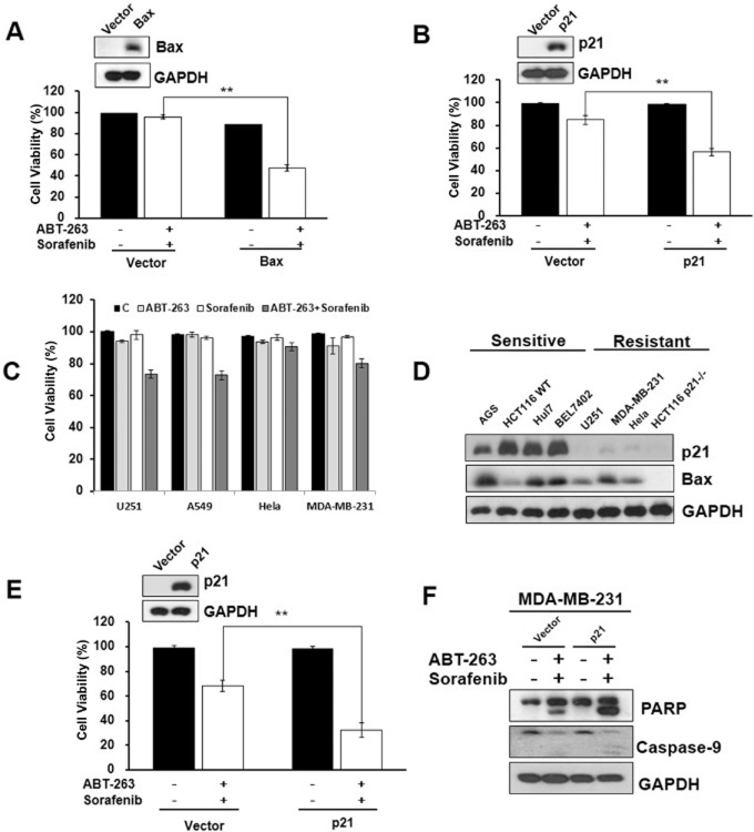
Key results: ABT-263 enhanced sorafenib-induced apoptosis while sparing non-tumourigenic cells. Although ABT-263 plus sorafenib significantly stimulated intracellular reactive oxygen species production and subsequent mitochondrial depolarization, this was not sufficient to trigger cell apoptosis. ABT-263 plus sorafenib significantly decreased Akt activity, which was, at least partly, involved in its effect on apoptosis. Bax and p21 (CIP1/WAF1) were shown to play a critical role in ABT-263 plus sorafenib-induced apoptosis. Combining sorafenib with ABT-263 dramatically increased its efficacy in vivo.

Conclusion and implications: The anti-tumour activity of ABT-263 plus sorafenib may involve the induction of intrinsic cell apoptosis via inhibition of Akt, and reduced Bax and p21 expression. Our findings offer a novel effective therapeutic strategy for tumour treatment.

Keywords: ABT-263; cancer; combination therapy; sorafenib.


Figures

Figure 1
Figure 1
The combination treatment of ABT-263 and sorafenib induces caspase-dependent apoptosis. The data represent the average of at least three independent experiments ± SD. **P < 0.01. (A) Huh7, HepG2, BEL7402, DLD1 and AGS cell lines were treated with 0.4 μM ABT-263 and 6 μM sorafenib either alone or in combination for 48 h, and the HCT116 cells were treated for 24 h, which were then assessed for cell viability using a trypan blue exclusion assay. (B) HEK293T, HFF and L02 cells were treated with 0.4 μM ABT-263 and 6 μM sorafenib either alone or in combination for 48 h. Cell viability was determined. (C) The cells were seeded in six-well plates and treated with ABT-263 and sorafenib, the HCT116 cells were treated for 24 h, and the BEL7402 and L02 cells were treated for 48 h. The attached cells were stained with crystal violet 10 days later. (D) FACS analysis of apoptosis following the treatment of ABT-263 (0.4 μM) and sorafenib (6 μM) either alone or in combination on the BEL7402, Huh7 and HepG2 cells for 48 h and the HCT116 cells for 24 h. The cells were dyed with 3 μL 10 mg·mL−1 PI and 10 μL 1 mg·mL−1 RNase, and the percentage of cells in sub-G1 determined. (E) The Huh7, BEL7402 and HCT116 cells were pretreated either with or without 50 μM z-VAD-fmk for 6 h, and then the trypan blue exclusion assay was performed to evaluate the cell viability after treatment with ABT-263 (0.4 μM) and sorafenib (6 μM) either alone or in combination. The percentage of cells dyed without trypan blue was determined.
Figure 2
Figure 2
Reactive oxygen species generation (ROS) and mitochondrial depolarization are involved but not required in ABT-263 and sorafenib combination-induced apoptosis. (A) BEL7402 cells were treated with ABT-263 and sorafenib either alone or in combination for 48 h and the intracellular ROS levels assessed. (B) BEL7402 cell viability was determined using the trypan blue exclusion assay in either the presence or absence of 15 mM NAC for 48 h. (C) BEL7402 cell viability was measured by trypan blue exclusion assay in either the presence or absence of 15 mM Tiron for 48 h. (D) To evaluate the time course of the mitochondrial membrane potential, BEL7402 cells were treated with ABT-263 and sorafenib either alone or in combination for 48 h followed by DCF fluorescence analysis. (E) BEL7402 cell viability was determined using the trypan blue exclusion assay in either the presence or absence of 2 μM CsA for 48 h.
Figure 3
Figure 3
ABT-263 and sorafenib combination-induced cell apoptosis correlates with Akt activity. (A) BEL7402 and HCT116 cells were treated with ABT-263 and sorafenib either alone or in combination, and then analysed via Western blot to examine the protein activation levels of p-Akt, T-Akt, p-ERK and T-ERK. (B) HCT116 cells transfected with either vehicle or constitutively active MEK (MEK CA) were incubated with a combination of ABT-263 (0.2 μM) and sorafenib (6 μM), and the cells were then collected and analysed by use of the trypan blue exclusion assay. (C) BEL7402 cells transfected with either constitutively active Akt (CA-Akt) or vehicle were incubated with the combination of 0.4 μM ABT-263 and 6 μM sorafenib for 48 h, and protein lysates were then prepared from the cells and analysed by Western blot. (D) Cell viability was determined following the treatment of ABT-263 and sorafenib in the cells transfected expressing either CA-Akt or the vehicle plasmid. (E) After pretreatment with 25 μM LY294002 for 1 h, the BEL7402 cells were treated with ABT-263 and sorafenib for 24 h, and cell viability was then determined. **P < 0.01.
Figure 4
Figure 4
Effect of Bax or p21 knockout on ABT-263 and sorafenib combination-induced apoptosis. (A) Cell viability was assessed in the HCT116 Bax-/- and HCT116 WT cells after the combination treatment of 0.2 μM ABT-263 and 6 μM sorafenib for 24 h. (B) Cell apoptosis was determined via FACS analysis by PI staining, and counting the percentage of cells in the sub-G1 phase. (C) Cell viability was determined with HCT116 p21-/- and HCT116 WT cells after the combination treatment of 0.2 μM ABT-263 and 6 μM sorafenib for 24 h. (D) Cell apoptosis was assessed via FACS. (E) Analysis of PARP, pro-caspase-3, caspase-8, caspsse-9 via Western blot. **P < 0.01.
Figure 5
Figure 5
Bax and p21 expression play a critical role in ABT-263 and sorafenib combination treatment-induced cell apoptosis. (A) HCT116 Bax-/- cells transfected with pUSE-GFP/pUSE-GFP-Bax for 48 h were incubated with ABT-263 and sorafenib for 24 h. The protein lysates prepared from the cells were then analysed by Western blot, and cell viability was determined. (B) HCT116 p21-/- cells transfected with pHAGE/pHAGE-p21 using the lentivirus-based stable transfection system were incubated with ABT-263 and sorafenib for 24 h. The protein lysates were analysed by Western blot, and cell viability was determined. (C) The U251, A549, Hela and MDA-MB-231 cell lines were treated with 0.4 μM ABT-263 and 6 μM sorafenib either alone or in combination for 48 h, followed by assessment for cell viability using the trypan blue exclusion assay. (D) The expression levels of p21 and Bax in ABT-263 and sorafenib combination treated-sensitive cells (AGS, HCT116WT, Huh7 and BEL7402) compared with the resistant cells (MDA-MB-231, Hela and HCT116 p21-/-) were determined by Western blot. (E) MDA-MB-231 cells transfected with pHAGE/pHAGE-p21 using the lentivirus-based stable transfection system were incubated with 0.4 μM ABT-263 and 6 μM sorafenib for 48 h, and cell viability was then determined. (F) MDA-MB-231 cell protein lysates were analysed by Western blot using antibodies targeting PARP, caspase-9 and GAPDH. **P < 0.01.
Figure 6
Figure 6
The combined treatment of ABT-263 and sorafenib demonstrated synergistic anti-tumour activity in an in vivo xenograft model. (A) HCT116 cells were inoculated into BALB/c mice (via s.c. injection) to establish a tumour model as indicated in the Materials and Methods section. Mice bearing tumours were randomly assigned into groups (six mice per group) and treated with vehicle, ABT-263 (100 mg·kg−1), sorafenib (25 mg·kg−1) either alone or as a combination of ABT-263 and sorafenib (ABT-263 100 mg·kg−1 and sorafenib 25 mg·kg−1) every other day. The tumour volume was then measured (the bars represented the means ± SD). (B) The tumours were dissected out and weighed after 20 days of treatment. (C) HCT116 transplanted tumours were dissected and analysed via TUNEL assay to examine the levels of apoptosis. The number of TUNEL-positive cells in five random (400×) fields was counted. (D) BALB/c mice (n = 6) were transplanted with HCT116 p21-/-cells and treated as above and then monitored for tumour development. (E) BALB/c mice (n = 7) were transplanted with HCT116 Bax-/-cells and treated as above and then monitored for tumour development. **P < 0.01.

Similar articles

Cited by

References

    1. Adams JM, Cory S. The Bcl-2 apoptotic switch in cancer development and therapy. Oncogene. 2007;26:1324–1337. - PMC - PubMed
    1. Alexander SPH, Benson HE, Faccenda E, Pawson AJ, Sharman JL, Spedding M, Peters JA, Harmar AJ, CGTP Collaborators The Concise Guide to PHARMACOLOGY 2013/14: Catalytic receptors. Br J Pharmacol. 2013a;170:1676–1705. - PMC - PubMed
    1. Bai D, Ueno L, Vogt PK. Akt-mediated regulation of NFkappaB and the essentialness of NFkappaB for the oncogenicity of PI3K and Akt. Int J Cancer. 2009;125:2863–2870. - PMC - PubMed
    1. Bayir H, Kagan VE. Bench-to-bedside review: mitochondrial injury, oxidative stress and apoptosis – there is nothing more practical than a good theory. Crit Care. 2008;12:206. - PMC - PubMed
    1. Bhattacharya S, Guo H, Ray RM, Johnson LR. Basic helix-loop-helix protein E47-mediated p21Waf1/Cip1 gene expression regulates apoptosis of intestinal epithelial cells. Biochem J. 2007;407:243–254. - PMC - PubMed

Publication types

MeSH terms