

3-D ultrastructure and collagen composition of healthy and overloaded human tendon: evidence of tenocyte and matrix buckling
- PMID: 24571576
- PMCID: PMC3981497
- DOI: 10.1111/joa.12164
3-D ultrastructure and collagen composition of healthy and overloaded human tendon: evidence of tenocyte and matrix buckling
Abstract
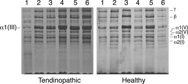
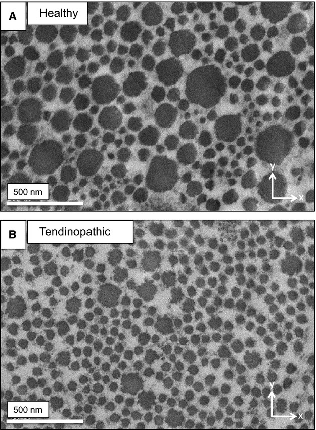
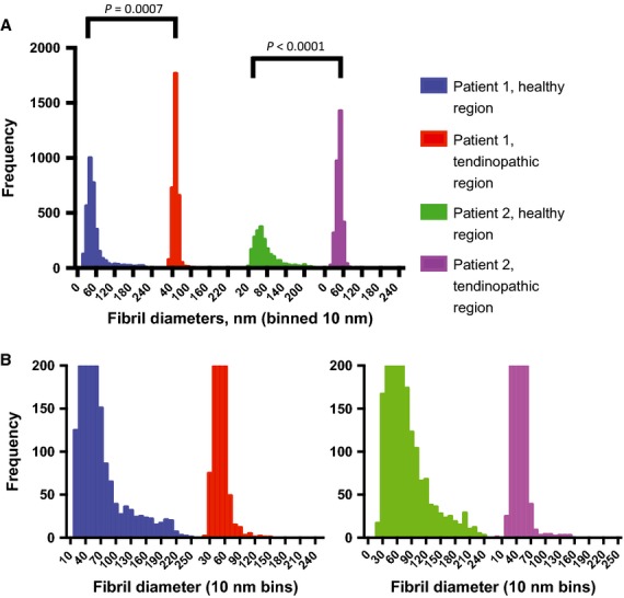
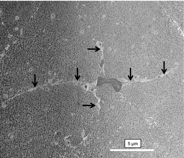
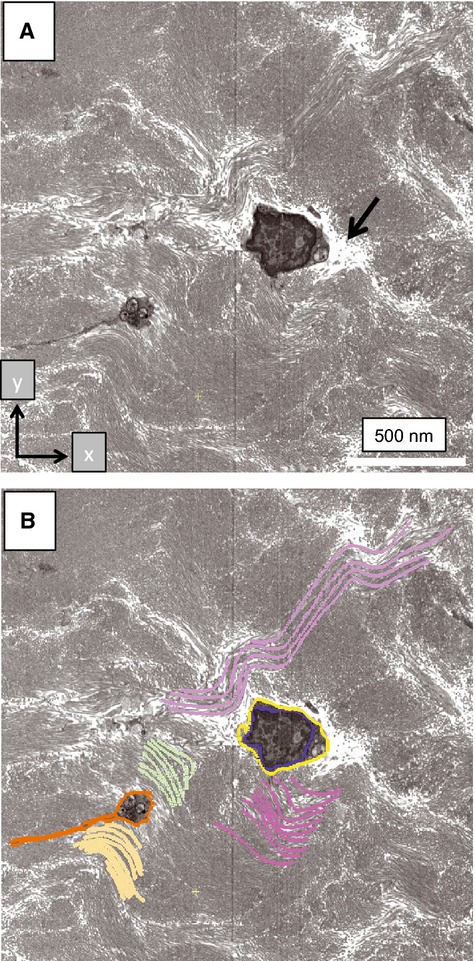
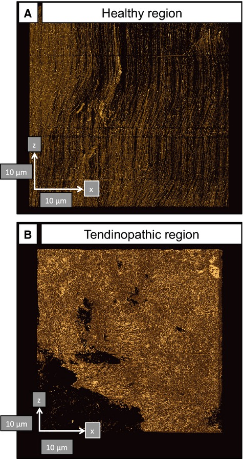
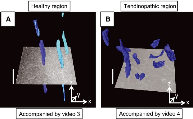
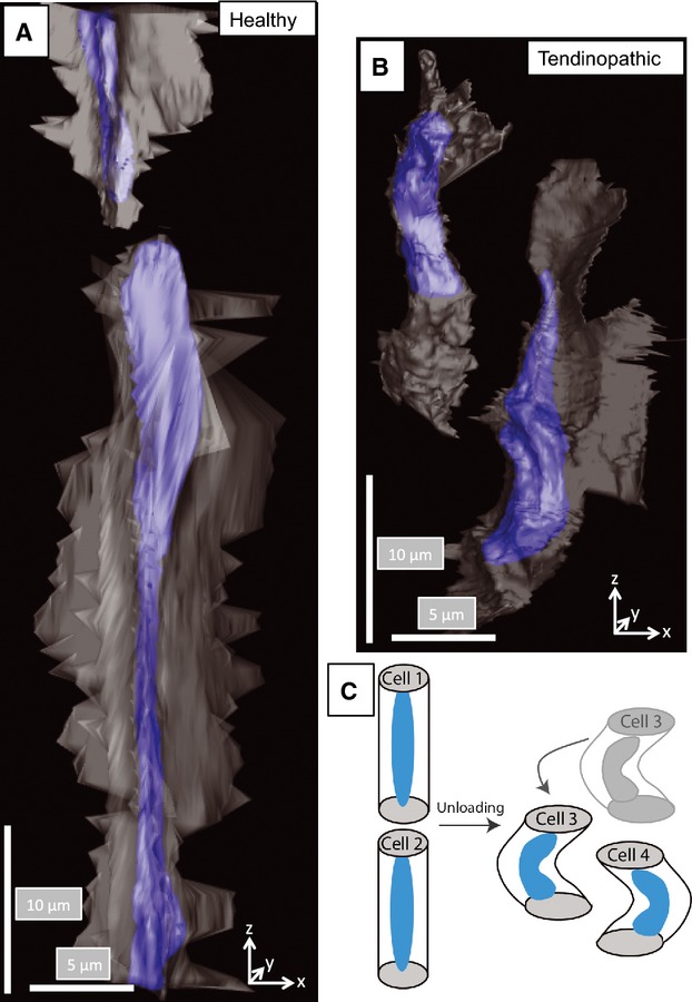
Achilles tendinopathies display focal tissue thickening with pain and ultrasonography changes. Whilst complete rupture might be expected to induce changes in tissue organization and protein composition, little is known about the consequences of non-rupture-associated tendinopathies, especially with regards to changes in the content of collagen type I and III (the major collagens in tendon), and changes in tendon fibroblast (tenocyte) shape and organization of the extracellular matrix (ECM). To gain new insights, we took biopsies from the tendinopathic region and flanking healthy region of Achilles tendons of six individuals with clinically diagnosed tendinopathy who had no evidence of cholesterol, uric acid and amyloid accumulation. Biochemical analyses of collagen III/I ratio were performed on all six individuals, and electron microscope analysis using transmission electron microscopy and serial block face-scanning electron microscopy were made on two individuals. In the tendinopathic regions, compared with the flanking healthy tissue, we observed: (i) an increase in the ratio of collagen III : I proteins; (ii) buckling of the collagen fascicles in the ECM; (iii) buckling of tenocytes and their nuclei; and (iv) an increase in the ratio of small-diameter : large-diameter collagen fibrils. In summary, load-induced non-rupture tendinopathy in humans is associated with localized biochemical changes, a shift from large- to small-diameter fibrils, buckling of the tendon ECM, and buckling of the cells and their nuclei.
Keywords: 3View®; collagen; cross-links; fibers; fibrils; serial block face-scanning electron microscopy.
© 2014 The Authors. Journal of Anatomy published by John Wiley & Sons Ltd on behalf of Anatomical Society.
Figures
References
-
- Craig AS, Parry DA. Growth and development of collagen fibrils in immature tissues from rat and sheep. Proc R Soc Lond B Biol Sci. 1981;212:85–92. - PubMed
-
- Fleischmajer R, MacDonald ED, Perlish JS, et al. Dermal collagen fibrils are hybrids of type I and type III collagen molecules. J Struct Biol. 1990a;105:162–169. - PubMed
MeSH terms
Substances
Grants and funding
LinkOut - more resources
Full Text Sources
Other Literature Sources
Medical
