. 2014 Aug;16(8):2568-90.
doi: 10.1111/1462-2920.12436. Epub 2014 Apr 2.

In situ visualization of newly synthesized proteins in environmental microbes using amino acid tagging and click chemistry

Affiliations

In situ visualization of newly synthesized proteins in environmental microbes using amino acid tagging and click chemistry

Roland Hatzenpichler et al. Environ Microbiol. 2014 Aug.

Abstract

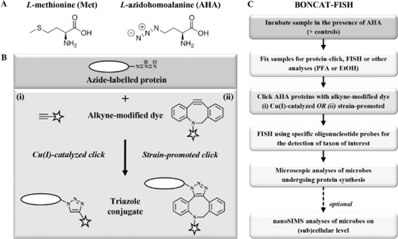
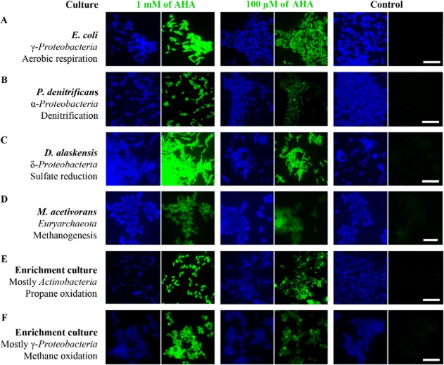
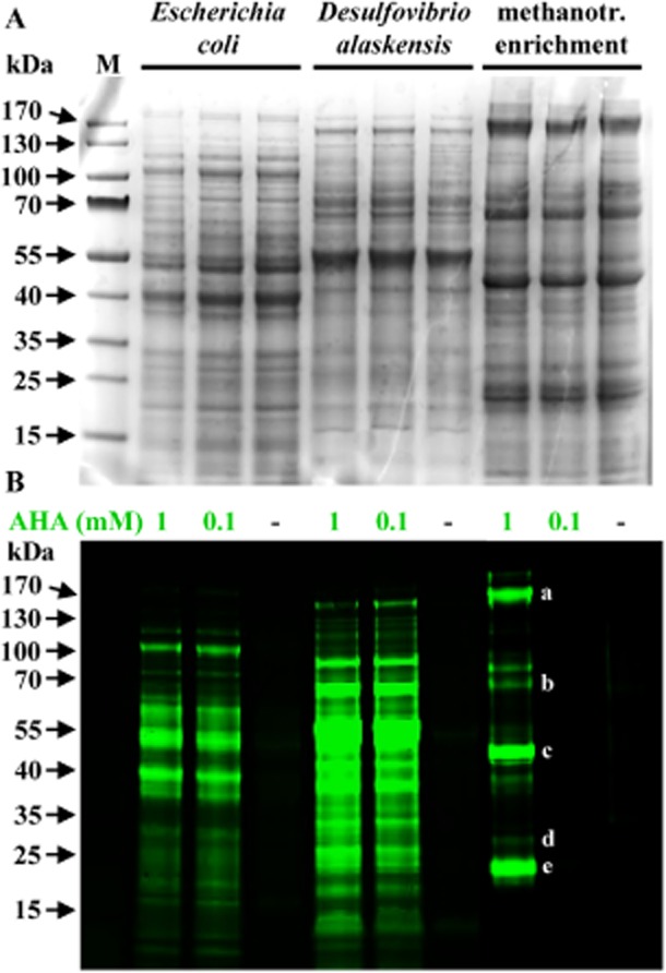
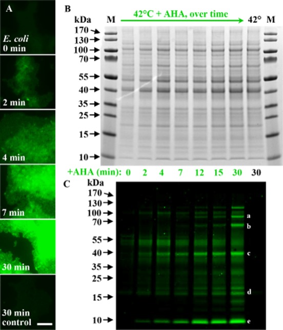
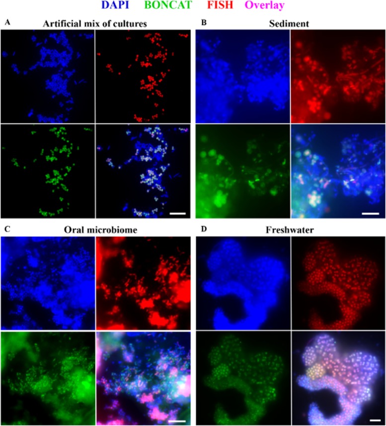
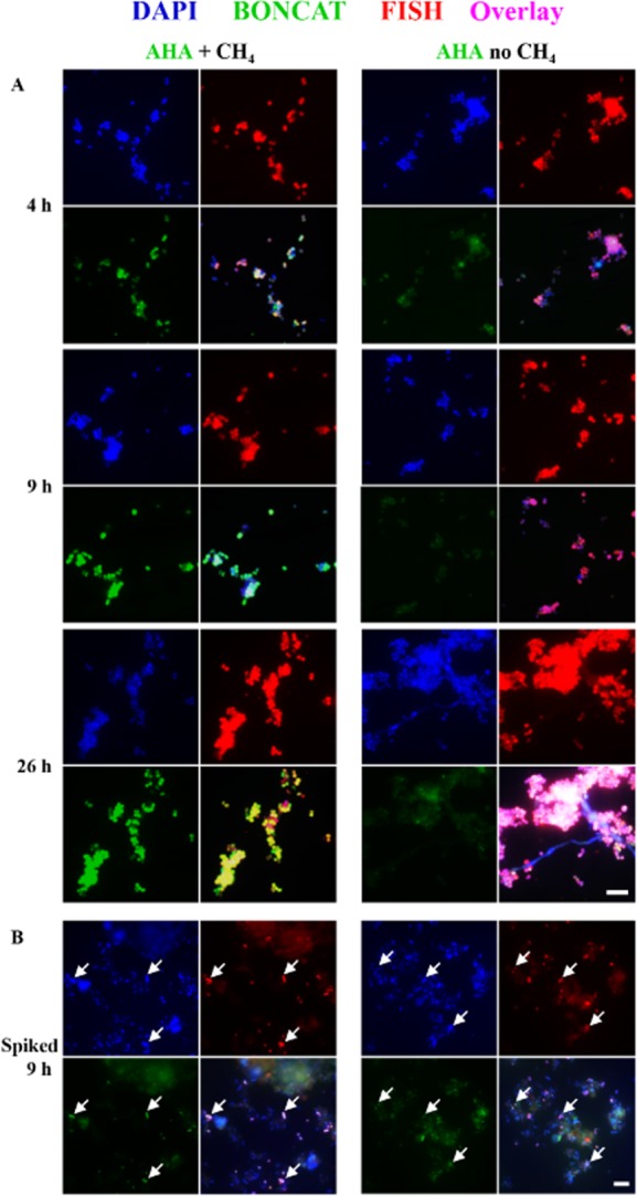
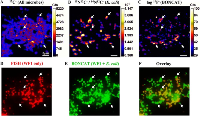
Here we describe the application of a new click chemistry method for fluorescent tracking of protein synthesis in individual microorganisms within environmental samples. This technique, termed bioorthogonal non-canonical amino acid tagging (BONCAT), is based on the in vivo incorporation of the non-canonical amino acid L-azidohomoalanine (AHA), a surrogate for l-methionine, followed by fluorescent labelling of AHA-containing cellular proteins by azide-alkyne click chemistry. BONCAT was evaluated with a range of phylogenetically and physiologically diverse archaeal and bacterial pure cultures and enrichments, and used to visualize translationally active cells within complex environmental samples including an oral biofilm, freshwater and anoxic sediment. We also developed combined assays that couple BONCAT with ribosomal RNA (rRNA)-targeted fluorescence in situ hybridization (FISH), enabling a direct link between taxonomic identity and translational activity. Using a methanotrophic enrichment culture incubated under different conditions, we demonstrate the potential of BONCAT-FISH to study microbial physiology in situ. A direct comparison of anabolic activity using BONCAT and stable isotope labelling by nano-scale secondary ion mass spectrometry ((15)NH(3) assimilation) for individual cells within a sediment-sourced enrichment culture showed concordance between AHA-positive cells and (15)N enrichment. BONCAT-FISH offers a fast, inexpensive and straightforward fluorescence microscopy method for studying the in situ activity of environmental microbes on a single-cell level.


Figures

Fig 1
Fig 1
Overview of the BONCAT method for visualizing newly synthesized proteins. A. Structures of Met and its surrogate AHA, which is incorporated into newly made peptides during translation. B. The click chemistry-mediated visualization of newly produced AHA-containing proteins can be achieved via either one of two strategies: (i) in a Cu(I)-catalyzed reaction a terminal alkyne that is coupled to a fluorescence dye (star) is linked to the azide group of AHA yielding a triazole (left side); (ii) conjugation can also be achieved via strain-promoted cycloaddition, a Cu-free variation of click chemistry (right side). C. Protocol for the combinatorial labelling of AHA-containing proteins via BONCAT and rRNA-targeted FISH.
Fig 2
Fig 2
Uptake and incorporation of AHA is independent of the physiological or phylogenetic background of the target organism. Different pure and enrichment cultures were incubated in the presence or absence of AHA. BONCAT signals (green) were taken at identical exposure times for individual series (i.e. 0.1 and 1 mM AHA plus control). Note that incubation conditions were different for the individual cultures, cells have contrasting levels of background fluorescence, and that different labelling strategies were used. Together, these issues limit the value of a direct comparison of signal intensities between different cultures. DAPI-staining is shown in blue. All scale bars equal 10 μm and apply to each set of images respectively. A–D. BONCAT-labelling of four bacterial and archaeal pure cultures. E–F. BONCAT-labelling of propane- and methane-oxidizing enrichment cultures.
Fig 3
Fig 3
AHA does not interfere with the cellular machinery. Visualization of new proteins from cultures of E. coli, Desulfovibrio alaskensis and a methanotrophic enrichment. A. Coomassie-stained protein band patterns of cultures that had been incubated in the absence (−) or presence of AHA are indistinguishable from each other, demonstrating that AHA does not interfere with the translational machinery. B. Newly made proteins in the same gel are identified via BONCAT. Please note that the incubation time for the methanotrophic culture exposed to 100 μM AHA was too short to yield new proteins in amounts high enough to be detectable via in-gel fluorescence. At the individual cell level, AHA uptake can, however, be easily demonstrated (Fig. 2F). Some of the most intensely labelled bands were cut from the gel and analyzed via mass spectrometry. The 20 most abundant proteins from the excised bands included: (a) the two large subunits of RNA-polymerase (150.4 and 155.4 kDa); (b) a hypothetical protein (67.8 kDa) as well as two homologs of the large subunits of methanol dehydrogenase (66.6 and 68.3 kDa); (c) PmoB, 45.6 kDa; (d) PmoA (28.4 kDa) and PmoC (29.1 kDa); (e) a formaldehyde activating enzyme (17.8 kDa), superoxide dismutase (21.1 kDa), and D-arabino-3-hexulose 6-phosphate formaldehyde lyase (21.8 kDa). Letters a–e denote the bands consistent with the molecular weights of these proteins. kDa, kiloDalton; M, marker.
Fig 4
Fig 4
The high sensitivity of BONCAT allows detection of newly synthesized proteins after only minutes of incubation. A. Fluorescence labelling of heat-shocked (42°C) E. coli cells grown in the presence of AHA increases over time. Already after 2 min of incubation, equivalent to about 2% of E. coli's generation time under the conditions used, labelled cells can be visualized. B, C. While no differences in the relative amounts of proteins can be observed via Coomassie-staining, the fluorescent tag conjugated to newly made proteins reveals that certain proteins are preferentially amplified with time. C. Some of the most intensely fluorescently labelled bands were cut from the gel and analyzed via mass spectrometry. The obtained proteins included: (a) DNA gyrase subunits A and B as well as chaperone protein ClpB (97.0, 89.9 and 95.6 kDa respectively); (b) chaperonin GroL, a heat-inducible Lysine-tRNA ligase and chaperone DnaK (57.3, 57.8 and 69.1 kDa respectively); (c) outer membrane protein A (37.2 kDa); (d) chaperone proteins Skp (17.7 kDa) and YajL (20.8 kDa); (e) nine ribosomal proteins of low molecular mass (≤ 10 kDa) and the highest abundant protein in our dataset, major outer membrane lipoprotein Lpp (8.3 kDa). Letters a–e indicate bands consistent with the molecular weights of these proteins. Control refers to 30 min incubation at 42°C in the absence of AHA. Fluorescent signals were recorded at identical exposure times. Scale bar equals 10 μm and applies to all images. kDa, kiloDalton; M, marker.
Fig 5
Fig 5
Visualization of newly synthesized proteins via BONCAT (green) in combination with rRNA-targeted FISH (red). A. An artificial mix of pure and enrichment cultures. The only microbe that had been incubated in the presence of AHA, a gamma proteobacterial methanotroph (Methylococcaceae sp. WF1), is identified via a species-specific FISH probe (MetI-444), demonstrating the feasibility of correlating translational activity with microbial identity. For details and a Cy5-probe image see Supporting Information Fig. S4. B–D. Many bacteria, identified by the general EUB338I-III FISH probe mix (B,C) or probe Gam42a (D), specific for gammaproteobacteria, are BONCAT-labelled, demonstrating in situ translational activity during time of incubation. Exposure times for click or FISH signals were identical for each sample series (i.e. AHA plus two controls), respectively. DAPI staining is shown in blue. For controls, see Supporting Information Figs. S4 and S6. All scale bars equal 10 μm.
Fig 6
Fig 6
Comparative BONCAT analyses of a methanotrophic enrichment culture in the absence and presence of methane. A. Click chemistry-mediated detection of AHA incorporation (green) reveals that a gammaproteobacterium (identified by FISH probe MetI-444; red) is highly active in the presence but not in the absence of methane. B. To test whether AHA-labelled WF1 cells (examples are pointed out by arrows) would be detectable in a complex samples, an aliquot of the culture was spiked into methane seep sediment and analyzed via BONCAT. Exposure settings for recording of BONCAT signals were identical for each image set (i.e. A, B). For images taking at different settings and controls, see Supporting Information Fig. S5. Scale bars equal 10 μm and apply to all images of the respective set.
Fig 7
Fig 7
Comparison of AHA labelling of newly synthesized proteins with cellular 15N-uptake. An artificial mix of several cultures was analyzed via BONCAT, FISH and nanoSIMS. Escherichia coli had been incubated in the presence of both 15NH4Cl and AHA, while a methanotroph was exposed to AHA only. Other microbes had been grown in the absence of AHA or 15NH4Cl. A species-specific FISH probe (MetI-444) is used to localize the methanotroph WF1 (D; red), while both WF1 as well as E. coli are fluorescently labelled by a fluorine-containing alkyne-dye (E; green). While all E. coli cells (examples are pointed out by arrows) are 15N- and 19F-labelled (B, C), the 19F-signal of methanotroph cells is indistinguishable from cells that had not been incubated in the presence of AHA (C). A second halogen-containing dye (see Supporting Information Fig. S1) could not be used due to problems removing unbound dye (see main text). Scale bars equal 5 μm. Abbreviations: Cts, counts; WF1, cells of Methylococcaceae sp. WF1. A–C. Elemental and isotopic mapping of an artificial mix of cultures via nanoSIMS. D–F. Correspondent FISH and BONCAT images of the same field of view.

References

    1. Adachi T, Yamada Y. Inoue I. Alternative method for selective reduction of unsaturated nucleoside azides to amines. Synthesis-Stuttgart. 1977;1977:45–46.
    1. Agard NJ, Prescher JA. Bertozzi CR. A strain-promoted [3 + 2] azide-alkyne cycloaddition for covalent modification of biomolecules in living systems. J Am Chem Soc. 2004;126:15046–15047. - PubMed
    1. Amann RI, Binder BJ, Olson RJ, Chisholm SW, Devereux R. Stahl DA. Combination of 16S rRNA-targeted oligonucleotide probes with flow cytometry for analyzing mixed microbial populations. Appl Environ Microbiol. 1990;56:1919–1925. - PMC - PubMed
    1. Amann RI, Zarda B, Stahl DA. Schleifer KH. Identification of individual prokaryotic cells by using enzyme-labeled, rRNA-targeted oligonucleotide probes. Appl Environ Microbiol. 1992;58:3007–3011. - PMC - PubMed
    1. Bagert JD, Xie YJ, Sweredoski MJ, Qi Y, Hess S, Schuman EM. Tirrell DA. Quantitative, time-resolved proteomic analysis by combining bioorthogonal noncanonical amino acid tagging and pulsed stable isotope labeling by amino acids in cell culture. Mol Cell Proteomics. 2014 doi: 10.1074/mcp.M113.031914 mcp.M113.031914. - DOI - PMC - PubMed

Publication types

MeSH terms