Correlation of EGFR, pEGFR and p16INK4 expressions and high risk HPV infection in HIV/AIDS-related squamous cell carcinoma of conjunctiva
- PMID: 24572046
- PMCID: PMC3996052
- DOI: 10.1186/1750-9378-9-7
Correlation of EGFR, pEGFR and p16INK4 expressions and high risk HPV infection in HIV/AIDS-related squamous cell carcinoma of conjunctiva
Abstract
Background: Squamous cell carcinoma of conjunctiva has increased tenfold in the era of HIV/AIDS. The disease pattern has also changed in Africa, affecting young persons, with peak age-specific incidence of 30-39 years, similar to that of Kaposi sarcoma, a well known HIV/AIDS defining neoplasm. In addition, the disease has assumed more aggressive clinical course. The contributing role of exposure to high risk HPV in the development of SCCC is still emerging.
Objective: The present study aimed to investigate if immunohistochemical expressions of EGFR, pEGFR and p16, could predict infection with high risk HPV in HIV-related SCCC.
Methods: FFPE tissue blocks of fifty-eight cases diagnosed on hematoxylin and eosin with SCCC between 2005-2011, and subsequently confirmed from medical records to be HIV positive at the department of human pathology, UoN/KNH, were used for the study. Immunohistochemistry was performed to assess the expressions of p16INK4A, EGFR and pEGFR. This was followed with semi-nested PCR based detection and sequencing of HPV genotypes. The sequences were compared with the GenBank database, and data analyzed for significant statistical correlations using SPSS 16.0. Ethical approval to conduct the study was obtained from KNH-ERC.
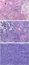
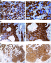
Results: Out of the fifty-eight cases of SCCC analyzed, twenty-nine (50%) had well differentiated (grade 1), twenty one (36.2%) moderately differentiated (grade 2) while eight (13.8%) had poorly differentiated (grade 3) tumours. Immunohistochemistry assay was done in all the fifty eight studied cases, of which thirty nine cases (67.2%) were positive for p16INK4A staining, forty eight cases (82.8%) for EGFR and fifty one cases (87.9%) showed positivity for p-EGFR. HPV DNA was detected in 4 out of 40 SCCC cases (10%) in which PCR was performed, with HPV16 being the only HPV sub-type detected. Significant statistical association was found between HPV detection and p16INK4 (p=0.000, at 99% C.I) and EGFR (p=0.028, at 95% C.I) expressions, but not pEGFR. In addition, the expressions of these biomarkers did not show any significant association with tumor grades.
Conclusion: This study points to an association of high risk HPV with over expressions of p16INK4A and EGFR proteins in AIDS-associated SCCC.
Figures


Similar articles
-
Human papillomavirus infection and squamous cell carcinoma of the conjunctiva.Br J Cancer. 2010 Jan 19;102(2):262-7. doi: 10.1038/sj.bjc.6605466. Epub 2009 Dec 8. Br J Cancer. 2010. PMID: 19997105 Free PMC article.
-
The Use of HPV16-E5, EGFR, and pEGFR as Prognostic Biomarkers for Oropharyngeal Cancer Patients.Front Oncol. 2018 Dec 11;8:589. doi: 10.3389/fonc.2018.00589. eCollection 2018. Front Oncol. 2018. PMID: 30619735 Free PMC article.
-
Overexpression of p16INK4 is a reliable marker of human papillomavirus-induced oral high-grade squamous dysplasia.Oral Surg Oral Med Oral Pathol Oral Radiol Endod. 2006 Jul;102(1):77-81. doi: 10.1016/j.tripleo.2005.11.028. Epub 2006 Apr 24. Oral Surg Oral Med Oral Pathol Oral Radiol Endod. 2006. PMID: 16831676
-
Human papillomaviruses in intraepithelial neoplasia and squamous cell carcinoma of the conjunctiva: a study from Mozambique.Eur J Cancer Prev. 2013 Nov;22(6):566-8. doi: 10.1097/CEJ.0b013e328363005d. Eur J Cancer Prev. 2013. PMID: 23752127 Free PMC article.
-
Report From the International Society of Urological Pathology (ISUP) Consultation Conference on Molecular Pathology of Urogenital Cancers V: Recommendations on the Use of Immunohistochemical and Molecular Biomarkers in Penile Cancer.Am J Surg Pathol. 2020 Jul;44(7):e80-e86. doi: 10.1097/PAS.0000000000001477. Am J Surg Pathol. 2020. PMID: 32235153
Cited by
-
The Interaction between Human Immunodeficiency Virus and Human Papillomaviruses in Heterosexuals in Africa.J Clin Med. 2015 Apr 2;4(4):579-92. doi: 10.3390/jcm4040579. J Clin Med. 2015. PMID: 26239348 Free PMC article. Review.
-
Human papilloma virus identification in ocular surface squamous neoplasia by p16 immunohistochemistry and DNA chip test: A strobe-compliant article.Medicine (Baltimore). 2019 Jan;98(2):e13944. doi: 10.1097/MD.0000000000013944. Medicine (Baltimore). 2019. PMID: 30633172 Free PMC article.
-
Clinical characteristics, HIV status, and molecular biomarkers in squamous cell carcinoma of the conjunctiva in Ghana.Health Sci Rep. 2019 Jan 10;2(2):e108. doi: 10.1002/hsr2.108. eCollection 2019 Feb. Health Sci Rep. 2019. PMID: 30809594 Free PMC article.
-
p16INK4A expression is frequently increased in periorbital and ocular squamous lesions.Diagn Pathol. 2015 Sep 24;10:175. doi: 10.1186/s13000-015-0396-8. Diagn Pathol. 2015. PMID: 26400483 Free PMC article.
-
Expression, intracellular localization, and mutation of EGFR in conjunctival squamous cell carcinoma and the association with prognosis and treatment.PLoS One. 2020 Aug 24;15(8):e0238120. doi: 10.1371/journal.pone.0238120. eCollection 2020. PLoS One. 2020. PMID: 32833992 Free PMC article.
References
-
- Margo C, Mack W, Guffey JM. Squamous cell carcinoma of the conjunctiva and human immunodeficiency virus infection. Arch Ophthalmol. 1996;114(3):349. - PubMed
-
- Sun EC, Fears TR, Goedert JJ. Epidemiology of squamous cell conjunctival cancer. Canc Epidemiol Biomark Prev Publ Am Assoc Canc Res Cosponsored Am Soc Prev Oncol. 1997;6(2):73–77. - PubMed
LinkOut - more resources
Full Text Sources
Other Literature Sources
Research Materials
Miscellaneous

