. 2014 May;111(10):2017-28.
doi: 10.1152/jn.00389.2013. Epub 2014 Feb 26.

Disturbances of motor unit rate modulation are prevalent in muscles of spastic-paretic stroke survivors

Affiliations

Disturbances of motor unit rate modulation are prevalent in muscles of spastic-paretic stroke survivors

C J Mottram et al. J Neurophysiol. 2014 May.

Abstract

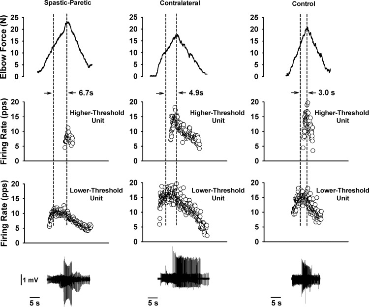
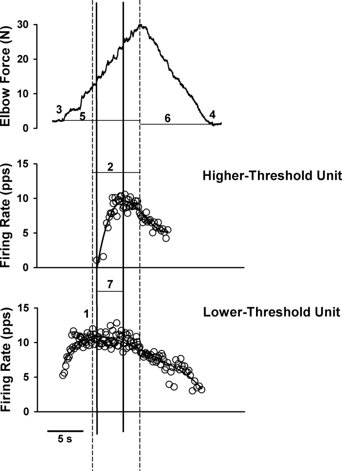
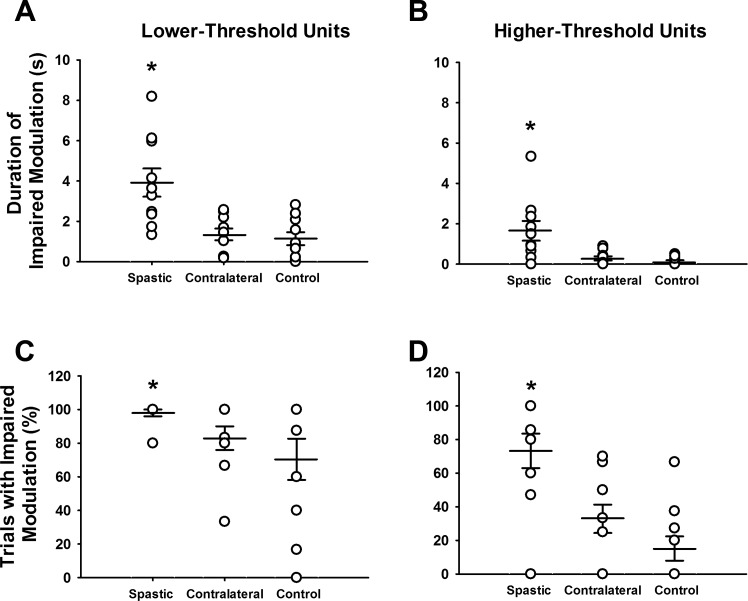
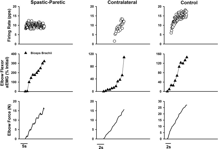
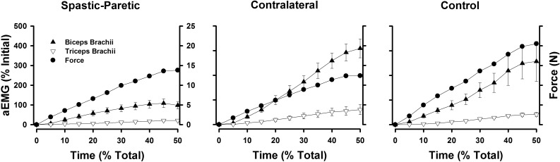
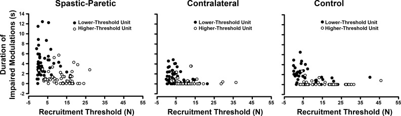
Stroke survivors often exhibit abnormally low motor unit firing rates during voluntary muscle activation. Our purpose was to assess the prevalence of saturation in motor unit firing rates in the spastic-paretic biceps brachii muscle of stroke survivors. To achieve this objective, we recorded the incidence and duration of impaired lower- and higher-threshold motor unit firing rate modulation in spastic-paretic, contralateral, and healthy control muscle during increases in isometric force generated by the elbow flexor muscles. Impaired firing was considered to have occurred when firing rate became constant (i.e., saturated), despite increasing force. The duration of impaired firing rate modulation in the lower-threshold unit was longer for spastic-paretic (3.9 ± 2.2 s) than for contralateral (1.4 ± 0.9 s; P < 0.001) and control (1.1 ± 1.0 s; P = 0.005) muscles. The duration of impaired firing rate modulation in the higher-threshold unit was also longer for the spastic-paretic (1.7 ± 1.6 s) than contralateral (0.3 ± 0.3 s; P = 0.007) and control (0.1 ± 0.2 s; P = 0.009) muscles. This impaired firing rate of the lower-threshold unit arose, despite an increase in the overall descending command, as shown by the recruitment of the higher-threshold unit during the time that the lower-threshold unit was saturating, and by the continuous increase in averages of the rectified EMG of the biceps brachii muscle throughout the rising phase of the contraction. These results suggest that impairments in firing rate modulation are prevalent in motor units of spastic-paretic muscle, even when the overall descending command to the muscle is increasing.

Keywords: PIC; motor unit; saturation; spasticity; stroke.


Figures

Fig. 1.
Fig. 1.
The duration of saturation in firing profiles was greater for the spastic-paretic (first column) than the contralateral (second column) or matched limb of a healthy control subject (third column). First column: two biceps brachii motor units recorded during a triangular isometric contraction with the elbow flexor muscles for the spastic-paretic limb of a stroke survivor. Bottom row denotes instantaneous firing frequency of a lower-threshold motor unit. Middle row denotes instantaneous frequency of a higher-threshold motor unit recorded during the contraction. Top row denotes force of elbow flexor muscles during the voluntary ramp contractions. Second and third columns: two biceps brachii motor units recorded from the contralateral limb of the stroke survivor and from the limb of an age-sex matched healthy control subject, respectively, during the same protocol as in the spastic-paretic limb. The duration of saturation in firing rate is shown between the vertical dashed lines for the respective limb types: spastic-paretic: 6.7 s; contralateral: 4.9 s; matched healthy control limb: 3.0 s. Note that the firing profiles of the higher-threshold motor units were increasing during the time that the lower threshold units were saturating, indicating an increase in descending drive to the respective motoneuron pools. Corresponding trains of motor unit action potentials for the lower- and higher-threshold motor units are shown for each respective muscle type. pps, Pulses per second.
Fig. 2.
Fig. 2.
Elbow force (top panel) and instantaneous firing frequency of a higher-threshold motor unit (middle panel) and a lower-threshold motor unit (bottom panel) during the triangular isometric contraction with the elbow flexor muscles. Adaptation in firing rate profiles was examined by the following measures. 1, Peak firing rates of the lower- and higher-threshold units were determined from the average of 3 spikes surrounding the first maximum firing rate attained (2 interspike intervals). Vertical dashed lines indicate the duration of impaired firing rate modulation (2; no increase or a decrease in firing rate despite increasing force). For the automated analyses to determine the duration of impaired firing rate modulation, a fifth-order polynomial was set to the firing profile of the motor units. Next, the lower-threshold peak firing rate and peak force were chosen (first and second dashed vertical lines, respectively). For the visual inspection, each profile was examined manually following the above automated analyses to ensure that the saturation in firing was adequately captured (duration between first and second dashed vertical lines). The incidence of impaired modulation in firing rate was determined from slope analyses of firing rates from the time at peak firing rate (first dashed vertical line) until the time at peak force (second dashed vertical line). Firing rate slopes that were ≤0.5 pps/s, despite increasing force, were considered as impaired rate modulation. Initial (3) and final (4) forces were calculated when the force left and returned to baseline after the rising and falling ramp contractions. The time to peak force (5) and time to final force (6) for each trial were determined for each subject. Because the force profile during the voluntary ramp contractions may not adequately represent the descending drive equally in the different muscle types, and because firing rates poststroke may be variable, we performed a secondary and more conservative analysis to determine the duration of impaired modulation in firing rates. Specifically, the duration of impaired modulation in firing rate of the lower-threshold unit was determined as the duration between the peak of the smoothed firing rate profile of the lower-threshold unit (first solid vertical line), and the peak of the smoothed firing rate of the higher-threshold unit (second solid vertical line). Slopes of the smoothed firing rate profiles of the lower-threshold unit between the two solid vertical lines (7) that were ≤0.5 pps/s were considered as impaired rate modulation. Note that the duration of impaired modulation in firing rate was ∼6 s when using the primary analysis method (duration between dashed vertical lines; 2), and ∼4 s when using the more conservative analysis method (duration between solid vertical lines; 7).
Fig. 3.
Fig. 3.
Duration of impaired modulation (A and B) and percentage of trials with impaired modulation (C and D) for the spastic-paretic, contralateral and control limb during the triangular isometric ramp contractions. A: the duration of impaired modulation in the lower-threshold unit within a trial was greater for the spastic-paretic (3.9 ± 2.2 s) than both the contralateral (1.4 ± 0.9 s; P < 0.001) and control (1.1 ± 1.0 s; P = 0.005) muscle. B: the duration of impaired modulation in the higher-threshold unit within a trial was also greater for the spastic-paretic (1.7 ± 1.6 s) than both the contralateral (0.3 ± 0.3 s; P = 0.007) and control (0.1 ± 0.2 s; P = 0.009) muscle. Each circle represents the average value from repeated trials of voluntary ramp contractions for each spastic-paretic and contralateral muscle of the 10 subjects, or the muscle of a healthy control subject; wider horizontal bars denote the average value for the group data; smaller horizontal bars denote 1 standard error of the mean (SEM) above and below mean values. C: the percentage of trials exhibiting impaired rate modulation (no increase or a decrease in firing rate, despite increasing force) in the lower-threshold unit within a trial during the ascending phase of the isometric ramp contractions was greater in the spastic-paretic (98.0 ± 6.3%) than both the contralateral (83.0 ± 22.2%; P = 0.004) and control (70.4 ± 38.8%; P = 0.002) muscle. D: the percentage of trials exhibiting impaired rate modulation in the higher-threshold unit within a trial during the ascending phase of the isometric ramp contractions was also greater in the spastic-paretic (73.3 ± 32.4%) than both the contralateral (32.8 ± 26.7%; P = 0.004) and control (15.1 ± 22.9%; P = 0.003) muscle. *P ≤ 0.009 compared with the contralateral and control limb.
Fig. 4.
Fig. 4.
Instantaneous frequency firing profiles of the lower-threshold motor unit (top row), biceps brachii (short head and long head combined; middle row), and force of the elbow flexor muscles (bottom row) during the ascending portion of the voluntary ramp contraction for the spastic-paretic, contralateral, and control muscle. Note that the force and surface averages of the rectified EMG (aEMG) of the biceps brachii are rising, despite flat firing rate profiles for the spastic-paretic muscle. Firing rate profiles are rising as expected for the contralateral and control muscle. Note that, in this spastic-paretic example, the duration of impaired modulation in firing is longer (∼13 s) than the average duration of impaired modulation (∼4 s) observed in spastic-paretic muscle. This is shown to provide an example of some of the longer durations of impaired modulation observed in this data set.
Fig. 5.
Fig. 5.
The aEMG of the biceps brachii (▲; biceps brachii short head and long head combined) and of the triceps brachii (▽) for the group data, expressed as a percentage of the initial aEMG values for the spastic-paretic, contralateral, and control muscle during the rising portion of the voluntary isometric ramp contraction. ●, The force of the elbow flexor muscles during the rising portion of the voluntary ramp contraction for the respective muscle types. For all muscle types, the force and aEMG of the elbow flexor muscles are rising during the ascending portion of the ramp contraction, whereas the aEMG of the antagonist triceps brachii muscle is fairly flat during the ascending portion of the ramp contraction. Values are means ± SE.
Fig. 6.
Fig. 6.
The duration of impaired modulation vs. the recruitment threshold for 51 motor unit pairs in the spastic-paretic muscle, 47 motor unit pairs in the contralateral muscle, and 44 motor unit pairs in the control muscle. ●, Lower-threshold motor units; ○, higher-threshold motor units. Note the longer duration of impaired modulation for both lower- and higher-threshold motor units in spastic-paretic muscle than contralateral or control muscle.

Similar articles

Cited by

References

    1. Avery RB, Johnston D. Multiple channel types contribute to the low-voltage-activated calcium current in hippocampal CA3 pyramidal neurons. J Neurosci 16: 5567–5582, 1996 - PMC - PubMed
    1. Bailey EF, Rice AD, Fuglevand AJ. Firing patterns of human genioglossus motor units during voluntary tongue movement. J Neurophysiol 97: 933–936, 2007 - PubMed
    1. Bennett DJ, Hultborn H, Fedirchuk B, Gorassini M. Short-term plasticity in hindlimb motoneurons of decerebrate cats. J Neurophysiol 80: 2038–2045, 1998a - PubMed
    1. Bennett DJ, Hultborn H, Fedirchuk B, Gorassini M. Synaptic activation of plateaus in hindlimb motoneurons of decerebrate cats. J Neurophysiol 80: 2023–2037, 1998b - PubMed
    1. Bennett DJ, Li Y, Harvey PJ, Gorassini M. Evidence for plateau potentials in tail motoneurons of awake chronic spinal rats with spasticity. J Neurophysiol 86: 1972–1982, 2001a - PubMed

Publication types

LinkOut - more resources