

Optical control of retrogradely infected neurons using drug-regulated "TLoop" lentiviral vectors
- PMID: 24572099
- PMCID: PMC4044340
- DOI: 10.1152/jn.00495.2013
Optical control of retrogradely infected neurons using drug-regulated "TLoop" lentiviral vectors
Abstract
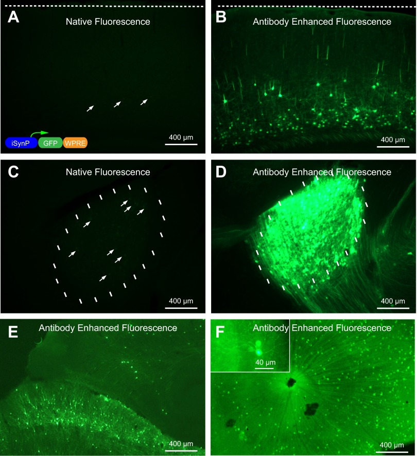
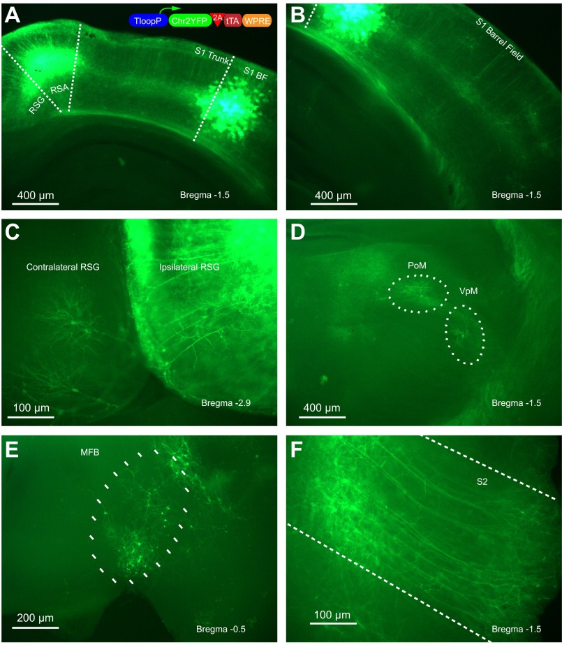
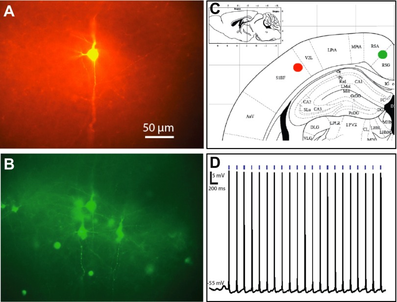
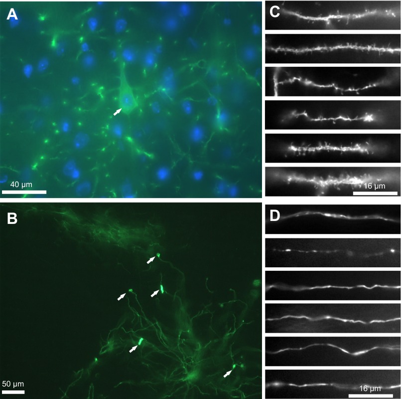
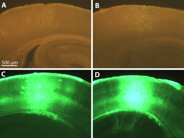
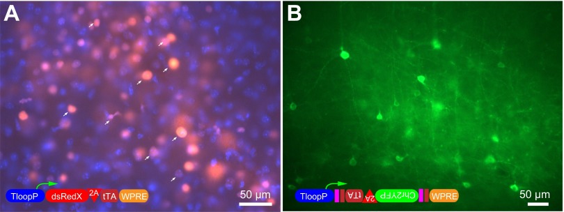
Many approaches that use viral vectors to deliver transgenes have limited transduction efficiency yet require high levels of transgene expression. In particular, infection via axon terminals is relatively inefficient but is a powerful means of achieving infection of specific neuron types. Combining this with optogenetic approaches requires high gene expression levels that are not typically achieved with nontoxic retrogradely infecting vectors. We generated rabies glycoprotein-pseudotyped lentiviral vectors that use a positive feedback loop composed of a Tet promoter driving both its own tetracycline-dependent transcription activator (tTA) ("TLoop") and channelrhodopsin-2-YFP (ChR2YFP). We show that TLoop vectors strongly express proteins in a drug-controllable manner in neurons that project to injection sites within the mouse brain. After initial infection, the virus travels retrogradely, stably integrates into the host genome, and expresses gene products. The expression is robust and allows optogenetic studies of neurons projecting to the location of virus injection, as demonstrated by fluorescence-targeted intracellular recordings. ChR2YFP expression did not cause observable signs of toxicity and continued for up to 6 mo after infection. Expression can be reversibly blocked by administration of doxycycline, if necessary, for expression of gene products that might be more toxic. Overall, we present a system that will allow researchers to achieve high levels of gene expression even in the face of inefficient viral transduction. The particular vectors that we demonstrate may enhance efforts to gain a precise understanding of the contributions of specific types of projection neurons to brain function.
Keywords: channelrhodopsin(H134R); equine infectious anemia virus; rabies glycoprotein; tTA.
Copyright © 2014 the American Physiological Society.
Figures
Similar articles
-
Strategies for targeting primate neural circuits with viral vectors.J Neurophysiol. 2016 Jul 1;116(1):122-34. doi: 10.1152/jn.00087.2016. Epub 2016 Apr 6. J Neurophysiol. 2016. PMID: 27052579 Free PMC article. Review.
-
Lentivirus-mediated transgene delivery to the hippocampus reveals sub-field specific differences in expression.BMC Neurosci. 2009 Jan 13;10:2. doi: 10.1186/1471-2202-10-2. BMC Neurosci. 2009. PMID: 19144149 Free PMC article.
-
Lentiviral vectors: regulated gene expression.Mol Ther. 2000 Jun;1(6):516-21. doi: 10.1006/mthe.2000.0083. Mol Ther. 2000. PMID: 10933976
-
Altering Entry Site Preference of Lentiviral Vectors into Neuronal Cells by Pseudotyping with Envelope Glycoproteins.Methods Mol Biol. 2016;1382:175-86. doi: 10.1007/978-1-4939-3271-9_12. Methods Mol Biol. 2016. PMID: 26611586
-
Pseudotyped lentiviral vectors for tract-targeting and application for the functional control of selective neural circuits.J Neurosci Methods. 2020 Oct 1;344:108854. doi: 10.1016/j.jneumeth.2020.108854. Epub 2020 Jul 11. J Neurosci Methods. 2020. PMID: 32663549 Review.
Cited by
-
Development of an Acute Method to Deliver Transgenes Into the Brains of Adult Xenopus laevis.Front Neural Circuits. 2018 Oct 26;12:92. doi: 10.3389/fncir.2018.00092. eCollection 2018. Front Neural Circuits. 2018. PMID: 30416430 Free PMC article.
-
Strategies for targeting primate neural circuits with viral vectors.J Neurophysiol. 2016 Jul 1;116(1):122-34. doi: 10.1152/jn.00087.2016. Epub 2016 Apr 6. J Neurophysiol. 2016. PMID: 27052579 Free PMC article. Review.
-
Optogenetic Tractography for anatomo-functional characterization of cortico-subcortical neural circuits in non-human primates.Sci Rep. 2018 Feb 20;8(1):3362. doi: 10.1038/s41598-018-21486-8. Sci Rep. 2018. PMID: 29463867 Free PMC article.
-
Not a one-trick pony: Diverse connectivity and functions of the rodent lateral geniculate complex.Vis Neurosci. 2017 Jan;34:E012. doi: 10.1017/S0952523817000098. Vis Neurosci. 2017. PMID: 28965517 Free PMC article. Review.
-
Flexible scaling and persistence of social vocal communication.Nature. 2021 May;593(7857):108-113. doi: 10.1038/s41586-021-03403-8. Epub 2021 Mar 31. Nature. 2021. PMID: 33790464 Free PMC article.
References
-
- Aronoff R, Matyas F, Mateo C, Ciron C, Schneider B, Petersen CC. Long-range connectivity of mouse primary somatosensory barrel cortex. Eur J Neurosci 31: 2221–2233, 2010 - PubMed
-
- Azzouz M, Ralph GS, Storkebaum E, Walmsley LE, Mitrophanous KA, Kingsman SM, Carmeliet P, Mazarakis ND. VEGF delivery with retrogradely transported lentivector prolongs survival in a mouse ALS model. Nature 429: 413–417, 2004 - PubMed
Publication types
MeSH terms
Substances
Grants and funding
LinkOut - more resources
Full Text Sources
Other Literature Sources
Research Materials