. 2014 May;22(5):940-51.
doi: 10.1038/mt.2014.31. Epub 2014 Feb 27.

Aptamer against mannose-capped lipoarabinomannan inhibits virulent Mycobacterium tuberculosis infection in mice and rhesus monkeys

Affiliations

Aptamer against mannose-capped lipoarabinomannan inhibits virulent Mycobacterium tuberculosis infection in mice and rhesus monkeys

Qin Pan et al. Mol Ther. 2014 May.

Abstract

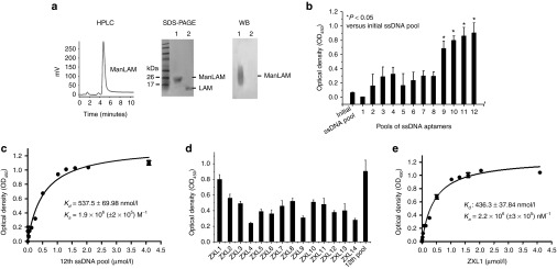
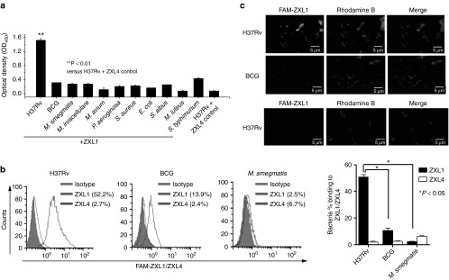
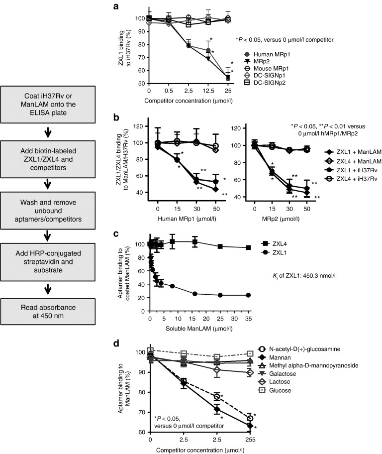
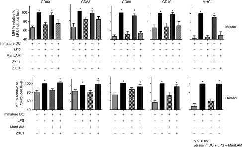
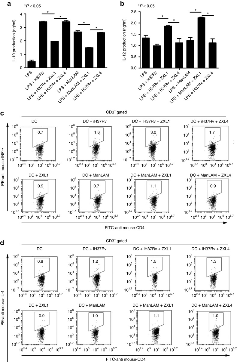
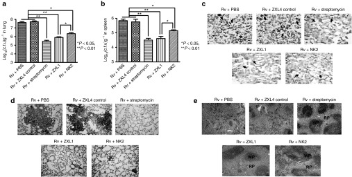
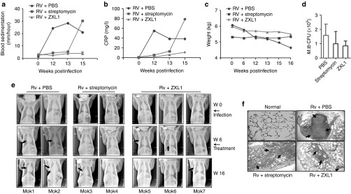
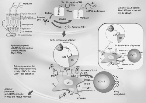
The major surface lipoglycan of Mycobacterium tuberculosis (M. tb), mannose-capped lipoarabinomannan (ManLAM), is an immunosuppressive epitope of M. tb. We used systematic evolution of ligands by exponential enrichment (SELEX) to generate an aptamer (ZXL1) that specifically bound to ManLAM from the virulent M. tb strain H37Rv. Aptamer ZXL1 had the highest binding affinity, with an equilibrium dissociation constant (Kd) of 436.3 ± 37.84 nmol/l, and competed with the mannose receptor for binding to ManLAM and M. tb H37Rv. ZXL1 significantly inhibited the ManLAM-induced immunosuppression of CD11c(+) dendritic cells (DCs) and enhanced the M. tb antigen-presenting activity of DCs for naive CD4(+) Th1 cell activation. More importantly, we demonstrated that injection of aptamer ZXL1 significantly reduced the progression of M. tb H37Rv infections and bacterial loads in lungs of mice and rhesus monkeys. These results suggest that the aptamer ZXL1 is a new potential antimycobacterial agent and tuberculosis vaccine immune adjuvant.


Figures

Figure 1
Figure 1
High-affinity aptamers for mannose-capped lipoarabinomannan (ManLAM) were generated by systematic evolution of ligands by exponential enrichment (SELEX). (a) Identification of the purified ManLAM from Mycobacterium tuberculosis (M. tb) H37Rv by high-performance liquid chromatography (HPLC) showing the final preparation of ManLAM, sodium dodecyl sulfate–polyacrylamide gel electrophoresis (silver staining), and western blot (WB) analyses. (b) After 12 rounds of screening against ManLAM, single-stranded DNA (ssDNA) pools from each round were analyzed for their binding to ManLAM by enzyme-linked oligonucleotide assay. Aptamer pools (2 µmol/l) were incubated in wells coated with ManLAM (40 µg/ml in phosphate-buffered saline). In the background control, 40 µg/ml of ManLAM was coated on the wells, but none of the ssDNA aptamers were added. For each sample, the optical density at 450 nm (OD450) of the background control was subtracted from the OD450 of experimental sample.*P < 0.05 versus initial ssDNA pool. All data are shown as the means ± SEMs (n = 3). (c) Binding of the 12th ssDNA pool to ManLAM. The 12th-round pool of ssDNA was incubated in wells coated with ManLAM. All data are shown as the means ± SEMs (n = 3). The Kd of 537.5 ± 69.98 nmol/l was determined as described in the Methods section. (d) Binding of the single aptamers ZXL1–ZXL14 to ManLAM. The single aptamers ZXL1–ZXL14 (2 µmol/l) were, respectively, incubated in wells coated with ManLAM. All data are shown as the means ± SEMs (n = 3). (e) Analysis of binding of ZXL1 to ManLAM. ZXL1 was added and incubated in the wells coated with ManLAM. All data are shown as the means ± SEMs (n = 3). The Kd of 436.3 ± 37.84 nmol/l was established as described in the Methods section.
Figure 2
Figure 2
The selected aptamer ZXL1 bound specifically to Mycobacterium tuberculosis (M. tb) H37Rv. (a) Binding of ZXL1 to bacteria was evaluated by enzyme-linked oligonucleotide assay. Biotin-labeled ZXL1 was incubated (2 µmol/l) in wells coated with 1 × 105 colony forming units (CFUs) of the bacteria. **P < 0.01 versus M. tb. H37Rv+ZXL4 control. All data are shown as the means ± SEMs (n = 3). (b) Binding of ZXL1 to M. tb H37Rv, Bacillus Calmette–Guérin (BCG), and M. smegmatis was evaluated by flow cytometry (FCM) analysis. Then, 1 × 107 CFUs of the bacteria were fixed with 70% ethanol and incubated with 6-carboxyfluorescein (FAM)-labeled ZXL1 (5 µmol/l) for FCM analysis. *P < 0.05 versus BCG and M. smegmatis. All data are shown as the means ± SEMs (n = 3). (c) Binding of ZXL1 to M. tb H37Rv and BCG was evaluated by confocal microscopy. Thus, 1 × 105 CFUs of the bacteria were coated onto slides and incubated with FAM-labeled ZXL1 (5 µmol/l) for confocal microscopy analysis.
Figure 3
Figure 3
ZXL1 competed with mannose receptor (MR) for binding to mannose-capped lipoarabinomannan (ManLAM) or Mycobacterium tuberculosis (M. tb) H37Rv. ManLAM (10 µg/ml in phosphate-buffered saline) or heat-inactivated M. tb H37Rv (1 × 106 colony forming units (CFUs)/ml) was coated on an enzyme-linked immunosorbent assay plate, and 2 µmol/l of biotin-labeled ZXL1 and competitors were added and incubated. In the background control, the ManLAM (or heat-inactivated M. tb H37Rv) was coated on the wells, but none of the single-stranded DNA aptamers were added. For each sample, the optical density at 450 nm (OD450) of the background control was subtracted from the OD450 of the experimental sample. (a) Human MRp1 and MRp2 inhibited binding of ZXL1 to M. tb H37Rv. Percentage binding: OD450 value of 0.5, 2.5, 12.5, or 25 µmol/l competitor group/OD450 value of 0 µmol/l competitor group. *P < 0.05 versus 0 µmol/l competitor group. All data are shown as the means ± SEMs (n = 3). (b) Human MRp1 and MRp2 inhibited binding of ZXL1 to ManLAM/M. tb H37Rv. Percentage binding: OD450 value of 15, 30, or 50 µmol/l human MRp1 (or MRp2) group/OD450 value of 0 µmol/l human MRp1 (or MRp2) group; *P < 0.05, **P < 0.01 versus 0 µmol/l human MRp1 (or MRp2) group. All data are shown as the means ± SEMs (n = 3). (c) Soluble ManLAM inhibited binding of ZXL1 to ManLAM coated on the well. Percentage binding: OD450 value of ManLAM competitor group/OD450 value of 0 µmol/l ManLAM competitor group. All data are shown as the means ± SEMs (n = 3). (d) Mannan and N-acetyl-D(+)-glucosamine inhibited binding of ZXL1 to ManLAM. Percentage binding: OD450 value of 2.5, 25, or 250 µmol/l carbohydrate competitor group/OD450 value of 0 µmol/l carbohydrate competitor group. *P < 0.05 versus 0 µmol/l carbohydrate competitor group. All data are shown as the means ± SEMs (n = 3).
Figure 4
Figure 4
ZXL1 enhanced the maturation of mouse and human dendritic cells (DCs). Immature DCs were stimulated with LPS (1 µg/ml) plus Mycobacterium tuberculosis (M. tb) H37Rv (1 × 107 colony forming units (CFUs) per 106 DCs)/mannose-capped lipoarabinomannan (ManLAM) (10 µg/ml) in the presence or absence of aptamer ZXL1 (5 µmol/l) for 24 hours. The expression of CD80, CD83, CD86, CD40, and major histocompatibility complex II (MHC-II) on DCs was determined by flow cytometry. Mean fluorescence intensities were normalized to the values obtained for stimulation with LPS alone and were expressed as percentages. *P < 0.05 versus immature DC + LPS + ManLAM. All data are shown as the means ± SEMs (n = 3).
Figure 5
Figure 5
ZXL1 enhanced antigen presentation by dendritic cells (DCs). (a,b) Immature mouse DCs (2 × 106 cells/ml) were cultured for 24 hours with LPS (1 µg/ml) or LPS plus iH37Rv (1 × 106 colony forming units (CFUs)/ml)/mannose-capped lipoarabinomannan (ManLAM) (10 µg/ml) in the presence or absence of ZXL1 (5 µmol/l). Supernatants were collected and analyzed for interleukin (IL)-10 and IL-12 production by enzyme-linked immunosorbent assay. (a) IL-10 secretion by Mycobacterium tuberculosis (M. tb) H37Rv/ManLAM-stimulated DCs. *P < 0.05 versus LPS+iH37Rv/ManLAM or LPS+iH37Rv/ManLAM+ZXL4. All data are shown as the means ± SEMs (n = 3). (b) IL-12 secretion by M. tb H37Rv/ManLAM-stimulated DCs. *P < 0.05 versus LPS+iH37Rv/ManLAM or LPS+iH37Rv/ManLAM+ZXL4. All data are shown as the means ± SEMs (n = 3). (c,d) Naive CD4+ T cells were purified from mouse splenocytes using antibody-coupled microbeads and a MACS separator. ZXL1-treated-M. tb H37Rv/ManLAM-pulsed DCs (0.5 × 106) were cocultured with 1 × 106 naive CD4+ T cells for 5 days. IFN-γ and IL-4 production by CD4+ T cells was determined by flow cytometry. (c) IFN-γ production by CD4+ T cells stimulated by DCs pulsed with ZXL1-treated-iH37Rv/ManLAM. (d) IL-4 production by CD4+ T cells stimulated by the DCs pulsed with ZXL1-treated-iH37Rv/ManLAM. IFN, interferon; PE, R-phycoerythrin.
Figure 6
Figure 6
ZXL1 prevented Mycobacterium tuberculosis (M. tb) H37Rv infection in mice. BALB/c mice were infected with M. tb H37Rv by an inhalation exposure system to deposit 100 CFUs of bacteria in the lungs. From day 3, ZXL1/NK2 were administered thrice i.v. into the mice once every 3 days (1 µmol/l × 3 per mouse) (n = 6 per group). Control treatments were i.v. injections with ZXL4 thrice (1 µmol/l × 3 per mouse) or i.m. injections streptomycin daily for a week (200 µg × 7 per mouse). (a) M. tb CFU assay in lung. After 30 days of M. tb infection, mice were sacrificed; the numbers of M. tb CFUs in lungs were counted. (b) M. tb CFU assay in spleen. (c) Ziehl-Neelsen acid-fast stain analysis. Lung tissue sections were analyzed with Ziehl-Neelsen acid-fast staining. Arrows indicate the positive bacteria (1,000×). (d) Histopathology. Lung tissues were stained with hematoxylin and eosin (H&E) and evaluated by light microscopy (100×). (e) Histopathology. Spleens were stained with H&E and evaluated by light microscopy (100×). PBS, phosphate-buffered saline.
Figure 7
Figure 7
ZXL1 prevented Mycobacterium tuberculosis (M. tb) H37Rv infection in rhesus monkeys. A total of seven rhesus monkeys were intratracheally infected with 100 colony-forming units (CFUs) of M. tb H37Rv at week 0. At week 6, infected monkeys were randomly divided into three groups as follows: two monkeys in the phosphate-buffered saline (PBS) group, two monkeys in the streptomycin group, and three monkeys in the ZXL1 group. For the ZXL1 group, monkeys were i.v. injected with ZXL1 (20 µmol/l or 100 µl/kg per monkey) three times per week for 4 weeks. For the streptomycin group, monkeys were i.m. injected with streptomycin (20 mg/kg per day) for 30 days. (a) Blood sedimentation determination. (b) C-reactive protein (CRP) determination. (c) Body weights of infected monkeys. (d) M. tb CFU assay. (e) Chest X-ray detection. The arrows indicate the pulmonary lesions. (f) Histopathology. Lung tissue sections were stained with hematoxylin and eosin and evaluated by light microscopy (100×). PBS, phosphate-buffered saline.
Figure 8
Figure 8
Putative model of aptamer ZXL1-mediated blocking of H37Rv mannose-capped lipoarabinomannan (ManLAM)-induced immunosuppression. DC, dendritic cell; IFN, interferon; IL, interleukin; MHC, major histocompatibility complex; MR, mannose receptor; M. tb, Mycobacterium tuberculosis; SELEX, systematic evolution of ligands by exponential enrichment; ssDNA, single-stranded DNA.

Similar articles

Cited by

References

    1. World Health Organization. 2011The burden of disease caused by TB Global tuberculosis control 2011. ( http://whqlibdoc.who.int/publications/2011/9789241564380_eng.pdf ).
    1. Svenson S, Källenius G, Pawlowski A, Hamasur B. Towards new tuberculosis vaccines. Hum Vaccin. 2010;6:309–317. - PubMed
    1. Colijn C, Cohen T, Ganesh A, Murray M. Spontaneous emergence of multiple drug resistance in tuberculosis before and during therapy. PLoS ONE. 2011;6:e18327. - PMC - PubMed
    1. Gengenbacher M, Kaufmann SH. Mycobacterium tuberculosis: success through dormancy. FEMS Microbiol Rev. 2012;36:514–532. - PMC - PubMed
    1. Mortellaro A, Robinson L, Ricciardi-Castagnoli P. Spotlight on Mycobacteria and dendritic cells: will novel targets to fight tuberculosis emerge. EMBO Mol Med. 2009;1:19–29. - PMC - PubMed

Publication types

MeSH terms

LinkOut - more resources