. 2014 Feb 27:5:3347.
doi: 10.1038/ncomms4347.

ATM specifically mediates repair of double-strand breaks with blocked DNA ends

Affiliations
Free PMC article

ATM specifically mediates repair of double-strand breaks with blocked DNA ends

Alejandro Álvarez-Quilón et al. Nat Commun. .
Free PMC article

Abstract

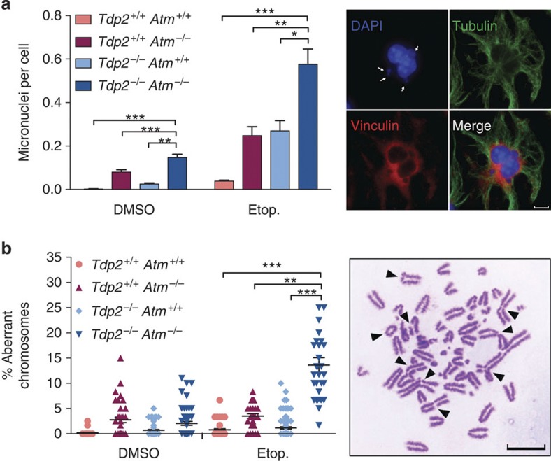
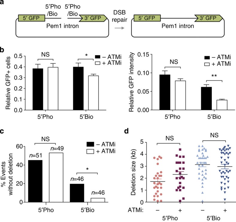
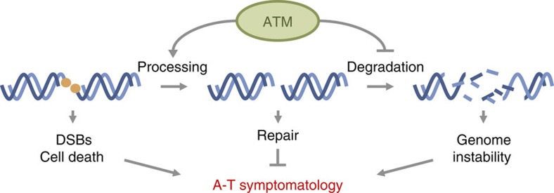
Ataxia telangiectasia is caused by mutations in ATM and represents a paradigm for cancer predisposition and neurodegenerative syndromes linked to deficiencies in the DNA-damage response. The role of ATM as a key regulator of signalling following DNA double-strand breaks (DSBs) has been dissected in extraordinary detail, but the impact of this process on DSB repair still remains controversial. Here we develop novel genetic and molecular tools to modify the structure of DSB ends and demonstrate that ATM is indeed required for efficient and accurate DSB repair, preventing cell death and genome instability, but exclusively when the ends are irreversibly blocked. We therefore identify the nature of ATM involvement in DSB repair, presenting blocked DNA ends as a possible pathogenic trigger of ataxia telangiectasia and related disorders.


Figures

Figure 1
Figure 1. Protein-blocked DSBs accumulate in Tdp2-deficient background upon TOP2 poison treatment.
(a) Scheme of the strategy used to generate breaks with specific structure. (b) SLOPE assay performed in Tdp2+/+ and Tdp2−/− confluency-arrested primary MEFs. Average±s.e.m. of signal from three independent experiments and statistical significance by two-way ANOVA test with Bonferroni post-test is shown (*P≤0.05; **P≤0.01; ***P≤0.001).
Figure 2
Figure 2. ATM facilitates repair of TOP2-blocked DSBs.
(a) Time course of γH2AX foci disappearance after 30 min 10 μM etoposide treatment and repair at different times following drug removal in Tdp2+/+ Atm+/+, Tdp2+/+ Atm−/−, Tdp2−/− Atm+/+ and Tdp2−/− Atm−/− confluency-arrested primary MEFs. Representative images of γH2AX foci (green) and DAPI counterstain (blue) for the 24 h repair time point are shown. Scale bar, 10 μm. (b) As above, in Tdp2+/+ and Tdp2−/− confluency-arrested primary MEFs with or without 10 μM ATM inhibitor (KU55933). (c) As above, in confluency-arrested primary fibroblast derived from A-T patients (AT) and wild-type controls (BJ1) depleted (shTDP2) or not for TDP2. In this case 20 μM etoposide treatment was applied. In all cases, average±s.e.m. of the percentage of foci remaining from at least three independent experiments and the minimal statistical significance between cells deficient in both TDP2 and ATM and all other cells by two-way ANOVA test with Bonferroni post-test is shown (*P≤0.05; **P≤0.01; ***P≤0.001).
Figure 3
Figure 3. Unrepaired TOP2-blocked DSBs are not associated with heterochromatin.
Confluency-arrested Atm−/− Tdp2−/− primary MEFs were treated as described in Fig. 1. (a) Percentage of γH2AX foci that are located within heterochromatin (Het) or euchromatin (Eu) following 0 and 24 h repair after etoposide treatment. A cutoff distance of 0.4 μM from the centroid of each focus to the nearest euchromatin/heterochromatin boundary was applied. Statistical significance by Chi Square test is indicated. (b) Distribution of distances between γH2AX foci and heterochromatin as determined above. Bins of 0.4 μm centred on the indicated distance were used for representation. (c) Example of the automatic calculation of distances between γH2AX foci (green) and densely DAPI-stained chromocenters (blue) used for the data shown above. Both a z-stack projection (left) and a 3D reconstitution (right) are shown.
Figure 4
Figure 4. ATM promotes survival upon induction of blocked DSBs.
(a) Clonogenic survival of the indicated transformed MEFs following acute treatment with the indicated concentrations of etoposide. Average±s.e.m. of at least three independent experiments and statistical significance at the highest indicated dose by one-way ANOVA with Bonferroni post-test are shown (*P≤0.05; **P≤0.01; ***P≤0.001). (b) As above, with or without incubation with 10 μM ATM inhibitor (KU55933).
Figure 5
Figure 5. ATM promotes the maintenance of genome integrity upon induction of blocked DSBs.
(a) Micronuclei were scored in the indicated binucleated transformed MEFs following acute treatment with 2.5 μM etoposide. Histogram bars (left) represent the average±s.e.m. of n≥600 cells from three independent experiments. Statistical significance by one-way ANOVA with Bonferroni post-test is indicated (*P≤0.05; **P≤0.01; ***P≤0.001). Representative image (right) of a treated Tpd2−/−Atm−/− cell containing multiple micronuclei is shown. Scale bar, 7.5 μm. (b) Chromosomal aberrations (both break and exchange type) were scored following acute treatment with 2.5 μM etoposide. Plot (left) shows the number of aberrations per 100 chromosomes from individual metaphase spreads obtained in at least 6 independent experiments. Average±s.e.m. and statistical significance by Kruskal-Wallis test with Dunn’s post-test is indicated (*P≤0.05; **P≤0.01; ***P≤0.001). Representative image (right) of a Tpd2−/− Atm−/− metaphase containing multiple chromosomal aberrations is shown. Scale bar, 10 μm.
Figure 6
Figure 6. ATM facilitates repair of biotin-blocked DSBs.
(a) Scheme of the modified pEGFP-Pem1 system harbouring 5′-phosphate or 5′-biotin ends (left) and the expected repair product (right). (b) HEK293T cells were transfected in the presence or absence of 10 μM ATM inhibitor (KU55933) with the substrates described above, and analysed by FACS for GFP-positive cells (left) and average GFP intensity (right). In both cases, data are relative to transfection with a control pEGFP-Pem1 circular plasmid. Average±s.e.m. of four independent experiments and statistical significance by two-way ANOVA with Bonferroni post-test are shown (*P≤0.05; **P≤0.01; ***P≤0.001). (c) Percentage of plasmid-repair events not associated with detectable sequence loss. Statistical significance by Chi Square test is shown (*P≤0.05; **P≤0.01; ***P≤0.001). (d) Deletion size in plasmid repair events. Substrates as described above. Average±s.e.m. and statistical significance by Kruskal-Wallis test with Dunn’s post-test is indicated.
Figure 7
Figure 7. Model for ATM involvement in blocked DSB repair.
ATM promotes processing of blocked DSBs and inhibits excessive degradation. This promotes efficient and accurate repair, preventing the accumulation of unrepaired DSBs, cell death and genome instability, which can trigger the development of A-T symptomatology.

References

    1. McKinnon P. J. & Caldecott K. W. DNA strand break repair and human genetic disease. Ann. Rev. Genomics. Hum. Genet. 8, 37–55 (2007). - PubMed
    1. Lavin M. F. Ataxia-telangiectasia: from a rare disorder to a paradigm for cell signalling and cancer. Nat. Rev. Mol. Cell. Biol. 9, 759–769 (2008). - PubMed
    1. McKinnon P. J. ATM and the molecular pathogenesis of ataxia telangiectasia. Ann. Rev. Pathol. 7, 303–321 (2012). - PubMed
    1. Shiloh Y. & Ziv Y. The ATM protein kinase: regulating the cellular response to genotoxic stress, and more. Nat. Rev. Mol. Cell. Biol. 14, 197–210 (2013). - PubMed
    1. Reynolds J. J. & Stewart G. S. A nervous predisposition to unrepaired DNA double strand breaks. DNA Repair (Amst) 12, 588–599 (2013). - PubMed

Publication types

MeSH terms

Substances