. 2014 Feb 26;34(9):3171-82.
doi: 10.1523/JNEUROSCI.3291-13.2014.

Proteasome modulates positive and negative translational regulators in long-term synaptic plasticity

Affiliations

Proteasome modulates positive and negative translational regulators in long-term synaptic plasticity

Chenghai Dong et al. J Neurosci. .

Abstract

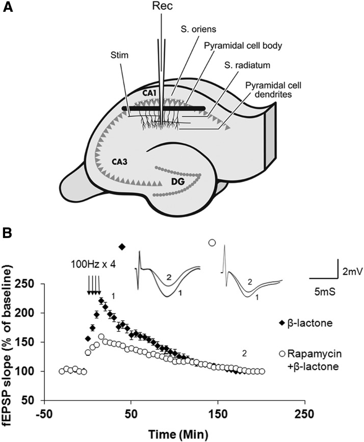
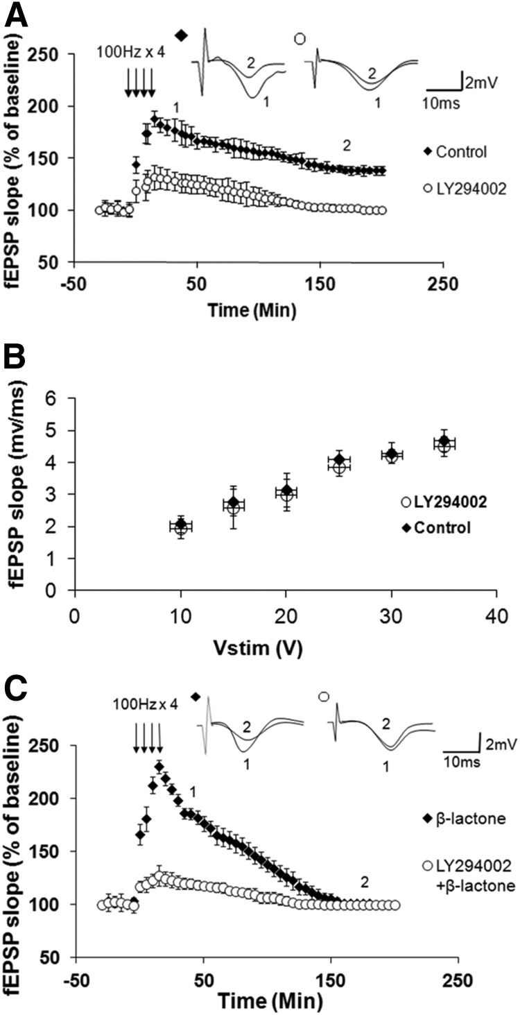
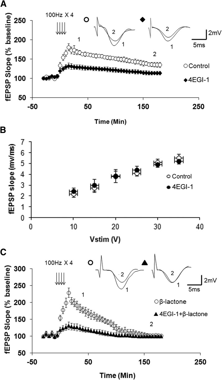
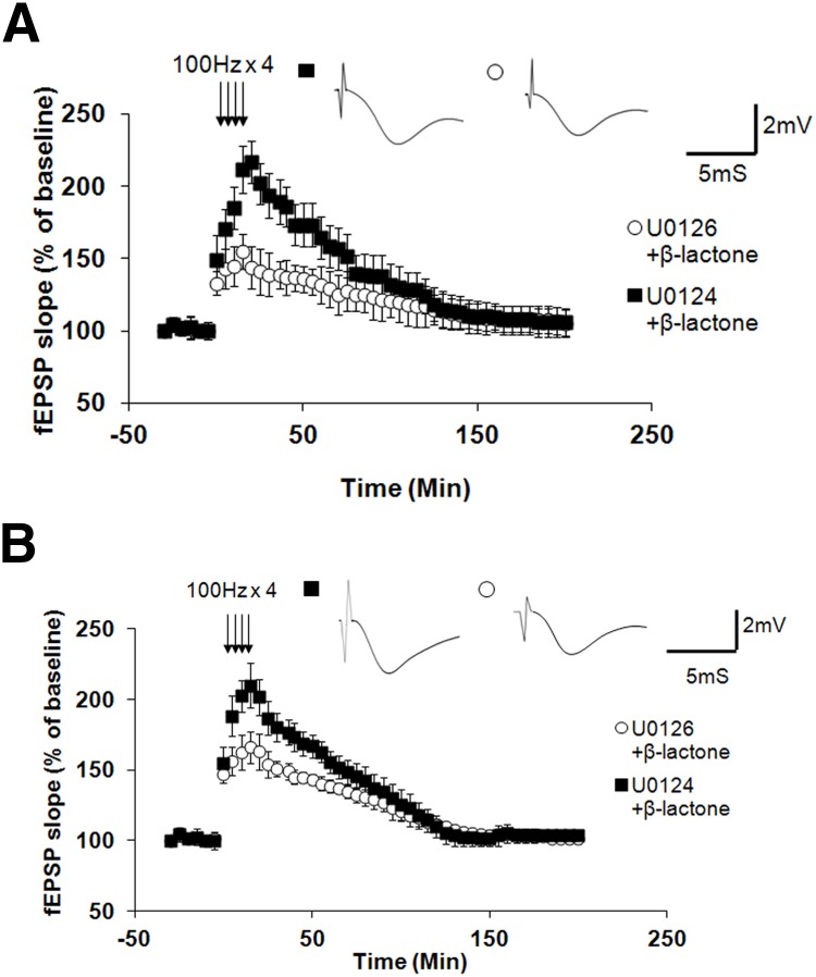
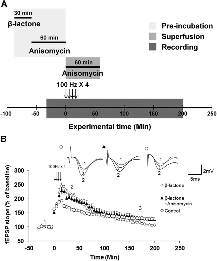
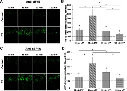
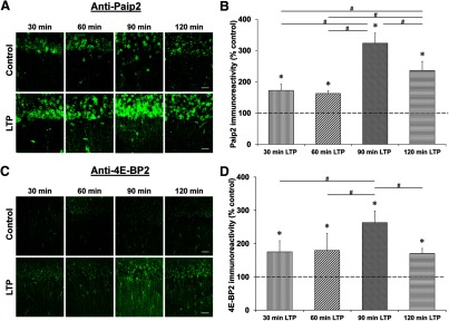
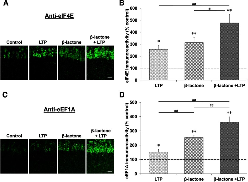
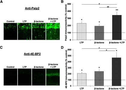
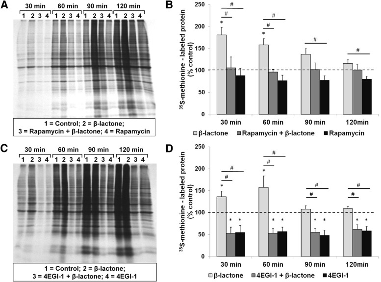
Proteolysis by the ubiquitin-proteasome pathway appears to have a complex role in synaptic plasticity, but its various functions remain to be elucidated. Using late phase long-term potentiation (L-LTP) in the hippocampus of the mouse as a model for long-term synaptic plasticity, we previously showed that inhibition of the proteasome enhances induction but blocks maintenance of L-LTP. In this study, we investigated the possible mechanisms by which proteasome inhibition has opposite effects on L-LTP induction and maintenance. Our results show that inhibiting phosphatidyl inositol-3 kinase or blocking the interaction between eukaryotic initiation factors 4E (eIF4E) and 4G (eIF4G) reduces the enhancement of L-LTP induction brought about by proteasome inhibition suggesting interplay between proteolysis and the signaling pathway mediated by mammalian target of rapamycin (mTOR). Also, proteasome inhibition leads to accumulation of translational activators in the mTOR pathway such as eIF4E and eukaryotic elongation factor 1A (eEF1A) early during L-LTP causing increased induction. Furthermore, inhibition of the proteasome causes a buildup of translational repressors, such as polyadenylate-binding protein interacting protein 2 (Paip2) and eukaryotic initiation factor 4E-binding protein 2 (4E-BP2), during late stages of L-LTP contributing to the blockade of L-LTP maintenance. Thus, the proteasome plays a critical role in regulating protein synthesis during L-LTP by tightly controlling translation. Our results provide novel mechanistic insights into the interplay between protein degradation and protein synthesis in long-term synaptic plasticity.

Keywords: activator; late-phase LTP; protein synthesis; proteolysis; repressor; ubiquitin.


Figures

Figure 1.
Figure 1.
Rapamycin pretreatment significantly reduces β-lactone-mediated enhancement in Ep-L-LTP in isolated dendrites. A, Schematic illustration of surgical isolation of dendrites (modified from Dong et al., 2008). A cut (black bar) is made just below the pyramidal cell body layer in the CA1 region. Recording electrode (Rec) is placed in the dendritic layer of pyramidal cells. Position of the stimulation electrode (Stim), and the layers stratum radiatum and stratum oriens are also indicated. DG, dentate gyrus. B, Treatment of cut slices with rapamycin before β-lactone treatment significantly (p < 0.01) diminishes the magnitude of Ep-L-LTP relative to β-lactone treatment alone. Inset, Representative traces taken at different time points (1 = 30 min; 2 = 180 min) for treatment with β-lactone alone and rapamycin + β-lactone.
Figure 2.
Figure 2.
PI3K inhibition reduces normal L-LTP and β-lactone-mediated enhancement of Ep-L-LTP. A, Pretreatment with PI3K inhibitor LY294002 significantly (p < 0.0001) reduces L-LTP. Inset, Representative traces taken at different time points (1 = 30 min; 2 = 180 min) for no treatment “Control” and treatment with LY294002. B, Normal basal synaptic transmission in LY294002-treated hippocampal slices (n = 7) compared with vehicle-treated control (n = 7) slices. The graph shows input–output curves of fEPSP slope (mV/ms) versus stimulus at the Schaffer collateral pathway upon treatment with the vehicle (DMSO) or LY294002. C, Incubation with LY294002 before β-lactone application significantly (p < 0.0001) decreases the enhancement in Ep-L-LTP. Inset, Representative traces taken at different time points (1 = 30 min; 2 = 180 min) for β-lactone alone and LY294002 + β-lactone.
Figure 3.
Figure 3.
An inhibitor of eIF4E-eIF4G interaction decreases normal L-LTP and β-lactone-mediated enhancement of Ep-L-LTP. A, Pretreatment with 4EGI-1, an inhibitor of eIF4E-eIF4G interaction, significantly (p < 0.0001) reduces L-LTP. Inset, Representative traces taken at different time points (1 = 30 min; 2 = 180 min) for no treatment “Control” and treatment with 4-EGI-1. B, Normal basal synaptic transmission in 4EGI-1-treated hippocampal slices (n = 8) compared with vehicle-treated control (n = 7) slices. The graph shows input–output curves of fEPSP slope (mV/ms) versus stimulus at the Schaffer collateral pathway upon treatment with the vehicle (DMSO) or 4EGI-1. C, Incubation with 4EGI-1 before β-lactone significantly (p < 0.0001) decreases the enhancement in Ep-L-LTP. Inset, Representative traces taken at different time points (1 = 30 min; 2 = 180 min) for β-lactone alone and 4EGI-1 + β-lactone.
Figure 4.
Figure 4.
ERK function is essential for β-lactone-mediated enhancement of Ep-L-LTP in intact slices as well as isolated dendrites. A, In intact slices, pre-incubation with MEK inhibitor U0126 before β-lactone treatment (but not with an inactive analog U0124) significantly (p < 0.001) reduces Ep-L-LTP. Individual traces taken at 30 min are shown in the inset. B, In isolated dendrites, pre-incubation with MEK inhibitor U0126 before β-lactone treatment (but not with an inactive analog U0124) significantly (p < 0.001) reduces Ep-L-LTP. Individual traces taken at 30 min are shown in the inset.
Figure 5.
Figure 5.
Reversal of proteasome inhibition-mediated decrease in L-LTP maintenance in isolated dendrites by subsequent inhibition of protein synthesis. A, Schematic outline of the experiment: hippocampal slices were pre-incubated with β-lactone for 30 min and then they were incubated with anisomycin for 60 min. Anisomycin was also added to the ACSF used for superfusion and maintained for 60 min after the first tetanus. B, Treatment with β-lactone alone (open diamonds) blocks L-LTP maintenance in isolated dendrites relative to L-LTP without any treatment, i.e., control (open circles). Application of anisomycin after β-lactone treatment (filled triangles) reverses blockade of L-LTP maintenance and restores maintenance to normal levels seen in L-LTP without any treatment, i.e., control (β-lactone vs β-lactone + anisomycin: p < 0.001; β-lactone + anisomycin vs Control: p = 0.168). Inset, Representative traces taken at different time points (1 = baseline, 2 = 30 min; 3 = 180 min) for β-lactone, β-lactone + anisomycin, and control.
Figure 6.
Figure 6.
Time course of eIF4E and eEF1A expression after L-LTP induction. Confocal images of eIF4E and eEF1A immunoreactivities in the CA1 region of hippocampal slices 30, 45, 90, and 120 min after the first tetanus and their respective time-matched controls (A and C). Scale bars: 20 μm. Quantification of eIF4E and eEF1A immunoreactivities relative to their time-matched controls shows a peak at 45 min for both proteins (B and D). *p < 0.05 compared with controls (depicted by a dashed line). #p < 0.05 comparison between two given time points as indicated by horizontal lines.
Figure 7.
Figure 7.
Time course of Paip2 and 4E-BP2 expression after L-LTP induction. Confocal images of Paip2 and 4E-BP2 immunoreactivities in the CA1 region of hippocampal slices 30, 60, 90, and 120 min after the first tetanus and their respective time-matched controls (A and C). Scale bars: 20 μm. Quantification of Paip2 and 4E-BP2 immunoreactivities relative to their time-matched controls shows a peak at 90 min for both proteins (B and D). *p < 0.05 compared with controls (depicted by a dashed line). #p < 0.05 comparison between two given time points as indicated by horizontal lines.
Figure 8.
Figure 8.
Proteasome inhibition causes accumulation of eIF4E and eEF1A early during L-LTP. Confocal images showing eIF4E (A) and eEF1A (C) immunoreactivities in the CA1 region of hippocampal slices: untreated (control), subjected to L-LTP induction, treated with β-lactone, and subjected to L-LTP induction after β-lactone treatment. L-LTP slices were fixed 30 min after the initial tetanus and the “Control” and “β-lactone” slices were time-matched to their respective L-LTP counterparts. Scale bars: 20 μm. Quantification of eIF4E (B) and eEF1A (D) immunoreactivities for the four experimental conditions is shown at right. *p < 0.05 and **p < 0.01 compared with controls (depicted by a dashed line). #p < 0.05 and ##p < 0.01 comparison between two given treatments as indicated by horizontal lines.
Figure 9.
Figure 9.
Proteasome inhibition causes accumulation of Paip2 and 4E-BP2 at late stages during L-LTP. Confocal images showing Paip2 (A) and 4E-BP2 (C) immunoreactivities in CA1 region of hippocampal slices: untreated (control), subjected to L-LTP induction, treated with β-lactone, and subjected to L-LTP induction after β-lactone treatment. L-LTP slices were fixed 60 min after the initial tetanus and the “Control” and “β-lactone” slices were time-matched to their respective L-LTP counterparts. Scale bars: 20 μm. Quantification of Paip2 (B) and 4E-BP2 (D) immunoreactivities for the four experimental conditions is shown at right. *p < 0.05 and **p < 0.01 compared with controls (depicted by a dashed line). #p < 0.05 and ##p < 0.01 comparison between two given treatments as indicated by horizontal lines.
Figure 10.
Figure 10.
β-Lactone enhances the quantity of newly translated proteins in hippocampal slices and the enhancement is blocked by rapamycin and 4EGI-1. Autoradiographs (A and C) showing incorporation of 35S-methionine at 30, 60, 90, and 120 min after initiation of metabolic labeling indicating the amount of newly synthesized proteins when the hippocampal slices are treated with β-lactone by itself or after prior treatment with rapamycin (A) or 4EGI-1 (C). Quantification shows that rapamycin (B) and 4EGI-1 (D) block the β-lactone-mediated increase in 35S-methionine-labeled proteins (lanes 2 compared with lanes 3 in A and C and second bars compared with first bars in B and D). Effect of rapamycin alone or 4EGI-1 alone on control slices (lanes 4 in A and C and third bars in B and D) is also shown. *p < 0.05 compared with controls (depicted by a dashed line). #p < 0.05 comparison between two given experimental conditions as indicated by horizontal lines.

Similar articles

Cited by

References

    1. Banko JL, Poulin F, Hou L, DeMaria CT, Sonenberg N, Klann E. The translation repressor 4E-BP2 is critical for eIF4F complex formation, synaptic plasticity, and memory in the hippocampus. J Neurosci. 2005;25:9581–9590. doi: 10.1523/JNEUROSCI.2423-05.2005. - DOI - PMC - PubMed
    1. Bolduc FV, Bell K, Cox H, Broadie KS, Tully T. Excess protein synthesis in Drosophila fragile X mutants impairs long-term memory. Nat Neurosci. 2008;11:1143–1145. doi: 10.1038/nn.2175. - DOI - PMC - PubMed
    1. Byrne JH, Baxter DA, Buonomano DV, Cleary LJ, Eskin A, Goldsmith JR, McClendon E, Nazif FA, Noel F, Scholz KP. Neural and molecular bases of nonassociative and associative learning in Aplysia. Ann N Y Acad Sci. 1991;627:124–149. doi: 10.1111/j.1749-6632.1991.tb25918.x. - DOI - PubMed
    1. Chain DG, Casadio A, Schacher S, Hegde AN, Valbrun M, Yamamoto N, Goldberg AL, Bartsch D, Kandel ER, Schwartz JH. Mechanisms for generating the autonomous cAMP-dependent protein kinase required for long-term facilitation in Aplysia. Neuron. 1999;22:147–156. doi: 10.1016/S0896-6273(00)80686-8. - DOI - PubMed
    1. Cracco JB, Serrano P, Moskowitz SI, Bergold PJ, Sacktor TC. Protein synthesis-dependent LTP in isolated dendrites of CA1 pyramidal cells. Hippocampus. 2005;15:551–556. doi: 10.1002/hipo.20078. - DOI - PubMed

Publication types

MeSH terms